IOS Press
Short Communication
Undetectable Levels of CSF Amyloid-

Peptide in a Patient with 17
-Hydroxysteroid
Dehydrogenase Deficiency
Carlos Ortez
a,b, Cristina Villar
a,b, Carmen Fons
a,b, Sof´ıa T. Duarte
a,c, Ana P´erez
a,b,
Judith Garc´ıa-Villoria
b,d, Antonia Ribes
b,d, Aida Ormaz´abal
b,e, Mercedes Casado
b,e,
Jaume Campistol
a,b, Maria Antonia Vilaseca
b,eand Angels Garc´ıa-Cazorla
a,b,∗ aDepartment of Neurology Hospital Sant Joan de D´eu, Barcelona, SpainbCIBER-ER (Biomedical Network Research Centre on Rare Diseases), Instituto de Salud Carlos III, Madrid, Spain
cNeuropaediatric Department, Hospital D. Estefˆania, CHLC, EPE and CEDOC, Faculdade de Ciˆencias M´edicas da Universidade Nova de Lisboa, Portugal
dSecci´on de Errores Cong´enitos del Metabolismo (IBC), Servicio de Bioqu´ımica y Gen´etica Molecular, Hospital Cl´ınic, Barcelona, Spain
eDepartment of Biochemistry, Hospital Sant Joan de D´eu, Barcelona, Spain
Accepted 24 June 2011
Abstract. 17-hydroxysteroid dehydrogenase 10 (HSD10) deficiency is a rare X-linked inborn error of isoleucine catabolism.
Although this protein has been genetically implicated in Alzheimer’s disease pathogenesis, studies of amyloid- peptide (A) in patients with HSD10 deficiency have not been previously reported. We found, in a severely affected child with HSD10 deficiency, undetectable levels of A in the cerebrospinal fluid, together with low expression of brain-derived neurotrophic factor, ␣-synuclein, and serotonin metabolites. Confirmation of these findings in other patients would help elucidating mechanisms of synaptic dysfunction in this disease, and highlight the role of A in both early and late periods of life.
Keywords: Amyloid- peptide, cerebrospinal fluid, childhood, HSD10 deficiency, inborn errors of metabolism, neurotransmit-ters, synaptic proteins
INTRODUCTION
17-hydroxysteroid dehydrogenase (HSD10) is a multifunctional mitochondrial enzyme with complex roles [1, 2]. HSD10 deficiency is an X-linked dis-ease (MIM300256) caused by mutations in HSD17B10
∗Correspondence to: Angels Garc´ıa-Cazorla, Neurology
Depart-ment, Hospital Sant Joan de Deu, Passeig Sant Joan de Deu, 2, 08950 Esplugues, Barcelona, Spain. Tel.: +34 93 280 4000; Fax: +34 93 203 3959; E-mail: [email protected].
gene [3]. This disease is characterized by a clinical picture very different from other organic acidurias: patients do not develop metabolic crises, but they follow a neurodegenerative course associated with mitochondrial dysfunction [4], progressive loss of skills, epilepsy, optic atrophy, retinopathy, deafness, and movement disorders [2].
HSD10 is a protein implicated in the pathogenesis of Alzheimer’s disease (AD) [5, 6]. Although HSD10 has an affinity for amyloid- peptide (A), no studies
regarding in vivo quantification of A have been reported in patients with HSD10 deficiency. Moreover there are no studies concerning normal cerebrospinal fluid (CSF) A values in the pediatric age. We aimed to analyze the expression of A in the CSF of an affected child, compared to a control pediatric popula-tion. In order to gather more information about synaptic mechanisms in this disease, neurotransmitters, brain-derived neurotrophic factor (BDNF; associated with dendritic growth, and serotoninergic transmission) and ␣-synuclein (AS; associated with neurotransmitter release, dopaminergic modulation and neurodegener-ation) were also included in our study.
PATIENT AND METHODS Clinical report
The patient is the first child of healthy non-consanguineous parents. Pregnancy, peripartum, and first year of life were uneventful (normal psychomo-tor development: he could walk with support and was able to say some words). At 13 months, and within the context of fever and diarrhea, he developed psychomotor regression (inability to sit unsupported, loss of normal use of hands), and disclosed abnor-mal fast erratic ocular movements, non-epileptic myoclonus, and irritability. Plasma ammonia and amino acids were normal but lactate concentration was up to 3 mmol/l (NV: 0.66–1.88 mM/l). Brain MRI was normal. Because of the initial symptoms, opsoclonus-myoclonus syndrome was suspected and dexamethasone (0.8 mg/kg/d IV for 6 days) was started. Although the patient showed slight improve-ment, ocular fundus revealed optic atrophy and a determination of urine organic acids revealed ele-vation of 3-hydroxy-2-methylbutyryic (48 mmol/mol creatinine; CV 5–12) and tiglylglycine (44 mmol/mol creatinine; CV <5). HSD10 activity in fibroblast was 0.5 nmols/mg protx min (C.V. 1.4± SD 0.43) and a new missense mutation was detected in HSD17B10 gen (c.628 C > T; p.P210 S) [7]. The mother was con-firmed to be a carrier. Despite low isoleucine diet, the global outcome was very poor. At 3 years the child was unable to walk due to severe motor dyspraxia and choreoathetosis. Abnormal ocular movements and severe cognitive delay were present. At 5 years of age, the patient died of pneumonia leading to sep-sis, multi-organ failure, and mitochondrial dysfunction (hyperlactacidemia, abundant Kreb’s cycle metabo-lites in urine). Autopsy was not authorized.
CSF samples
CSF samples from our patient and controls were obtained by lumbar puncture as previously described [8]. First ten drops were used for routine cytochem-ical/microbiological studies and the rest immediately stored at−80◦C until further analysis. The study of controls was performed in 30 subjects (age range: 21 days–5 years; average: 0.9 years, 16 boys, 14 girls) whose CSF samples were submitted under suspicion of central nervous system infection. Exclusion crite-ria were diagnosis of viral or bactecrite-rial meningitis, a chronic neurological condition, hyperproteinorrachia, and hematic or xanthochromic CSF.
Biogenic amine metabolites (3-orthomethyldopa, 3-methoxy-4-hydroxyphenylglycol, HVA, 5hydrox-ytryptophan, and 5-HIAA) and pterins (neopterin and biopterin) were analyzed by HPLC with electrochemi-cal and fluorescence detection. Results were compared with our reference values [8].
Western blot analysis was performed for each protein (BDNF, A, AS). Twenty L of CSF was loaded on to the gel and proteins were separated on 10% sodium dodecyl sulphate-polyacrylamide gels and transferred to polyvinylidene difluoride (PVDF) membranes (Amersham™ Hybond™–ECL; GE Healthcare). Membranes were incubated in TBST buffer (0.02 M Tris-base, pH 7.6, 0.8% NaCl, 0.1% Tween 20) supplemented with 5% dried skimmed milk for 60 min to block non-specific binding. Anti-BDNF extracellular loop (1 : 500; Santa Cruz Biotechnology
®), anti-A (1 : 500; Santa Cruz Biotechnology®) and
Anti AS (1 : 500; Santa Cruz Biotechnology®) antibod-ies were added, and the preparations were incubated at 4◦C overnight. The membranes were washed three times with TBST buffer and then incubated with appro-priate anti-rabbit (1 : 3,000; Promega®) or anti-mouse (1 : 5,000; Promega®) IgG secondary antibodies at room temperature for 1 h. The blots were then washed six times with TBST and prepared with ECL (Pierce® ECL Western Blotting Substract; Thermo Scientific) for developing. Relative levels of each protein were quantified by measuring optical densities (OD) of the corresponding bands with Quantity One®V 4.3.1
soft-ware.
CSF total protein concentration was measured by standard automated procedures in an Architect ci8200 analyzer (Abbott, USA).
Samples were obtained in accordance with the Helsinki Declaration of 1964, as revised in 2000. The ethical committee of the Hospital Sant Joan de D´eu approved the study. Statistical analysis (linear
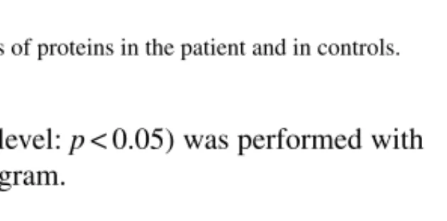
regres-Fig. 1. Western blots of proteins in the patient and in controls.
sion; significance level: p < 0.05) was performed with the SPSS 19.0 program.
RESULTS
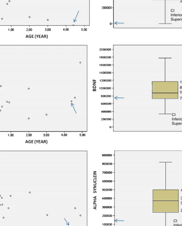
In our patient, CSF 5-hydroxyindolacetic acid concentration was low (133 nmol/L; C.V. 170–490) whereas dopamine metabolites were within normal limits (HVA: 360 nmol/L; C.V.: 344–906). BDNF, A, and AS were detected in the CSF of the patient and controls, at the expected molecular weight (Fig. 1). CSF total protein concentration values in patients and controls were within normal limits according to dif-ferent age ranges [9]. Linear regression showed no correlation between age and any of the studied proteins (A: p:0,1; BDNF: p:0,2; AS: p:0,1). The expression of BDNF and AS in the CSF of our patient showed low values with respect to the control group, particularly when compared with age-matched controls (Fig. 2.). The expression of A was undetectable in the patient whereas it was clearly present in all subjects of the con-trol group. Given its negativity, it was repeated twice in two CSF samples (at 3 and 4.5 years), disclosing the same result.
DISCUSSION
HSD10 deficiency is a neurodegenerative disease with a complex pathophysiology. Furthermore, HSD10 is a protein that may mediate the neurodegeneration of AD through its apparent capacity to bind A (1). Due to this affinity, we hypothesized that CSF A con-centration in patients with HSD10 deficiency could be abnormal and contribute to the neurobiology of this disorder. The most striking feature of this study is the lack of expression of A in this patient in several samples obtained at different ages.
HSD10 catalyzes the conversion of 2-methyl-3-hydroxybutyryl-CoA to 2-methylacetoacetyl-CoA in
the isoleucine degradation pathway, and is active against a broad range of substrates in diverse pathways such as steroid metabolism, GABAAreceptors, and the
oxidation of other substrates (hydroxyacyl-CoAs, 2-methyl-3-hydroxyacyl-coas) [1, 2]. Consequences of HSD17B10 gene mutations seem to be unrelated to accumulation of toxic metabolites in the isoleucine pathway but rather are caused by a non-enzymatic effect triggering mitochondrial disintegration and apoptosis [4].
Several studies indicate that mitochondria plays an important role in the development of AD, and this effect could be caused by a direct interaction of A with MHBD (2-methyl-3-hydroxybutyryl-CoA dehydroge-nase) [5–7]. HSD10 has a unique loop D (residues 95–114) that binds A [1]. In AD brain tissue, A accumulates in mitochondria. Conversely its concen-tration is low in CSF. HSD17B10 gene mutation in our patient could have modified this binding and pro-mote post-synaptic A trapping, thereby explaining the apparent absence of A in the CSF.
Little is known about the neurobiology of A in childhood. Recent studies suggest that A and its precursor protein (APP) may play important roles in development such as promoting synapse elimina-tion [10, 11], pruning neurites [12], and restricting mature forms of LTP in glutamatergic synapses [13]. In the rodent hippocampus, A has a maximum expres-sion during the period of most intense synaptogenesis and synaptic elimination [14]. Therefore, persistently low synaptic levels of A in our patient could have impaired synaptic balance and glutamatergic transmis-sion. Furthermore, increased post-synaptic A levels could have disrupted synaptic plasticity and promoted apoptosis [15–18].
BDNF is associated with dendritic growth, synap-tic transmission [19], and the development of the GABAergic and monoaminergic system [20]. In our patient, BDNF low expression argues in favor of low dendritic density and support reduced CSF con-centration of 5-HIAA, as this neurotrophin regulates serotonin system development. Concerning AS, this is a presynaptic protein that binds to the SNARE com-plex [21] and is involved in regulation of vesicle pools. In fact, its deletion causes a reduction in the reserve pool size [22], leading to impaired long-term poten-tiation and synaptic plasticity. Synaptic accumulation of AS has been related to dopaminergic loss [23]. In our patient AS showed very low expression which perhaps might explain why CSF dopaminergic metabo-lites were not reduced as expected at high AS synaptic levels.
283760,00 540028,0 0 (median) 817576,50 CI Inferior limit: 400212,98 Superior limit: 625938,46 728771,00 877842,00 (median) 1174881,00 CI Inferior limit :802434,03 Superior limit: 1065826,48 CI Inferior limit: 294214,19 Superior limit: 426154,98 237305,00 372819,0 0 (median) 499797,50
Fig. 2. Distribution of A, BDNF and AS values in the patient (arrows) and in controls. Units on the left are optical densities. CI: confidence interval.
In summary, we report a patient with HSD10 defi-ciency, undetectable CSF A expression, and low BDNF and AS levels, which probably disrupted critical developmental functions, contributing to impaired synaptic plasticity, low serotoninergic
trans-mission and apparently preserved dopaminergic func-tion.
Confirmation of these findings in other patients would help elucidating mechanisms of synaptic dys-function in this disease, and highlight the need for
further studies to better understanding of the role of A in both early and late periods of life.
ACKNOWLEDGMENTS
CIBERER is an initiative of the ISCIII (MICINN, Spain). This study was funded by the grant FIS PS09/01132. CO is supported by a grant from the Caja Navarra.
Authors’ disclosures available online (http://www.j-alz.com/disclosures/view.php?id=924).
REFERENCES
[1] Yang SY, He XY, Miller D (2007) HSD17B10: A gene involved in cognitive function through metabolism of isoleucine and neuroactive steroids. Mol Genet Metab 92, 36-42.
[2] Perez-Cerd´a C, Garc´ıa-Villoria J, Ofman R, Ruiz-Sala P, Garc´ıa-Silva M, Dalmau J, Ugarte M, Ribes A (2005) 2-methyl-3-hydroxybutyryl-CoA dehydrogenase (MHBD) deficiency: An X-linked inborn error of isoleucine metabolism that may mimic a mitochondrial disease. Pediatr Res 58, 488-491.
[3] Ofman R, Ruiter JP, Feenstra M, Duran M, Poll-The BT, Zschocke J, Ensenauer R, Lehnert W, Sass JO, Sperl W, Wanders RJ (2003) 2-Methyl-3-hydroxybutyryl-CoA dehy-drogenase deficiency is caused by mutations in the HADH2 gene. Am J Hum Genet 72, 1300-1307.
[4] Rauschenberger K, Sch¨oler K, Sass JO, Sauer S, Djuric Z, Rumig C, Wolf NI, Okun JG, K¨olker S, Schwarz H, Fis-cher C, Grziwa B, Runz H, N¨umann A, Shafqat N, Kavanagh KL, H¨ammerling G, Wanders RJ, Shield JP, Wendel U, Stern D, Nawroth P, Hoffmann GF, Bartram CR, Arnold B, Bier-haus A, Oppermann U, Steinbeisser H, Zschocke J (2010) A non-enzymatic function of 17beta-hydroxysteroid dehydro-genase type 10 is required for mitochondrial integrity and cell survival. EMBO Mol Med 2, 51-62.
[5] Lustbader JW, Cirilli M, Lin C, Xu HW, Takuma K, Wang N, Caspersen C, Chen X, Pollak S, Chaney M, Trinchese F, Liu S, Gunn-Moore F, Lue LF, Walker DG, Kuppusamy P, Zewier ZL, Arancio O, Stern D, Yan SS, Wu H (2004) ABAD directly links Abeta to mitochondrial toxicity in Alzheimer’s disease. Science 16, 448-452.
[6] Yan SD, Fu J, Soto C, Chen X, Zhu H, Al Mohanna F, Collison K, Zhu A, Stern E, Saido T, Tohyama M, Ogawa S, Roher A, Stern D (1997) An intracellular protein that binds amyloid peptide and mediates neurotoxicity in Alzheimer’s disease. Nature 389, 689-695.
[7] Garc´ıa-Villoria J, Navarro-Sastre A, Fons C, P´erez-Cerd´a C, Baldellou A, Fuentes-Castell´o MA, Gonz´alez I, Hern´andez-Gonzalez A, Fern´andez C, Campistol J, Delpiccolo C, Cort´es N, Messeguer A, Briones P, Ribes A (2009) Study of patients and carriers with 2-methyl-3-hydroxybutyryl-CoA dehydro-genase (MHBD) deficiency: Difficulties in the diagnosis. Clin Biochem 42, 27-33.
[8] Ormazabal A, Garc´ıa-Cazorla A, Fernandez Y, Fernandez-Alvarez E, Campistol J, Artuch R (2005) HLPC with
electrochemical and fluorescence detection procedures for the diagnosis of inborn errors of biogenic amines and pterins. J Neurosci Meth 142, 153-158.
[9] Biou D, Benoist JF, Nguyen-Thi Xuan Huong C, Morel P, Marchand M (2000) Cerebrospinal fluid protein concen-trations in children: Age-related values in patients without disorders of the central nervous system. Clin Chem 46, 399-403.
[10] Zetterberg H, Blennow K, Hanse E (2010) Amyloid beta and APP as biomarkers for Alzheimer’s disease. Exp Gerontol 45, 23-29.
[11] Priller C, Bauer T, Mitteregger G, Krebs B, Kretzschmar HA, Herms J (2006) Synapse formation and function is modulated by the amyloid precursor protein. J Neurosci 26, 7212-7221. [12] Nikolaev A, McLaughlin T, O’Leary DD, Tessier-Lavigne M (2009) APP binds DR6 to trigger axon pruning and neuron death via distinct caspases. Nature 19, 981-989.
[13] Townsend M, Qu Y, Gray A, Wu Z, Seto T, Hutton M, Shearman MS, Middleton RE (2010) Oral treatment with a gamma-secretase inhibitor improves long-term potentiation in a mouse model of Alzheimer’s disease. J Pharmacol Exp Ther 333, 110-119.
[14] Chiocco MJ, Lamb BT (2007) Spatial and temporal control of age-related APP processing in genomic-based beta-secretase transgenic mice. Neurobiol Aging 28, 75-84.
[15] Lambert MP, Barlow AK, Chromy BA (1998) Diffusible, non-fibrillar ligands derived from Abeta1–42 are potent central nervous system neurotoxins. Proc Natl Acad Sci U S A 95, 6448-6453.
[16] Walsh DM, Klyubin I, Fadeeva JV, Cullen WK, Anwyl R, Wolfe MS, Rowan MJ, Selkoe DJ (2002) Naturally secreted oligomers of amyloid beta protein potently inhibit hippocam-pal long-term potentiation in vivo. Nature 416, 535-539. [17] Chui DH, Dobo E, Makifuchi T, Akiyama H, Kawakatsu S,
Petit A, Checler F, Araki W, Takahashi K, Tabira T (2001) Apoptotic neurons in Alzheimer’s disease frequently show intracellular Abeta42 labeling. J Alzheimers Dis 3, 231-239. [18] Schmitz C, Rutten BP, Pielen A (2004) Hippocampal neuron
loss exceeds amyloid plaque load in a transgenic mouse model of Alzheimer’s disease. Am J Pathol 164, 1495-1502. [19] Huang Z, Shimazu K, Woo NH, Zang K, M¨uller U, Lu B,
Reichardt LF (2006) Distinct roles of the beta 1-class inte-grins at the developing and the mature hippocampal excitatory synapse. J Neurosci 25, 11208-11219.
[20] Aguado F, Carmona MA, Pozas E, Aguil´o A, Mart´ınez-Guijarro FJ, Alcantara S, Borrell V, Yuste R, Iba˜nez CF, Soriano E (2003) BDNF regulates spontaneous correlated activity at early developmental stages by increasing synap-togenesis and expression of the K+/Cl- co-transporter KCC2. Development 130, 1267-1280.
[21] Burr´e J, Sharma M, Tsetsenis T, Buchman V, Etherton MR, S¨udhof TC (2010) Alpha-synuclein promotes SNARE-complex assembly in vivo and in vitro. Science 329, 1663-1667.
[22] Murphy DD, Rueter SM, Trojanowski JQ, Lee VM (2000) Synucleins are developmentally expressed, and alpha-synuclein regulates the size of the presynaptic vesicular pool in primary hippocampal neurons. J Neurosci 20, 3214-3220. [23] Garcia-Reitb¨ock P, Anichtchik O, Bellucci A, Iovino M, Ballini C, Fineberg E, Ghetti B, Della Corte L, Spano P, Tofaris GK, Goedert M, Spillantini MG (2010) SNARE pro-tein redistribution and synaptic failure in a transgenic mouse model of Parkinson’s disease. Brain 133, 2032-2044.