Co ntro l o f ge ne e xpre ssio n
in Trypano so m atidae
Departamento de Bioquímica e Imunologia,
Universidade Federal de Minas Gerais, Belo Horizonte, MG, Brasil S.M.R. Teixeira
Abstract
The study of mechanisms which control gene expression in trypanoso-matids has developed at an increasing rate since 1989 when the first successful DNA transfection experiments were reported. Using pri-marily Trypanosoma brucei as a model, several groups have begun to elucidate the basic control mechanisms and to define the cellular factors involved in mRNA transcription, processing and translation in these parasites. This review focuses on the most recent studies regard-ing a subset of genes that are expressed differentially durregard-ing the life cycle of three groups of parasites. In addition to T. brucei, I will address studies on gene regulation in a few species of Leishmania and the results obtained by a much more limited group of laboratories studying gene expression in Trypanosoma cruzi. It is becoming evi-dent that the regulatory strategies chosen by different species of trypanosomatids are not similar, and that for these very successful parasites it is probably advantageous to employ multiple mechanisms simultaneously. In addition, with the increasing numbers of parasite genes that have now been submitted to molecular dissection, it is also becoming evident that, among the various strategies for gene expres-sion control, there is a predominance of regulatory pathways acting at the post-transcriptional level.
Co rre spo nde nce
S.M.R. Teixeira
Departamento de Bioquímica e Imunologia, ICB, UFMG Av. Antonio Carlos, 6627 30161-970 Belo Horizonte, MG Brasil
Fax: + 55-31-441-5963 E-mail: santuzat@ icb.ufmg.br
Research supported by CNPq and FAPEMIG.
Received May 26, 1998 Accepted August 26, 1998
Ke y wo rds
•Trypanosom a •Leishm ania •Gene expression •RNA
•Antigenic variation •VSG
Intro ductio n: ge ne ral m e chanism s o f ge ne e xpre ssio n in trypano so matids
In the Trypanosomatidae family, the gen-era Trypanosoma and Leishmania consist of several species of protozoa that are unicellu-lar and uniflagellated. These organisms are digenetic parasites whose life cycles present multiple differentiation forms that alternate between invertebrate and vertebrate hosts. T. brucei, T. cruzi and a few species of Leish-mania are the model systems used in the studies described here. They belong to an
ancient group of lower eukaryotes which, although we tend to consider them to be closely related organisms, are in fact highly divergent evolutionarily (1).
resulted in drastic reduction of vectorial and transfusional transmission in five Latin Ameri-can countries, prevention and control of these infectious diseases by standard methods of breaking chains of transmissions is still an attainable goal. Nevertheless, there continues to be a need for basic research to develop new prophylactic tools and to determine more spe-cific targets for therapy.
In addition to being of medical impor-tance, trypanosomes and Leishmania have attracted the attention of molecular parasi-tologists because of recent studies revealing peculiar aspects of their biology. Knowing that the Trypanosomatidae family diverged very early in the eukaryotic lineage, we should understand their aberrant behaviors: their rules for molecular and cellular biology are quite distinct when compared to higher eu-karyotes. As most textbooks describe, in eukaryotes protein-coding genes are scribed into monocistronic pre-mRNA tran-scripts containing exons (coding sequences) and introns (mostly non-coding sequences) that are processed into mature mRNAs through cis-splicing reactions. RNA poly-merase II is the enzyme responsible for the transcription of protein-coding genes, whereas RNA polymerase I transcribes ribo-somal RNA. In trypanosomatids, however, transcription is polycistronic, there are no introns and, therefore, no cis-splicing reac-tions. Processing of pre-mRNA into single-gene units is effected by trans-splicing reac-tions, a process that has been found to oper-ate only in trypanosomatids, Euglena and in
nematode and trematode worms (2). In addi-tion to these differences, other cellular and molecular novelties were discovered in try-panosomes: i) during cell division, chromo-some condensation and nuclear membrane disruption do not occur; ii) peculiar organelles are found, such as glycosomes which con-centrate the enzymes and the intermediates of the glycolytic pathway; iii) an extensive post-transcriptional modification of mito-chondrial RNA known as RNA editing is
required for the correct expression of mito-chondrial enzymes, and, finally, iv) tran-scription of protein-coding genes can be achieved by RNA polymerase I (or an en-zyme with similar properties). Because of these differences between trypanosomatids and higher eukaryotes, we shall first see how the general process of gene transcription and mRNA maturation takes place before study-ing how gene regulation occurs.
Since the primary transcripts of most pro-tein-coding genes that have been studied so far are polycistronic, cleavage of the pre-mRNA has to occur in the nucleus in order to produce monocistronic mRNAs. Further-more, for efficient translation by the ribo-somes, the addition of a methylated G nucle-otide or cap at the 5' end and the poly(A) tail at the 3' end of each mRNA is essential. In trypanosomatids, no consensus signal se-quence for polyadenylation has been identi-fied and the cap has a unique, highly methyl-ated structure (3). In fact, the 5' cap is part of the 39-nucleotide miniexon (or spliced leader, SL) that is joined to the 5' end of every mRNA in a transesterification reaction called
trans-splicing. Recent studies have provided several lines of evidence demonstrating that miniexon/cap addition and polyadenylation are not independent events, but, instead, are part of a cut-and-paste mechanism that occurs concomitantly or immediately after transcription (Figure 1). Coupling between
trans-splicing and polyadenylation was first described by LeBowitz et al. (4) who dem-onstrated that poly(A) selection is governed by the location of the splice acceptor site of the downstream gene in the polycistronic primary transcript of the DHFR-TS locus of
Leishmania major. Soon thereafter other groups showed that this was also the case for
and the site for polyadenylation became ac-cepted as a solution to this problem. Several experiments using deletion mutants have tested this hypothesis and have shown the importance of intergenic regions in govern-ing trans-splicing and polyadenylation in T. brucei. In addition to the correct distance,
the presence of a polypyrimidine-rich motif is also crucial, since only AG dinucleotides situated downstream from a polypyrimidine tract are used as splice acceptor site (Figure 1) (4-6,8).
Early transfection experiments of trypa-nosomes and Leishmania were performed in the absence of information regarding the sequences required for gene expression in these organisms (9). For a recent review on genetic transformation in protozoa, see
ref-erence 10. Based on the studies described above, the primary rules are now well estab-lished: in order to have a foreign gene ex-pressed in trypanosomatids, SL addition sites must be present both upstream (for trans -splicing) and downstream (for polyadenyla-tion) from the gene. On the other hand, the need for a promoter may depend on which trypanosomatid is involved. While in T. brucei, studies of genes encoding two
abun-dant surface proteins, variant surface glyco-protein (VSG) and PARP (see below), have provided strong promoters which were used to create powerful expression vectors, in T. cruzi and some species of Leishmania, we and others have been very successful in ob-taining expression of foreign genes without promoters (10).
SL genes gene cluster A/B
SL RNA
Transcription pre-mRNA
processing enzymes
poly(A)
Trans-splicing and polyadenylation
mature mRNAs
Figure 1 - The general process of mRNA synthesis in trypano-somatids. M ost trypanosome and Leishmania genes charac-terized thus far are organized in tandem repeats as indicated by the shaded boxes (representing tw o copies of an A gene and one copy of a B gene). At an-ot her chrom osom al locat ion there are several hundred tan-dem direct repeats encoding SL RNAs (dark boxes). In some cases, a single promoter is pres-ent upstream of the first gene in the cluster w hereas in the SL gene cluster each repeat is pre-sumed to bear a transcriptional promoter (small flags). After transcription, the polycistronic pre-m RNA is processed by
trans-splicing and polyadenyla-tion, w hich require signals pres-ent w ithin the intergenic re-gions: a polypyrimidine-rich se-quence or (CT)n (small boxes),
the spliced leader addition site or AG (arrow heads), and a poly-adenylat ion addit ion sit e or poly(A) (arrow s). M ature mRNAs w hich consist of the coding re-gion and 5’UTR (shaded boxes), the SL sequence (dark boxes), 3’UTR and poly(A) tail (thin lines w ith small “ A” letters) are rep-resented by the diagram at the bottom of the figure.
7
77
7
AAAAAAA
AAAAAA
7
{
B{
Apre-mRNA
(CT)n A
...
...
...
..
B A
A
(AG)
B
A ...
Antige nic variatio n as a mo de l syste m to study ge ne e xpre ssio n co ntro l in T. b ruce i
Most of the studies on the molecular biology of trypanosomatids were conducted by parasitologists in the United States and in Europe who were curious about the process of antigenic variation. Antigenic variation, perhaps one of the most interesting recent discoveries in the parasitology field, is a powerful survival strategy devised by Afri-can trypanosomes which allows them to es-cape the immunological attack of the host. The T. brucei life cycle,unlike T. cruzi and
Leishmania, does not have an intracellular stage. It has two developmental forms: procyclics, which multiply in the insect mid-gut, and bloodstream forms, which multiply in the mammalian host where they are able to survive because of antigenic variation.
Antigenic variation started to be under-stood at the molecular level about 20 years ago. However, the first observations point-ing to this phenomenon were made in 1910 (11) when the number of parasites was de-termined in blood samples from patients with sleeping sickness: there was a dramatic rise and fall in the number of trypanosomes, with one peak of parasitemia followed by another peak every one to two weeks. In 1975, George Cross (12) isolated a surface protein known to be the single major protein present in the bloodstream forms of T. brucei
and named it variant surface glycoprotein (VSG). When the N-terminal sequences of VSGs isolated from four cloned trypano-some populations derived from a single rab-bit infected with T. brucei were compared, it became clear that each peak of parasitemia corresponded to a trypanosome population expressing a different VSG on their surface (13). The conclusion was obvious: after host antibodies against one type of VSG mole-cule are produced and the immune system destroys 99% of the trypanosome popula-tion, one of the few surviving parasites
changes the type of VSG it expresses and, with a new surface coat, begins reproducing as if it had never been seen by host immune defenses. This game continues to be played with the immune system always running one step behind the parasite. Each time a VSG switch occurs, 107 identical new proteins are
added as a molecular shield to the surface of the parasite. Estimated switching rates as high as 10-2 cells per generation have been
reported in recently transmitted trypanosome isolates (14). Recently, various aspects of antigenic variation have been reviewed (14, 15).
ES, that can extend for 50-60 kb and con-tains about 10 other genes (18-20). The prod-ucts of these genes, called ESAG for expres-sion site-associated genes, have no func-tional relationship to VSG, but an important function has been assigned to at least two of them: the T. brucei heterodimeric transferrin
receptor is encoded by ESAG6 and ESAG7 (21). In every telomere that has been charac-terized so far, a resident VSG gene has been found. Since some VSG genes are also found internally in the chromosomes, some type of DNA rearrangement must occur (such as the ones originally described by Williams) in order to activate these internal genes. But because not all VSGs located in telomeres are being expressed in one cell, there must also exist some type of in situ gene activa-tion/inactivation to differentiate these telo-meric ES. So, it appears that a VSG gene can be turned on in two basic ways: either it is already in a telomeric ES that needs to be activated in situ (concomitantly with the inactivation of another ES), or it is an inter-nal gene that needs to be copied and moved near a telomere where it can replace an active gene.
Understanding these mechanisms, par-ticularly the in situ switch, has been a diffi-cult task. A model to explain the activation/ inactivation of one particular ES in blood-stream trypanosomes, comparable to silenc-ing mechanisms described in yeast, proposes
a significant role for telomeric sequences (22). Moreover, the role of a newly identi-fied nucleotide ß-D-glucosyl-hydroxy-methyluracil, called J, that is found pre-dominantly in telomeric repeats near silent VSG genes,is also being investigated (17). The cloning of promoter regions from active VSG genes, which is crucial for the understanding of the mechanism of VSG regulation, was not an easy experiment: the region researchers were looking for was in fact located 60 kb upstream of a VSG gene (14,15,23). Before a VSG promoter was cloned, the work from Borsts group (23) provided the first evidence for the presence of a distant single promoter transcribing sev-eral genes in what is now known as a blood-stream form VSG transcription unit (Figure 2). More recently, it has been found that in a special class of VSG genes, i.e., those that are expressed by metacyclic trypanosomes, the promoter is located immediately upstream of the VSG gene, resulting in an unusual trypanosome monocistronic mRNA (24). After sequencing several VSG promoters, another unusual discovery was made: they do not possess any of the typical sequences present in eukaryotic RNA pol II promoters, such as the TATA box. Moreover, nuclear run-on transcription assays showed that VSG gene promoters are functionally more re-lated to RNA pol I promoters since they are 100% active in the presence of 1 mg/ml α
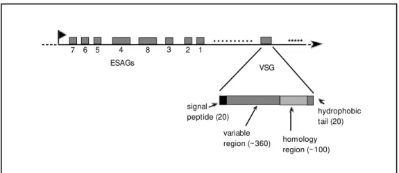
-Figure 2 - The T. brucei VSG mol-ecule and a VSG gene expres-sion site. Studies of several vsg
loci indicate that expression sites of bloodstream forms are organized similarly to the sche-matic representation show n, in w hich shaded boxes represent one VSG gene and several ESAGs. The flag indicates the promoter, the numbers below the boxes define each ESAG, the small dots and asterisks rep-resent the 70-bp (or 76-bp) re-peats and telomeric hexanucle-otide repeats, respectively, and the arrow head indicates the chromosome terminus. The dis-tance betw een the VSG gene and the start of the repetitive telomeric sequence is 1-3 kb. The enlargement below fea-tures a diagram w ith the differ-ent regions of a nascdiffer-ent VSG (numbers in parentheses indi-cate the number of amino acids in each portion of the molecule).
7 6 5 4 8 3 2 1
ESAGs VSG
signal peptide (20)
variable
region (~360) homology region (~100)
*****
...
amanitin (3). Thus, it appears that T. brucei
has a unique system that allows transcription of protein-coding genes to be initiated by RNA polymerase I or an RNA polymerase I-like enzyme. This is only possible in trypa-nosomes because maturation of the 5' end (and the addition of cap) is independent of the RNA polymerase. Contrary to the gen-eral process of eukaryote transcription, where the enzymes required for the capping pro-cess are thought to be associated with RNA polymerase II, in trypanosomes the trans -splicing mechanism will provide the SL se-quence that already contains a cap structure at its 5' end.
It has been only recently that experi-ments with stable transfected trypanosomes have begun to provide convincing evidence that the control of VSG gene expression occurs at several levels including transcrip-tion initiatranscrip-tion. By inserting a VSG promoter linked to a selectable marker into a chromo-somal location, instead of episomes, or by tagging an expression site with a reporter gene, it was possible to follow the activity of a given VSG promoter when the trypano-some is induced to differentiate, or when it spontaneously undergoes a VSG switch (22,25,26). The control of VSG gene expres-sion during the parasite life cycle has been more extensively investigated using nuclear run-on assays. Several groups have shown that, although VSG mRNAs are not detected in procyclic forms, transcription initiation does occur at a low level from many, if not all, ES promoters (27-29). This is consistent with the observation of CAT or luciferase expression under the control of VSG gene promoters cloned in episomal vectors in tran-siently transfected procyclic parasites (29,30). However, when VSG promoters were further analyzed within their chromo-somal context, instead of plasmid vectors, it was found that the transcription elongation step is subjected to stage-specific regulation, i.e., the active VSG gene is fully transcribed in bloodstream parasites but most
transcrip-tional activity stops within 1 kb of the VSG promoter in procyclics (27-29).
Po st-transcriptio nal m e chanism s as the m ain m e tho d o f co ntro lling ge ne e xpre ssio n in T. b ruce i and
Le ishm ania spp
In T. brucei, in addition to VSG genes, a second group of genes encoding highly abun-dant surface glycoproteins has been used as a model to explain the mechanisms of gene expression control. When bloodstream try-panosomes differentiate into procyclics, the VSG coat is rapidly replaced by a new sur-face coat composed of small, acidic proteins named procyclins or procyclic acid repeti-tive proteins (PARP) (for a review, see Ref. 31). PARP genes comprise a polymorphic gene family that is found at two genetic loci (α and ß) in the genome of several strains of
T. brucei. Two or three PARP genes are found at each locus, together with a PARP-associated gene (PAG). Like the majority of trypanosome genes, PARP genes are part of a polycistronic transcription unit, and like VSG genes, PARP genes are transcribed by an α-amanitin-resistant RNA polymerase (32). However, sequence comparison shows that the PARP promoters and VSG promot-ers that have been characterized so far bear little resemblance to each other, or to the classical RNA polymerase I rRNA promoter (3).
transcribed at low levels in bloodstream para-sites indicating that some post-transcription mechanisms must take place in order to pre-vent the accumulation of PARP mRNA. Moreover, although PARP and PAG genes are derived from the same polycistronic tran-scription unit, there is a 100-fold difference in the steady-state levels when PARP and PAG mRNAs are compared (31). Several possibilities have been considered to explain this difference in the accumulation of mRNAs, including differences in trans- splic-ing efficiency and the presence of regulatory sequences in the 3'-untranslated regions (UTR) affecting the steady-state levels of these mRNAs. The study of regulatory ele-ments derived from the 3'UTR has been extensively explored (28,31,34,35). Using transient transfections, Roditis group (35) has demonstrated that a 16-mer sequence which is predicted to form a stem-loop struc-ture in the 3'UTR of PARP mRNAs can confer regulation of a CAT reporter gene on procyclics. Deletion of this element causes a 12-fold drop in CAT expression but no sig-nificant difference in the levels of total RNA, suggesting control at the translational level. Interestingly, the same sequence, with the same predicted secondary structure, has also been found in the analogous gene of another trypanosome species, T. congolense. More recently, a systematic analysis of the entire 297-base 3'UTR revealed three additional elements which are involved in post-tran-scriptional regulation of PARP genes, all three of them having an effect on the rate of mRNA turnover (34). It seems that, by em-ploying several layers of regulation, these parasites can ensure that the rapid changes associated with transmission between insect vector and mammalian host are followed by an instant reprogramming of genetic expres-sion.
When studies performed with T. brucei
are compared with the results obtained with other trypanosomatids, the basic observa-tions remain. However, there are no reports
describing transcriptional regulation affect-ing gene expression in Leishmania or T. cruzi. In fact, there is not a single report describing promoter elements for protein-coding genes in these organisms. Most of the work in Leishmania species has been fo-cused on one of the following gene families, the first three of which are developmentally regulated during the parasite life cycle: the α and ß tubulin genes (36), the surface metal-loprotease gp63 or msp genes (37), the amas-tigote-specific A2 genes (38) and heat-shock protein genes (39). The initial studies per-formed by Dyann Wirths group (9) describ-ing a transfection system for L. enrietti used a plasmid containing the intergenic region of the α-tubulin cluster flanking a CAT gene. Their prediction was that there might exist at least two potential sites for transcription ini-tiation in a gene cluster: one iniini-tiation site adjacent to each gene, or a single promoter upstream from the first gene in the cluster. When tests for the presence of intergenic promoters in the tubulin cluster were per-formed using transient transfections there was a surprise: only a very short sequence derived from the tubulin locus was neces-sary for CAT expression and that sequence basically contained an SL addition site (9,40). It was proposed that the promoter function was being provided by plasmid DNA se-quences and the tubulin sese-quences were just providing SL acceptor sites for the process-ing of the mRNA. Since then, additional attempts to identify promoter regions within
Ge ne manipulatio n in T. cruz i: the de ve lo pme nt o f ve cto rs and transfe ctio n pro to co ls
The field of molecular genetics was pro-gressing rapidly, with several new reports about gene expression in T. brucei and Leish-mania being published, when, in 1991, the first transfection experiment of T. cruzi was reported. Using a vector containing a seg-ment of the SL gene placed upstream of the bacterial CAT gene, Lu and Buck (41) were able to detect CAT activity in transfected epimastigotes. Although those experiments were later called transient transfections, CAT expression reached a maximum after 48 h but remained unchanged at 72 h and 96 h, indicating that the plasmid was being main-tained with some stability inside the cells. In 1992, Kelly et al. (42) described an im-proved vector named pTEX which was con-structed using flanking sequences derived from the two tandemly repeated glyceralde-hyde-3-phosphate dehydrogenase (gapdh) genes. To help cloning and to allow selecting transformants, a multiple cloning site and the neo gene as a drug resistance marker were included. As was reported in similar experiments with T. brucei and Leishmania, the plasmid replicates episomally in the form of large concatemers and remains stable in the presence of drug selection. More impor-tantly, similar to the results observed in sev-eral Leishmania species, the copy number
and the level of expression can be modulated by changing drug concentration (42).
The results obtained by Kellys group and later by other investigators using the pTEX vector suggested that, regarding the mechanisms of gene expression following plasmid transfections, T. cruzi has more in common with Leishmania than with T. brucei. Whereas stable transfectants of T. cruzi and Leishmania were easy to obtain using episo-mal vectors, with T. brucei this was rarely achieved. In fact, pTEX can be used to trans-fect several species of Leishmania, but not T.
brucei or Crithidia fasciculata (42). More
surprisingly, expression of foreign genes in
T. cruzi and Leishmania was achieved using episomal vectors containing no promoter el-ements. As is the case for Leishmania, we can also speculate that transcription initiates randomly within the plasmid vector and the long transcript generated is processed ac-cording to the rules of trans-splicing and polyadenylation. And once again, similar to
Leishmania, attempts to identify an RNA polymerase II promoter in T. cruzi using the plasmid transfection approach have failed. In a series of experiments aimed at identify-ing the promoter region of the T. cruzi amastin gene cluster (see below), we constructed a variety of plasmids containing various DNA fragments derived from the intergenic region between two copies of amastin genes as well as the 5'-flanking region of the cluster placed in front of the luciferase reporter gene (Fig-ure 3) (43). When luciferase expression in epimastigotes transfected with these con-structs was compared, the results were quite similar, about 700-fold above background levels. Moreover, the levels of luciferase activity resulting from transfections with these constructs were also similar to the levels found in cells transfected with the plasmid pLST, where most of the trypano-some sequences were deleted. In this dele-tion, only a 73-bp fragment containing basi-cally trans-splicing signals of amastin mRNA
the time, a group of investigators was in the process of characterizing the T. cruzi rRNA promoter. Dietrich et al. (44) have deter-mined the sequence of the intergenic spacer of an rRNA gene and mapped the putative transcription start site. When we inserted a 580-bp fragment containing this sequence in front of the amastin SL addition site, in the proper orientation within the pLST vector, luciferase activity resulting from transfected epimastigotes was 270,000-fold above back-ground (or 2,000-fold greater than that gen-erated with pLST). With this improved vec-tor we were able, for the first time, to obtain luciferase expression in transiently trans-fected amastigotes (43). At the same time, Tyler-Cross et al. (45) also described a tran-sient expression system mediated by the T. cruzi rRNA promoter and showed that the promoter fragment is highly efficient at driv-ing CAT expression in transiently transfected epimastigotes. More recently, Martínez-Calvillo et al. (46) described a pTEX deriva-tive, called pRIBOTEX, where the upstream region of gapdh was replaced by another rRNA gene spacer fragment bearing a previ-ously mapped transcription start site. T. cruzi
cultures transiently transfected with pRIBOTEX express a CAT activity 16,000-fold greater than the activity generated with the original pTEX vector. More importantly, since that vector contains a selectable marker that allows generation of stable transfectants, the presence of a strong promoter permitted a more rapid drug selection time, with G418-resistant clones being obtained 2 weeks ear-lier.
The only other T. cruzi promoter that has been characterized is the SL gene promoter, but the activity of this promoter, as described by Nunes et al. (47), was unexpectedly low. However, these investigators reported a ma-jor finding about the activity of the SL and rRNA promoters: promoters isolated from a
T. cruzi strain classified as group I, as de-fined by Souto et al. (48), are efficiently expressed only in T. cruzi isolates classified as belonging to the same group (49). The description of two major lineages of T. cruzi
strains, based on the analysis of several nuclear markers, has now been strongly rein-forced by the functional specificity of the SL and rRNA promoters. Given the well-docu-mented species-specific pattern of the rRNA
Figure 3 - A model for the regula-tion of amastin gene expression in T. cruzi. The amastin/tuzin gene cluster is show n, w ith dark and gray boxes representing copies of tuzin and am astin genes, respectively. Intergenic regions are show n as thin lines (The diagram including the re-striction sites w as modeled af-ter Teixeira et al. (43)). Polycis-tronic transcription, w hich oc-curs at the same rates in amasti-gotes and epimastiamasti-gotes, gener-ates a pre-mRNA (w avy lanes) that needs to be processed in order to produce mature mRNA encoding amastin (gray box) and tuzin (dark box) proteins. The dif-ferential accumulation of these mRNAs in the tw o forms of the parasite’s life cycle may be de-pendent upon at least tw o fac-tors: higher efficiency of amastin mRNA processing and increased stability of the mRNA in amasti-gotes. An RNA-binding protein, w hich is more abundant in amas-tigotes, is proposed to be in-volved in the regulatory pathw ay through specific binding at the 3’UTR of amastin mRNA (gray line).
gene cluster
Epimastigotes
Transcription
RNA processing
mRNAs
High efficiency of amastin mRNA
trans-splicing and increased stability
pre-mRNA ^^^^^^^^^^^^^^^^^ ^^^^^^^^^^^^^^^^^^^^
B P E B BI S BEBIS B BIE S
1 kb
Increased expression of amastin
promoter in higher eukaryotes, these results represent functional evidence for ancient evo-lutionary divergence among T. cruzi strains. In addition to episomal transfections, stable transformants of T. cruzi can also be generated with high efficiency as a result of integration of the plasmid vector by homolo-gous recombination. Using either episomal vectors or DNA integration protocols of trans-fection, several groups have reported the expression of foreign genes in T. cruzi, in-cluding mammalian cytokines such as inter-leukin-2 and gamma-interferon (50), the T. brucei GPI-phospholipase C (51), and the green fluorescent protein (GFP) from algae (Teixeira SMR, unpublished data). The use of GFP-fusion proteins or epitope tagging has great potential as a method for subcellu-lar localization, as recently reported (52). Likewise, stable transfectants were also gen-erated in order to overexpress T. cruzi genes such as cruzipain, a cysteine proteinase (53). With this methodology, gene disruption could also be achieved by targeted integration of a DNA segment containing a selectable marker. A few groups have exploited this new technology as a tool for functional a-nalysis of T. cruzi genes. That number re-mains rather low not only for T. cruzi but for trypanosomatids in general, mainly because in these organisms genes are usually present as multicopy families, making gene knock-out experiments very difficult. In 1993, just after Hariharan et al. (54) described plasmid integration into the polyubiquitin gene lo-cus, disrupting one of the polyubiquitin genes, Otsu et al. (55) and Cooper et al. (56) reported on target gene replacement result-ing in disruption of sresult-ingle copy genes of T. cruzi. Deletion of the gene encoding the immunodominant T. cruzi surface glycopro-tein GP72 by target integration caused a dramatic morphological change in which the normal attachment of the flagellum to the parasite cell membrane was lost (56). Al-though this experiment has established that gene knock-out is a useful tool for functional
studies of genes in T. cruzi, other
method-ologies, such as the use of anti-sense RNA and, more recently, ribozymes, may also contribute to addressing these questions in cases where multicopy genes are present.
Re gulato ry e le me nts co ntro lling the e xpre ssio n o f stage -spe cific ge ne s in
T. cruz i
One of the beauties of working with T. cruzi as well as other trypanosomatids is that
they represent excellent models to study the genetic control of cell differentiation. Com-pared to other trypanosomatids, regulatory mechanisms of gene expression during the life cycle of T. cruzi have been much less
explored. Albeit the molecular tools are now available for in depth studies of life cycle stage-specific regulation of gene expression in T. cruzi, only two groups have taken on the task of identifying regulatory elements within T. cruzi genes. The strategies are
quite straightforward: after defining which DNA fragments are to be analyzed, we can use reporter genes and transfection proto-cols similarly to the approaches that have been successfully applied in the studies of T. brucei and Leishmania genes.
Working with John Donelson at the Uni-versity of Iowa, a few years ago I decided to investigate the processes responsible for the control of genes that are expressed preferen-tially in amastigotes, the intracellular stage of the T. cruzi life cycle. Using the
genes, which we named tuzin (43,57) (Fig-ure 3). DNA sequence analysis predicted that amastins are hydrophobic proteins of 174 amino acids localized at the surface of amastigotes. Northern blot analysis indicated that amastin mRNAs are 60-fold more abun-dant in amastigotes compared to their levels in the epimastigote or trypomastigote stage. Analysis of similar Northern blots probed with tubulin genes showed that tubulin ex-pression is also under a strict stage-specific control, being expressed at much higher lev-els in epimastigotes compared to other forms (Teixeira SMR, unpublished data). In con-trast to amastin and tubulin genes, expres-sion of tuzin genes, which code for a much less abundant hydrophilic protein, is not regu-lated during the parasites life cycle. Al-though amastin and tuzin genes are tran-scribed by the same RNA polymerase into the same polycistronic pre-mRNA, mature tuzin mRNAs accumulate at low, constant levels, equivalent to the levels of amastin mRNA in epimastigotes (43). As a first step towards the identification of elements in-volved in the control of expression of the amastin/tuzin gene cluster we demonstrated, using nuclear run-on experiments, that tran-scription initiation remains constant when nuclei from epimastigotes and amastigotes are compared. To investigate if the 3' or 5'UTRs and intergenic regions were involved, fragments containing different regions of the cluster were examined using transient transfections. With luciferase as a reporter gene we compared luciferase expression in transfected epimastigotes and amastigotes, normalized to a control plasmid. Our results indicated a significant regulatory role of the amastin 3'UTR: luciferase activity appeared to be 36-fold higher in amastigotes when these cells were transfected with a construct containing this fragment inserted downstream of the luciferase gene (43). We further ex-amined the presence of regulatory elements located in that region using an approach similar to the linker scanner assay where we
sequentially replaced contiguous 70-bp seg-ments of the 630-bp 3'UTR with random sequences derived from synthetic oligonucle-otides. We also obtained evidence for the existence of an RNA-binding protein that recognizes these sequences in the amastin 3'UTR (59). More recently, we have ob-tained evidence indicating that differences in 5'UTR are also important for determining the levels of amastin and tuzin mRNAs (Teixeira SMR, Kirchhoff LV and Donelson JE, unpublished data). Taken together, these results are consistent with the regulatory model shown in Figure 3: several factors acting at the post-transcriptional level may be responsible for determining the differen-tial expression of amastin and tuzin genes in
T. cruzi. After we described the effect of the amastin 3'UTR, Nozaki and Cross (60) re-ported a similar study where the 3'UTR from various stage-specific and constitutive T. cruzi genes were tested using stable trans-fection experiments with the luciferase re-porter gene. Their results confirmed our find-ings with the amastin 3'UTR, i.e., luciferase expression increases 16-fold in amastigotes while decreasing 14-fold when epimastigotes were transfected. In contrast, the presence of the 3'UTR from two other stage-specific genes, gp72 and gp85, showed a much smaller modulatory activity than amastin 3'UTR. Various exciting experiments are currently in progress and we expect that we will soon have a better understanding of the mechanisms involving the 3'UTR sequences and other factors that might be responsible for determining the level at which a particu-lar mRNA accumulates during the parasites life cycle. So far, preliminary evidence indi-cates the efficiency of the processing step and the mechanisms determining the mRNA half-life.
Co ncluding re marks
their favorite parasite since the electron mi-croscope was invented. Genetic transforma-tion, or DNA transfectransforma-tion, has produced not just an extraordinary amount of new infor-mation about the biology of these organisms but has given us the possibility of manipulat-ing a significant part of their genomes. More-over, the new era of molecular parasitology can go hand-in-hand with the fast growing developments achieved with parasite genome projects. Above all, the excitement that each new report or technical breakthrough brings to the field is especially welcome because it
can drive an important fraction of the future generation of researchers into a field that needs all the help we can get. As everyone agrees, understanding post-transcriptional control in trypanosomes may not solve Chagas disease, but will certainly contri-bute to making us more prepared to fight it.
Ackno wle dgm e nts
I thank Dr. Gregory Kitten and Dr. Maria de Fátima Horta for discussion and com-ments on the manuscript.
Re fe re nce s
1. Fernandes AP, Nelson K & Beverley SM (1993). Evolution of nuclear ribosomal RNAs in kinetoplastid protozoa: perspec-tives on the age and origins of parasitism.
Proceedings of the National Academy of Sciences, USA, 90: 11608-11612. 2. Donelson JE & Zeng W (1990). A
com-parison of trans-RNA splicing in trypano-somes and nematodes. Parasitology To-day, 6: 327-334.
3. Lee M GS & Van der Ploeg LHT (1997). Transcription of protein-coding genes in trypanosomes by RNA polymerase I. An-nual Review of M icrobiology, 51: 463-489. 4. LeBow itz JH, Smith HQ, Rusche L & Beverley SM (1993). Coupling of poly(A) site selection and trans-splicing in Leish-mania. Genes and Development, 7: 996-1007.
5. M atthew s KR, Tschudi C & Ullu E (1994). A common pyrimidine-rich motif governs
trans-splicing and polyadenylation of tu-bulin polycistronic pre-mRNA in trypano-somes. Genes and Development, 8: 491-501.
6. Schurch N, Hehl A, Vassella E, Braun R & Roditi I (1994). Accurate polyadenylation of procyclin mRNAs in Trypanosom a brucei is determined by pyrimidine-rich elements in the intergenic regions. M o-lecular and Cellular Biology, 14: 3668-3675.
7. Huang J & Van der Ploeg LHT (1991). M aturation of polycistronic pre-mRNA in
Trypanosoma brucei: analysis of trans -splicing and poly(A) addition at nascent RNA transcripts from the hsp70 locus.
M olecular and Cellular Biology, 11: 3180-3190.
8. Huang J & Van der Ploeg LHT (1991). Requirement of a polypyrimidine tract for
trans-splicing in trypanosomes: discrimi-nating the PARP promoter from immedi-ately adjacent 3' splice acceptor site.
EM BO Journal, 10: 3877-3885.
9. Laban A & Wirth D (1989). Transfection of
Leishmania enriettii and expression of chloramphenicol acetyltransferase gene.
Proceedings of the National Academy of Sciences, USA, 86: 9119-9123.
10. Kelly JM (1997). Genetic transformation of parasitic protozoa. Advances in Parasi-tology, 39: 227-270.
11. Ross R & Thomson D (1910). A case of sleeping sickness studied by precise enu-merative methods. Regular periodic in-crease in the parasites disclosed. Pro-ceedings of the Royal Society of London, 82: 411-415.
12. Cross GAM (1975). Identification, purifi-cation and properties of clone-specific gly-coprotein antigens constituting the sur-face coat of Trypanosoma brucei. Parasi-tology, 71: 393-417.
13. Bridgen PJ, Cross GAM & Bridgen J (1976). N-terminal amino acid sequences of variant-specific antigens from Trypano-soma brucei. Nature, 263: 613-614. 14. Cross GAM (1996). Antigenic variation in
trypanosomes: secrets surface slow ly.
BioEssays, 18: 283-291.
15. Borst P, Rudenko G, Taylor M C, Blundell PA, Van Leeuw en F, Bitter W, Cross M & M cCulloch R (1996). Antigenic variation in trypanosomes. Archives of M edical Re-search, 27: 379-388.
16. Williams RO, Young JR & M ajiva PAO (1979). Genomic rearrangements
corre-lated w ith antigenic variation in Trypano-soma brucei. Nature, 282: 847-849. 17. Van Leeuw en F, Taylor M C, M ondragon
A, M oreau H, Gibson W, Kieft R & Borst P (1998). Beta-D-glucosyl-hydroxymethylu-racil is a conserved DNA modification in kinetoplastid protozoans and is abundant in their telomeres. Proceedings of the National Academy of Sciences, USA, 95: 2366-2371.
18. Cully DF, Ip HS & Cross GAM (1985). Coordinate transcription of variant surface glycoprotein genes and an expression site associated gene family in Trypanosoma brucei. Cell, 42: 173-182.
19. Gibbs CP & Cross GAM (1988). Cloning and transcription analysis of a variant sur-face glycoprotein gene expression site in
Trypanosoma brucei. M olecular and Bio-chemical Parasitology, 28: 197-206. 20. Kooter JM , Van der Speck HJ, Wagter R,
Oliveira CE, Van der Hoeven F, Johnson PJ & Borst P (1987). The anatomy and transcription of a telomeric expression site for variant-specific surface antigens in Trypanosoma brucei. Cell, 51: 261-272. 21. Salm on D, Geuskens M , Hanocq F, Hanocq-Quertier J, Nolan D, Ruben L & Pays E (1994). A novel heterodimeric transferrin receptor encoded by a pair of VSG expression site-associated genes in
T. brucei. Cell, 78: 75-86.
22. Rudenko G, Cross M & Borst P (1998). Changing the end: antigenic variation or-chestrated at the telomeres of African try-panosomes. Trends in M icrobiology, 6: 113-116.
irradia-tion of T. brucei provides evidence for a multicistronic transcription unit including a VSG gene. Cell, 51: 273-281.
24. Alarcon CM , Son HJ, Hall T & Donelson JE (1994). A monocistronic transcript for a trypanosome variant surface glycoprotein.
M olecular and Cellular Biology, 14: 5579-5591.
25. Rudenko G, Blundell PA, Dirks-M ulder A, Kieft R & Borst P (1995). A ribosomal DNA promoter replacing the promoter of a telomeric VSG gene expression site can be efficiently sw itched on and off in T. brucei. Cell, 83: 547-553.
26. Horn D & Cross GAM (1995). A develop-mentally regulated position effect at a telomeric locus in Trypanosoma brucei.
Cell, 83: 555-561.
27. Graham SV (1995). M echanisms of stage-regulated gene expression in kinetoplas-tida. Parasitology Today, 11: 217-223. 28. Vanhamme L & Pays E (1995). Control of
gene expression in trypanosomes. M icro-biological Review s, 59: 223-240. 29. Pays E, Coquelet H, Tebabi P, Pays A,
Jefferies D, Steinert M , Koening E, Will-iams RO & Roditi I (1990). Trypanosoma brucei: constitutive activity of the VSG and procyclin gene promoters. EM BO Journal, 9: 3145-3151.
30. Jefferies D, Tebabi P & Pays E (1991). Transient activity assays of the Trypano-soma brucei variant surface glycoprotein gene promoter: control of gene expres-sion at the post-transcriptional level. M o-lecular and Cellular Biology, 11: 338-343. 31. Roditi I (1996). The VSG-procyclin sw itch.
Parasitology Today, 12: 47-49.
32. Rudenko G, Le Blancq S, Smith J, Gw o-Shu-Lee M , Rattray A & Van der Ploeg LHT (1990). Procyclic acidic repetitive pro-tein (PARP) genes located in an unusually small α-amanitin-resistant transcription unit: PARP promoter activity assayed by transient DNA transfection of Trypanoso-ma brucei. M olecular and Cellular Biol-ogy, 10: 3492-3504.
33. Roditi I, Schurch N, Furger A & Ruep S (1996). Trust is good, control is better: the philosophy behind the regulation of gene expression by trypanosomes. M emórias do Instituto Osw aldo Cruz, 91 (Suppl): 50-51.
34. Furger A, Schurch N, Kurath U & Roditi I (1997). Elements in the 3' untranslated region of procyclin mRNA regulate ex-pression in insect form of Trypanosoma brucei by modulating RNA stability and translation. M olecular and Cellular Biol-ogy, 17: 4372-4380.
35. Hehl A, Vassella E, Braun R & Roditi I
(1994). A conserved stem-loop structure in the 3' untranslated region of procyclin mRNAs regulates expression in Trypano-soma brucei. Proceedings of the National Academy of Sciences, USA, 91: 370-374. 36. Coulson RM R, Connor V, Chen JC & Ajioka J (1996). Differential expression of
Leishmania major ß-tubulin genes during the acquisition of promastigote infectiv-ity. M olecular and Biochemical Parasitol-ogy, 82: 227-236.
37. Ramamoorthy R, Sw ihart K, M cCoy JJ, Wilson M E & Donelson JE (1995). Inter-genic regions betw een tandem gp63 genes influence the differential expres-sion of gp63 RNAs in Leishmania chagasi.
Journal of Biological Chem istry, 270: 12133-12139.
38. Charest H, Zhang W & M atlashew ski G (1996). The developmental expression of
Leishmania donovani A2 amastigote-spe-cific genes is post-transcriptionally medi-ated and involves elements locmedi-ated in the 3' UTR region. Journal of Biological Chemistry, 271: 17081-17090.
39. Argaman M , Aly R & Shapira M (1994). Expression of heat shock protein 83 in
Leishmania is regulated post-transcrip-tionally. M olecular and Biochemical Para-sitology, 64: 95-110.
40. Curotto de Lafaille M A, Laban A & Wirth D (1992). Gene expression in Leishmania: analysis of essential 5' DNA sequences.
Proceedings of the National Academy of Sciences, USA, 89: 2703-2707.
41. Lu H & Buck GA (1991). Expression of exogenous gene in Trypanosoma cruzi
epimastigotes. M olecular and Biochemi-cal Parasitology, 44: 109-114.
42. Kelly JM , Ward HM , M iles M A & Kendall G (1992). A shuttle vector w hich facili-tates the expression of transfected genes in Trypanosoma cruzi and Leishmania do-novani. Nucleic Acids Research, 20: 3963-3969.
43. Teixeira SM R, Kirchhoff LV & Donelson JE (1995). Post-transcriptional elements regulating expression of mRNAs from the amastin/tuzin gene cluster of Trypanoso-ma cruzi. Journal of Biological Chemistry, 270: 22586-22594.
44. Dietrich P, Soares M B, Affonso M HT & Floeter-Winter L (1993). The Trypanoso-ma cruzi ribosomal RNA-encoding genes: analysis of promoter and upstream inter-genic spacer sequences. Gene, 125: 103-107.
45. Tyler-Cross RE, Short SL, Floeter-Winter L & Buck GA (1995). Transient expression mediated by the Trypanosoma cruzi rRNA promoter. M olecular and Biochem ical
Parasitology, 72: 23-31.
46. M artínez-Calvillo S, Lopez I & Hernandez R (1997). pRIBOTEX expression vector: a pTEX derivative for a rapid selection of
Trypanosoma cruzi transfectants. Gene, 199: 71-76.
47. Nunes LC, Carvalho M RC, Shakarian AM & Buck GA (1997). The transcription pro-moter of the spliced leader gene from
Trypanosoma cruzi. Gene, 188: 157-168. 48. Souto RP, Fernandes O, M acedo AM ,
Campbell DA & Zingales B (1996). DNA markers define tw o major phylogenetic lineages of Trypanosoma cruzi. M olecular and Biochemical Parasitology, 83: 141-152.
49. Nunes LC, Carvalho M RC & Buck GA (1997). Trypanosoma cruzi strains parti-tion into tw o groups based on the struc-ture and function of the spliced leader RNA and rRNA gene promoters. M olecu-lar and Biochemical Parasitology, 86: 211-224.
50. La Flamme AC, Buckner FS, Sw indle J, Ajioka J & Van Voorhis WC (1995). Ex-pression of mammalian cytokines by Try-panosoma cruzi indicates unique signal sequence requirements and processing.
M olecular and Biochemical Parasitology, 75: 25-31.
51. Garg N, Tarleton RL & M ensa-Wilmot K (1997). Proteins w ith GPI signal se-quences have divergent fates during a GPI deficiency. GPIs are essential for nuclear division in Trypanosoma cruzi.
Journal of Biological Chem istry, 272: 12482-12491.
52. Tibbetts RS, Klein KG & Engman DM (1995). A rapid method for protein local-ization in trypanosomes. Experimental Parasitology, 80: 572-574.
53. Tomas AM , M iles M A & Kelly JM (1997). Overexpression of cruzipain, the major cysteine proteinase of Trypanosoma cruzi, is associated w ith enhanced metacyclo-genesis. European Journal of Biochemis-try, 244: 596-603.
54. Hariharan S, Ajioka J & Sw indle J (1993). Stable transformation of Trypanosoma cruzi: inactivation of the PUB12.5 polyu-biquitin gene by targeted gene disruption.
M olecular and Biochemical Parasitology, 57: 15-30.
55. Otsu K, Donelson JE & Kirchhoff LV (1993). Interruption of a Trypanosoma cruzi gene encoding a protein containing 14-amino acid repeats by targeted inser-tion of the neomycin phosphotransferase gene. M olecular and Biochemical Parasi-tology, 57: 317-330.
(1993). Deletion of an immunodominant
Trypanosoma cruzi surface glycoprotein disrupts flagellum-cell adhesion. Journal of Cell Biology, 122: 149-156.
57. Teixeira SM R, Russell D, Kirchhoff LV & Donelson JE (1994). A differentially ex-pressed gene family encoding “ amastin” , a surface protein of Trypanosoma cruzi
amastigotes. Journal of Biological
Chem-istry, 269: 20509-20516.
58. Ürményi T, de Castro FT, Carvalho JF, de Souza W & Rondinelli E (1992). Transcrip-tional and post-transcripTranscrip-tional control of tubulin gene expression in Trypanosoma cruzi. DNA and Cell Biology, 11: 101-109. 59. Teixeira SM R, Coughlin BC, Kirchhoff LV & Donelson JE (1996). 5' and 3' regula-tory RNA sequences in the
developmen-tally regulated amastin/tuzin gene cluster of Trypanosom a cruzi. M em órias do Instituto Osw aldo Cruz, 91 (Suppl):49. 60. Nozaki T & Cross GAM (1995). Effects of
3' untranslated and intergenic regions on gene expression in Trypanosoma cruzi.