Directly Transforming PCR-Amplified DNA Fragments
into Plant Cells Is a Versatile System That Facilitates the
Transient Expression Assay
Yuming Lu, Xi Chen, Yuxuan Wu, Yanping Wang, Yuqing He, Yan Wu*
State Key Laboratory of Hybrid Rice, Department of Cell and Developmental Biology, College of Life Sciences, Wuhan University, Wuhan, People’s Republic of China
Abstract
A circular plasmid containing a gene coding sequence has been broadly used for studying gene regulation in cells. However, to accommodate a quick screen plasmid construction and preparation can be time consuming. Here we report a PCR amplified dsDNA fragments (PCR-fragments) based transient expression system (PCR-TES) for suiting in the study of gene regulation in plant cells. Instead of transforming plasmids into plant cells, transient expression of PCR-fragments can be applicable. The transformation efficiency and expression property of PCR-fragments are comparable to transformation using plasmids. We analyzed the transformation efficiency in PCR-TES at transcription and protein levels. Our results indicate that the PCR-TES is as versatile as the conventional transformation system using plasmid DNA. Through reconstitutingPYR1 -mediated ABA signaling pathway inArabidopsismesophyll protoplasts, we were not only validating the practicality of PCR-TES but also screening potential candidates of CDPK family members which might be involved in the ABA signaling. Moreover, we determined that phosphorylation of ABF2 by CPK4 could be mediated by ABA-induced PYR1 and ABI1, demonstrating a crucial role of CDPKs in the ABA signaling. In summary, PCR-TES can be applicable to facilitate analyzing gene regulation and for the screen of putative regulatory molecules at the high throughput level in plant cells.
Citation:Lu Y, Chen X, Wu Y, Wang Y, He Y, et al. (2013) Directly Transforming PCR-Amplified DNA Fragments into Plant Cells Is a Versatile System That Facilitates the Transient Expression Assay. PLoS ONE 8(2): e57171. doi:10.1371/journal.pone.0057171
Editor:Lam-Son Phan Tran, RIKEN Plant Science Center, Japan
ReceivedSeptember 25, 2012;AcceptedJanuary 17, 2013;PublishedFebruary 26, 2013
Copyright:ß2013 Lu et al. This is an open-access article distributed under the terms of the Creative Commons Attribution License, which permits unrestricted use, distribution, and reproduction in any medium, provided the original author and source are credited.
Funding:This work was supported by grants from the National Natural Science Foundation of China (http://www.nsfc.gov.cn/Portal0/default152.htm) to Y. Wu (30770459, 90817013). The funder had no role in study design, data collection and analysis, decision to publish, or preparation of the manuscript.
Competing Interests:The authors have declared that no competing interests exist.
* E-mail: [email protected]
Introduction
Transient expression system is an important research approach for conducting cell-based assays in plant and animal cells. In comparison with stable transformation system a transient expres-sion assay is of quick analyzing advantage, which may not interfere with the stability of host genome [1,2]; therefore, it is widely used for studying transient activities of genes in cells [3–6].
In order to analyze the function of a gene in plant cells, a number of strategies of transient expressions are commonly used in laboratories. For instance, microinjection enables delivery of molecules into single cells with a set of microinjector [7]. Biolistic bombardment allows delivery of foreign DNA into cells to achieve transient and stable transformations [8,9]. Agrobacteria mediated transformation method has been applied for introducing plasmid DNA into plant cells, thus, stable transgenic plants can be generated [5,10]. In addition, the polyethylene glycol (PEG) mediated transformation serves as an efficient system for analyzing gene regulation at the single cell level [6,11–13]. For example, using the mesophyll protoplasts transfection approach, the reconstitution of ABA receptor PYR/RCAR dependent signaling pathway is determined [11]. In response to ABA, PYR/RCAR recruits ABI1 (phosphotase 2C), resulting in the activation of SnRK2.6 kinase and ABA responsive transcription factors including ABF2 in plant cells [11]. The interaction between CPK23 (one of calcium-dependent protein kinases, CDPKs) and SLAC1 (a slow-anion channel) is also demonstrated in the
transient expression assay with mesophyll protoplasts [13]. Moreover, the importance of CDPKs’ activities in the innate immune signaling pathways has also been concluded with transient expression assay in protoplasts ofArabidopsis[12].
as vital as the plasmid transformation system, which was tested in PEG-mediated protoplasts transformation as well as in biolistic bombardment transformation with leave tissues. Transformation of PCR-fragments can be time-saving and efficient for the preliminary evaluation of a gene function. Based on PCR-TES, we screened 24 members of CDPK family, and determined that CPK4may play as a putative component in Ca2+
-dependent ABA signaling pathway inArabidopsis.
Results
Transient Expression of PCR-fragments in Epidermal Cells The components designed in the cassette of a PCR-fragment include a promoter, such as CaMV 35S promoter [16] or UBQ10 promoter [17], coding sequences of the gene of interest (CDS), and the terminator (NOS) (Figure 1A). Initially, PCR-fragments were generated through PCR amplification from the template plasmid DNA which contains the backbone of vectorpUC18. The pair of universal primers PVU-F (located at 180 bp upstream of the promoter) and PVU-R (located at 100 bp downstream of NOS terminator) was used (Figure 1A). Amplified PCR-fragments were briefly purified with the extraction of phenol/chloroform and precipitated in ethanol [18]. In the first task, we prepared PCR-fragments of 35S-GFP-NOSfrom plasmidp35S-GFP. To remove residues of template plasmid DNA from the PCR-fragments, we purified PCR-fragments 35S-GFP-NOS through agarose gel extraction. Then, purified PCR-fragments 35S-GFP-NOS were transformed into epidermal cells of Onion peels with the biolistic bombardment delivery system. To compare transformation efficiency, plasmid DNA p35S-GFP was transformed in parallel. For a negative control, we transformed PCR-fragments generated from the same template plasmid but not containing GFP sequences. GFP signal was compared in transformations with PCR-fragments and with plasmid DNA. Results showed that GFP fluorescent signal was detected in the transformation with 35S-GFP-NOS or p35S-GFP, no GFP signal was detectable in the negative control (Figure S1). These results indicated that PCR-fragment based transient expression system (PCR-TES) could be used as an alternative way to assess gene expression in plant cells. Next, we compared the efficiency between PCR-fragments transformation and plasmids transformation. PCR-fragment 35S-GFP-NOS and plasmid p35S-Cherry were cotransformed into epidermal cells ofOnionpeels. Results showed that cells expressing Cherry fluorescent signal (red) were also displaying GFP fluores-cent signal (green) (Figure 1B), suggesting that transformation with PCR-fragments, instead of plasmid DNA, could be a substantial approach for transiently assessing a gene expression in plant cells. In attempted to examine the applicability of PCR-TES for analyzing subcellular localizations of genes, three cellular markers were tested in PCR-TES. PCR-fragments of YFP-mTn (for showing actin filaments) [19], NAG-YFP (for labeling golgi apparatus) [20] andER-YFP(for showing endoplasmic reticulum) [21] were cotransformed with their correspondent plasmids (p35S-CFP-mTn, p35S-NAG-CFP and p35S-ER-CFP) into epidermal cells of Onion peels and Arabidopsis leaves, respectively. Results demonstrated that PCR-fragments (showing YFP fluorescence) of each cellular marker were expressed as efficient as their correspondent plasmids (showing CFP fluorescence) at their cellular locations (Figure 1C).
Transient Expression of PCR-fragments in Protoplasts The transient expression approach to analyze a gene expression in mesophyll protoplasts ofArabidopsishas been efficiently applied for the cell-based assay [6]. To test the practicality of PCR-TES in
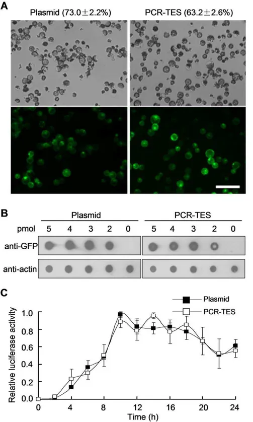
protoplasts, we compared transformations with plasmid DNA p35S-GFP and with PCR-fragment 35S-GFP-NOS in Col meso-phyll protoplasts, respectively. Similar transformation efficiency was obtained with both transformation systems (Figure 2A, Figure S2A). Reproducible results were scored in repeated experiments. For instance, 63.262.6% transformation efficiency was obtained in PCR-TES whereas 73.062.2% transformation efficiency was shown in transformation with plasmid DNA (Table S1). To further confirm the transformation efficiency at protein level the dot blot analysis was performed (Figure 2B). We titrated out the amount of transformed PCR-fragments35S-GFP-NOSand plasmid DNA in a serial concentration (0 pmol, 2 pmol, 3 pmol, 4 pmol, and 5 pmol). After transformation and incubation for 8 hours, cell lysates were extracted and detected with anti-GFP antibody. Results showed the fact that expression level of GFP protein in PCR-TES and in plasmid transformation system was comparable (Figure 2B). In addition, we tested PCR-fragments directly generated using the fusion PCR method [22]. As shown in Figure S2B, the transformation of PCR-fragments35S-GFP-NOS gener-ated through fusion PCR was comparable to that of using PCR-fragments 35S-GFP-NOSamplified from template plasmid p35S-GFP (Figure S2B). To attest PCR-fragments that directly generated from genomic DNA without plasmid construction, we amplified the PCR-fragmentSnRK2.6(G-SnRK2.6), containing its native promoter sequence, from genomic DNA of 2-week-old Arabidopsis seedlings (Figure S2C). The PCR-fragmentsSnRK2.6 (G-SnRK2.6) were transformed into mesophyll protoplasts of Col, and results showed that increased expression level ofSnRK2.6was detected in the protoplasts transformed with PCR-fragments SnRK2.6(G-SnRK2.6). In addition, the improved expression level of RD29B(an ABA responsive gene) was scored with and without ABA (5mM) treatment; the enhanced relative activity (LUC/ GUS) of RD29B-LUC was also measured (Figure S2C). Taken together, these data demonstrate that PCR-TES is applicable for examining a gene expression cassette (with its native promoter) without plasmid construction. To further explore how effective of PCR-TES at the protein level, we validated PCR-TES by transforming PCR-fragments of myc-tagging PYR1. PCR-frag-ments 35S-myc-PYR1-NOS were expressed abundantly in proto-plasts, as efficient as that shown in the transformation with plasmid p35S-myc-PYR1(Figure S2D). Collectively, these results provide the evidences in that PCR-TES could be practical for speedily examining gene expression in protoplasts.
To evaluate how stable PCR-fragments might be sustained in plant cells after transformation, we quantified activity of luciferase reporter (LUC) timely. Equal amount of PCR-fragments UBQ-LUC-NOSand plasmid DNA pUBQ-LUC was respectively trans-formed into mesophyll protoplasts. Results indicated that compa-rable expression patterns were produced in both transformation systems (Figure 2C). The LUC activity was detectable after transformation for 24 hours, indicating that PCR-fragments were as stable as plasmid DNA after being transformed into protoplasts.
Evaluations of PCR-TES
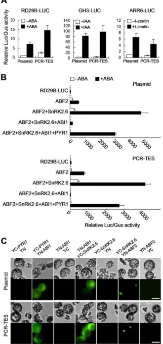
GUS activity was quantified. Despite of transforming with PCR-fragments or with plasmid DNA, similar expression patterns were scored (Figure 3A). Hence, these data support our suggestion on that PCR-TES might be used for assessing the expression level of a gene in plant cells. Thus, we designed experiments to reassemble the ABA signal transduction pathway that has been reported previously [11]. Equal amount of plasmids and PCR-fragments were transformed into Col mesophyll protoplasts, respectively. The relative LUC/GUS activity was quantified and consistent results were shown in both transformation systems (Figure 3B). In the presence of ABA, SnRK2.6 activated ABF2 was inhibited when ABI1 was added to the system; however, the inhibitory effect of ABI1 could be relieved by PYR1 (Figure 3B). To confirm this data, we examined the protein-protein interactions using TES and plasmid transformation system, respectively. PCR-fragments ofYC-PYR1 andYN-ABI1, YN-ABI1andYC-SnRK2.6, YC-SnRK2.6 and YN-ABF2 were transformed into protoplasts, respectively. The recombinant YFP signal was analyzed in each group of transformation (Figure 3C). In parallel, PCR-fragments of35S-YC-NOS(YC) and35S-YN-NOS(YN) were transformed into protoplasts and used for the negative control experiment (Figure 3C). Similar results were obtained in transformations with correspondent plasmids (Figure 3C). Together, our data are supportive to the fact that PCR-TES could facilitate the assessment of signaling molecules.
Screen of CDPK Candidates Involving in the ABA Signaling
In order to examine how practical of PCR-TES could be, we designed a functional screen looking for CDPK (calcium-dependent protein kinase) family members which might play the role in the ABA signaling. We tested 24 members of CDPK which were reported to show predominant expression patterns in Arabidopsis leaves [12]. The constitutive activation form of each CDPK (CPKac) was generated by deleting the C-terminal Ca2+ regulatory domain and auto-inhibitory domain [12]. PCR-fragments composing c-myc tagged CPKac (Figure S3A) were transformed into protoplasts along with plasmid pRD29B-LUC (ABA-responsive reporter construct). In the absence of ABA, 9 out of 24CPKac, such asCPKac4,CPKac5,CPKac6,CPKac7,CPKac10, CPKac11,CPKac12,CPKac26andCPKac30, could trigger activity of RD29B-LUC; more than five-fold increase in relative activity of RD29B-LUCwas scored (Figure 4A).
It has been reported that some family members of CDPKs can phosphorylate ABF1 and ABF4 [23]. We attempted to test the role of screened 9 CPKac candidates in the regulation of ABF2, a bZIP transcription factor that has been reported to be one of players in response to ABA [24]. Results showed that synergistic effects in activating RD29B-LUC were produced when ABF2 was coex-pressed. Significant increase in relative activity ofRD29B-LUCwas observed in coexpressions with CPKac4, CPKac7, CPKac10 and CPKac30(Figure 4B). To explore the correlation between CPKac and ABF2, we examined interactions between ABF2 and CPKac4, CPKac7, CPKac10, or CPKac30, respectively. The control experiment was to analyze the interaction between SnRK2.6 and ABF2 [11]. As results, each examined CPKac exhibited ability
to interact with ABF2 in the nucleus. (Figure 4C and Figure S3B). Thus, our data implicated the notion that the regulation of CPKac to ABF2 (or other bZIP transcriptional factors) might be executed in the nucleus. Additionally, we attested the interaction of ABI1 and CPKac, and results suggest that CPKac4 is possible to interact with ABI1 (Figure 4C). To verify the reliability of results from PCR-TES we repeated all analyses in transformations with plasmids DNA. Not surprising, consistent results were reproduced (Figure S3C). Overall, our results not only prove the credibility of PCR-TES, but also suggest that CPKac4 might be a target or serves as an interactive molecule to ABI1.
Reconstitution of CPK4-mediated ABA Signaling Pathway with PCR-TES
To determine the role ofCPK4in the ABA signal transduction pathway, we focused on examining the full length CPK4. Consistent result was obtained in PCR-TES and plasmid transformation system. The interaction between CPK4 and ABF2 was observed in protoplasts (Figure 5A). In ABA treatment, CPK4andABF2could synergistically affectRD29B-LUCactivity. The relative activity (LUC/GUS) ofRD29B-LUC was enhanced more than 2000-fold in the presence of ABA. In contrast, the relative activity (LUC/GUS) of RD29B-LUC was strikingly inhibited by ABI1; however, the inhibitory effect of ABI1 could be rescued by PYR1 upon ABA treatment (Figure 5B). These results were further confirmed in transformation with correspon-dent plasmids (Figure 5B). To investigate the role of CPK4 in modulatingABF2activity while responding to ABA, recombinant proteins of His-PYR1, His-ABF2, His-CPK4 and GST-ABI1 were prepared (Figure S4) and analyzed (Figure 5C). Undertaken the in vitrokinase assay, we found out that CPK4 could phosphorylate ABF2. The ABI1 inhibitory effect on CPK4 kinase activity could be partially compromised by PYR1 and eventually reversed by ABA (Figure 5C). In addition, we noticed that self-phosphorylation of CPK4 was phenomenal when ABI1 was present in the system (Figure 5C, lane 3), which was similar to that observed in analyzing CPK23 [13]. Taken together, our results provide supportive evidences on that PCR-TES is applicable for rapidly screening molecular candidates and for analyzing molecular mechanisms underlying the ABA signal transduction.
Discussion
PCR-TES can be a Pivotal Tool for the Cell-based Assay For studying regulations of genes in plant cells, diverse approaches are undertaken at desired circumstances. In this report, we introduce a simple system that can be used for quickly examining a gene function in plant cells. We demonstrated that PCR-TES (PCR-fragment based transient expression system) could facilitate preliminary screening for putative candidates in the transient expression assay. The transformation efficiency of PCR-TES can be comparable to that of conventional plasmid transformation.
Figure 2. Transient expression of PCR-fragments inArabidopsismesophyll protoplasts.(A) Protoplasts were transfected with plasmid p35S-GFP(Plasmid) or with PCR-fragments35S-GFP-NOS(PCR-TES). Transformation efficiencies were determined by analyzing the protoplasts with fluorescence after incubation for overnight. Data represent the means6SEM from repeated experiment (n = 4). Scale bar, 200mm. (B) Comparisons of transformation efficiencies at the protein level with the dot blot assay. A serial concentrations (5 pmol, 4 pmol, 3 pmol, 2 pmol and 0 pmol) of p35S-GFP(Plasmid) or PCR-fragments of35S-GFP-NOS(PCR-TES) were used to titrate out the transformation efficiency. Dots were blotted with anti-GFP antibody. The anti-actin shows the internal control. (C) Analysis on the stability of PCR-fragments in cells. Equal amount ofpUBQ10-LUC(Plasmid) or PCR-fragments ofUBQ10-LUC(PCR-TES) was transformed into 36104protoplasts and aliquot into 12 samples. Protoplasts were harvested in 2 hours apart and the luciferase activity was measured. The relative luciferase activity was calculated as the ratio against to the maximum value. Data represent the means6SEM from repeated experiment (n = 3).
doi:10.1371/journal.pone.0057171.g002
plasmid or from a source of DNA library through fusion PCR or from a source of genomic DNA into plant cells could shorten experimental procedures, because we do not have to construct or purify plasmids before conducting a preliminary screen. In conventional plasmid transformation system, over 50% transfor-mation efficiency is required for obtaining a reliable analytic result [6]. Traditional CsCl-gradient purification is used to be the way to purify large amount of high quality plasmid DNA; however it can be time consuming and troublesome [6]. Using routine plasmid preparation kit, one may be able to prepare large amount of plasmid DNA, but it might result in low transformation efficiency and unsatisfied for obtaining steady results [25] (Figure S2A). One of the plasmid midi-preparation kits (Plasmid Plus, QIAGEN, Germany) has been suggested for purifying high quality plasmids [25]. However, comparing to directly transforming PCR-frag-ments it would multiply experimental procedures, especially when we need to handle dozens of samples for preliminary screening. Thus, PCR-TES may serve as an alternative way to speedy up screen of putative molecules before doing extensive characteriza-tions. In addition, in some cases when the cloned gene is harmful toE. coligrowth, plasmid construction or propagation inE. colican be problematic [26,27]. Under these circumstances, the PCR-TES can be a way used for bypassing this problem.
Direct delivery of PCR-fragments into cells is time saving. After brief purification to remove solvents of PCR reaction, PCR-fragments are ready for transformation. PCR-TES might be feasible for conducting high throughput screening; at least, it is usable for analyzing molecular candidates. In our analyses, PCR-fragments were amplified from the template plasmid DNA. Contamination of template plasmid DNA was a concern. We clarified this fear by comparing transformations with PCR-fragments prepared from non-coding sequences of the same template plasmid. Results were cleared out this concern (Figure S1). Another argument would be the stability of transformed PCR-fragments in cells. To address this point we examined transfor-mation efficiency at the protein level and found out that PCR-fragments were as effective as plasmid DNA in plant cells after transformation (Figure 2 and Table S1). In general, the accuracy of plasmid constructs has to be confirmed by DNA sequencing. Only the confirmed DNA sequences can be constructed into a vector for making the correct plasmid. If the mutated DNA sequences were inserted into a vector and then transformed into a carrier such asE. coli, the resulted bacterial colonies would be harboring the mutations; as a consequence, all the propagated plasmids from that single colony of E. coli would also carry mutations which may cause a general effect. In PCR-TES, however, PCR-fragments are amplified from a correct plasmid template, a source of DNA library through fusion PCR [22] or a source of genomic DNA with its native promoter; among the millions of PCR fragments in a PCR reaction, mutated PCR fragments may be minor. Nobody can ‘‘assume’’ that such mutations will never cause a general effect. Therefore, we always compare results from the PCR-TES to those from the conven-tional plasmid transformations (Figure 3, Figure 5 and Figure S2). Our data indicate that consistent results were always produced in both systems.
Although PCR-TES may facilitate massive screening for candidates, its application for stable transformation is unsatisfied. If a large-scale transformation is needed, PCR-TES might not be satisfied for conducting such experiment in considering the cost of DNA polymerases for amplification of PCR-fragments. A com-promise way one might chose is to scale up the PCR reaction. In fact, we found out that 100–200mg PCR-fragments could be easily generated by scale up the PCR reaction at a relatively lower cost. Figure 3. Comparisons of transformations with plasmid DNA
and PCR fragments.(A) PCR-fragments (RD29B-LUC-NOS, GH3-LUC-NOS and ARR6-LUC-NOS) were transformed into protoplasts, respec-tively. Their correspondent plasmids were used for positive control experiments. Plasmid pUBQ10-GUSwas cotransfected as the internal control (ABA, 5mM; IAA, 1mM; t-zeatin, 10 nM). Data represent the means6SEM from repeated experiments (n = 3). (B) The reconstitution of ABA signaling pathway with PCR-TES. Mesophyll protoplasts of Col were transformed with plasmids (Plasmid), or with PCR-fragments (PCR-TES). PlasmidspRD29B-LUCandpUBQ10-GUSwere used as the ABA-responsive reporter and the internal control, respectively. After transformation, protoplasts were incubated for 5 hours in the absence of ABA (2ABA) or in the presence of 5mM ABA (+ABA). Data represent the means6SEM from repeated experiments (n = 4). (C) Interactions of PYR1-ABI1, ABI1-SnRK2.6, and SnRK2.6-ABF2 were analyzed with BiFC assay. Plasmids (p35S-YC-PYR1, p35S-ABI1-YN, p35S-YC-SnRK2.6 and p35S-YN-ABF2) and correspondent PCR-fragments were transformed into protoplasts. YC: C-terminal of YFP; YN: N-terminal of YFP. Scale bar, 30mm.
In our point of view, combining PCR-TES and plasmid transformation system may accelerate our pace in studying gene regulation in plant cells.
Application of PCR-TES in Speeding Up Screen of Putative ABA Responsive CDPKs
Thirty-four family members of calcium-dependent protein kinases (CDPKs) are found in Arabidopsisgenome. Some CDPK members involving in ABA- and/or abiotic-signal transduction pathways have been determined. For example,CPK10,CPK23and CPK32 are involved in ABA- or drought-responses [28–30]. In addition,CPK3andCPK6are key regulators in the ABA-regulated stomatal movement. ABA induced stomatal closure and activation of slow-type anion channels are inhibited incpk3andcpk6mutants [31]. Functional as kinases, CPK4, CPK11 and CPK32 can regulate activity of ABF1 or ABF4, the bZIP transcription factor that responds to ABA through phosphorylation modification [23,28]. Moreover, CPK21 and CPK23 can modulate activity of the anion channel protein SLAC1 in ABA-induced stomatal movement [13]. In this report we revealed the potential role of CPK7 and CPK26 in ABA response which was not reported in previous studies [23,28–32]. In our screen, we could determine that CPK7 and CPK26 were able to triggerRD29B-LUCactivity using PCR-TES (Figure 4A), demonstrating that PCR-TES can be a practical tool for quickly testing putative signaling molecules. In PCR-TES, we were able to determine the functional specificity of individual CDPK in regulation of ABF2 activity. Although CPK4 and CPK11 both share high identity in amino acid sequences, only CPK4 (not CPK11) could effectively influence ABF2 activity (Figure 4B). In contrast to other CDPKs, CPK28 was likely to play an opposite role in regulating ABF2 activity (Figure 4B); suggesting that the subgroup IV CDPK members (CPK16, CPK18, and CPK28) [33] may play differential role in comparison to other CDPK members. The interaction of CPK23 and ABI1 is shown with BiFC assay inXenopus oocytes[13]. Here we could demonstrate that interaction between CPK4 and ABI1 was observed in protoplasts in PCR-TES (Figure 4C and Figure 5A). In addition, CPK4 could synergistically induce theRD29B-LUCactivity in the presence of ABF2 (Figure 5B). These results support the importance of CDPKs in the ABA signal transduction pathway.
Upon ABA stimulation, the receptor PYR/RCAR recruits ABI1 (PP2C) [34,35], thus the suppression of ABI1 on kinase SnRK2.6 can be relieved; in turn, the ABA signaling is relayedvia modulating ABF2 activity [11]. Based on this theory, we were able to reconstitute the ABA response in protoplasts with PCR-TES. We attested the correlation between phosphorylation of CPK4 and activation of ABF2in vivoandin vitro. Moreover, we unveiled the negative regulation of ABI1 to CPK4 kinase activity, which is similar to the negative regulatory fashion of ABI1 to SnRK2.6 [11]. Again, the data from reconstitution of the ABA signaling in protoplasts and thein vitrokinase assay are evidently confirmed the practicality of PCR-TES.
Materials and Methods
PCR-fragments Preparation
PCR-fragments were generated in three different ways. First, PCR-fragments were directly generated from correspondent plasmids with the universal primer pair PVU-F (59 -CTGGCAC-GACAGGTTTCCCGACT-39) and PVU-R (59 -GGCGAAAGGGGGATGTGCTGCAA-39) through PCR am-plification in 35-cycles, and the polymerase KOD-Plus-Neo (Toyobo, Japan) was used. Produced PCR-fragments were purified by following the protocol of phenol/chloroform extraction and ethanol precipitation [18]. Briefly, equal volume of phenol/ chloroform (1:1) was added to the PCR mixture. The aqueous phase was collected after centrifugation (12,000 g) for 3 minutes and then mixed with 1/10 volume of sodium acetate (3 M, pH 5.2) and 2.5 volume of ice-cold 100% ethanol. After centrifugation (12,000 g) for 5 minutes, the DNA of PCR-fragments was precipitated. Through a brief washing with 70% ethanol, the DNA pellet of PCR-fragments was dissolved in water and quantified. In general, about 10–15mg PCR-fragments could be yielded from a 50ml PCR reaction and the final concentration of purified PCR-fragments was adjusted to 1mg/ml in water or TE buffer and stored at 220uC. To remove residues of template plasmid DNA from the fragments, we purified PCR-fragments through agarose gel extraction (TIANgel Midi Purifi-cation Kit, Tiangen, China). Next, PCR-fragments were produced using fusion PCR by following the method ascribed in the previous report [22] with minor modifications. In brief, fragments35Sand NOSwere amplified from plasmidp35S-MCS(kindly provided by Dr. Benedikt Kost, University of Erlangen-Nuremberg, Germany) using primer pairs Fu35S-F/Fu35S-R and FuNOS-F/FuNOS-R, respectively. CDS sequence of GFP was amplified from plasmid p35S-GFP, that was generated by inserting GFP fragment into p35S-MCS, using primer pair FuGFP-F/FuGFP-R. After agarose gel extraction, PCR-fragments 35S-GFP-NOS were assembled using the two-step fusion PCR method [22]. Purified PCR-fragments could be stored up to six months in TE buffer at 220uC. The third way to make PCR-fragments is directly amplifying a gene fragment from the genomic DNA that was extracted from 2-week-old seedlings of Arabidopsis. For instance, PCR-fragments ofSnRK2.6(G-SnRK2.6, with its native promoter) were thus generated. All primer sequences for producing PCR-fragments are listed in Table S2.
Biolistic Bombardment Assay
PCR-fragments or plasmids were delivered through the biolistic bombardment method (Bio-Rad, USA). Young leaves from 3-week-old Arabidopsis plants or epidermal peels from Onion were used in this assay. In each experiment, 2mg PCR-fragments and/ or 2mg plasmid DNA were mixed with 60 mg/ml gold particles (diameter in 1.0mm, Bio-Rad) in 25ml coating buffer that contains 1 M CaCl2, and 16 mM spermidine. The detailed protocol for the
biolistic bombardment was described in previous reports [8,9]. Figure 4. Functional analyses on CDPKs involving in the ABA signaling pathway.(A) Screening for the CPKac that may activate ABA-responsiveRD29B-LUC. Protoplasts of Col were transformed with PCR-fragments of individual CPKac. Transformation of35S-YFP-NOSwas used as the control (ctrl). PlasmidspRD29B-LUCandpUBQ10-GUSwere used as the ABA-responsive reporter and the internal control, respectively. Relative LUC/ GUS activity was measured after 5-hour incubation. The anti-myc antibody was used to show the expression level of CPKac. Arrow indicates non-specific protein band detected by anti-myc antibody. Asterisks (*) indicate 9 CPKac candidates possessed over five-fold expression levels of relative LUC/GUS activity. Data represent the means6SEM from repeated experiments (n = 4). (B) Analysis on CPKac and ABF2 in triggeringRD29B-LUCactivity in protoplasts. PCR-fragments ofABF2andCPKacwere cotransfected to mesophyll protoplasts isolated from Col leaves. PlasmidspRD29B-LUCand pUBQ10-GUS were used as the ABA-responsive reporter and internal control, respectively. Data represent the means6SEM from repeated experiments (n = 3). (C) BiFC assay to examine the interactions of CPKac and ABF2 or ABI1 in PCR-TES. PCR-fragments (YN-ABF2, YN-ABI1 and YC-CPKac) were transformed into protoplasts. YC: C-terminal of YFP; YN: N-terminal of YFP Scale bar, 30mm.
After bombardment transformed leaves were incubated for overnight under light conditions at 23uC and observed with NIKON TE-2000U fluorescent microscope (Nikon, Japan) and Olympus Confocal Laser Scanning Microscope FV1000 (Olym-pus, Japan).
Transient Expression Assay in Mesophyll Protoplasts Protoplasts were isolated from leaves of 4-week-old Col plants and transformed using the method ascribed in the previous report [6] with some modifications, in order to suite for transforming PCR-fragments. First, 16104 protoplasts in 50ml volume were mixed with plasmids DNA (in total 10mg) or PCR-fragments (in total 10mg); then, gently mixed with 60ml PEG solution (40% W/ V PEG-4000, 0.2 M mannitol, 100 mM CaCl2). After 10 minutes
incubation at room temperature, 240ml W5 solution (2 mM MES at pH 5.7, 54 mM NaCl, 125 mM CaCl2, 5 mM KCl) was added
to the mixture to stop the transformation. Protoplasts were resuspended in 250ml WI solution (4 mM MES at pH 5.7, 0.5 M mannitol, 20 mM KCl) and incubated at 23uC.
For the luciferase assay, protoplasts were harvested after 5-hour incubation under light conditions at 23uC with or without stimuli (5mM ABA, or 1mM IAA, or 10 nM t-zeatin). The activities of LUC and GUS were measured with the GloMax-Multi Jr Single Tube Multimode Reader (Promega, USA) by following the protocol described previously [6]. In each sample, 1mg plasmid pUBQ10-GUS was used as an internal control and 4mg reporter plasmids or reporter PCR-fragments were used. All experiments were repeated at least three times. For BiFC assays, protoplasts were transformed with plasmids (5mg) or PCR-fragments (5mg) and incubated for overnight at 23uC. Images were acquired with NIKON TE-2000U fluorescent microscope (Nikon, Japan).
Protein blot
For dot blot assay, the lysate of protoplasts was extracted from transformed protoplasts in 20ml lysis buffer (25 mM Tris-phosphate at pH 7.8, 2 mM DTT, 2 mM 1,2-diaminocyclohex-ane-N,N,N’,N’-tetraacetic acid, 10% glycerol, 1% Triton X-100). Then, extracted total protein was dotted and blotted on Hybond ECL Nitrocellulose Membrane (GE Healthcare) by following the manufacturer’s protocols. GFP was detected with antibody anti-GFP (Proteintech Group) whereas anti-actin (Proteintech Group) was used as the internal control. For western blot, the protoplast lysate was directly mixed with 50ml loading buffer and separated with 12% SDS-PAGE gel. Antibody of c-myc (Sigma, USA) was used to detect the protein expression level.
In vitroKinase Assay
Recombinant proteins of His-PYR1, GST-ABI1, His-CPK4 and His-ABF2 were respectively expressed inE. coliand purified. Thein vitrokinase assay was carried out according to the method described previously [12,13] with some modifications. First, His-PYR1 (2mg) and GST-ABI1 (2mg) were incubated together with or without 1mM ABA at room temperature (25uC) for 5 minutes. Afterwards, His-CPK4 (1mg) and His-ABF2 (5mg) were added to the 20ml reaction system (20 mM Tris-HCl at pH7.4, 100 mM NaCl, 12 mM MgCl2, 1 mM CaCl2, 1mM ATP, 5mCi of [c-32P]
ATP, and 1 mM DTT). After incubation for 30 minutes at 30uC, the reaction was stopped by adding 20ml 2X loading buffer and separated with 12% SDS-PAGE gel. Radioactive intensities of
c-32P were measured with Typhoon 9200 Imager (GE healthcare, USA).
Plasmids Construction
Plasmid p35S-MCS-Myc was constructed by inserting blunt ended fragment of c-myc tag into vectorp35S-MCSthat contains a 35S promoter, multiple cloning sites (MCS) and a NOS terminator (kindly provided by Dr. Benedikt Kost, University of Erlangen-Nuremberg, Germany). Plasmid p35S-MCS-YFP or p35S-MCS-CFPwas also constructed by inserting CFP or YFP into vector p35S-MCS. Plasmids p35S-YFP-mTn and p35S-CFP-mTn were constructed by modifying plasmidGFP-mTn[19] through replac-ing GFP to CFP or YFP. Plasmidp35S-NAG-YFPwas generated by replacing the CFP to YFP based on plasmid p35S-NAG-CFP (kindly provided by Dr. Jian Xu, Huazhong Agricultural University, China) [20]. Plasmid p35S-ER-CFP and p35S-ER-YFP was constructed by inserting the sequence of ER signal peptide [21] to p35S-MCS-YFP or p35S-MCS-CFP. Promoter UBQ10was amplified from plasmidpUBQ10-GUS[17] and then inserted into theLUCempty vector that was generated by deleting promoterRD29Afragment from plasmidpRD29A-LUC, therefore pUBQ-LUCwas constructed. Plasmids pUBQ10-GUS, pGH3-LUC andpARR6-LUC were both obtained from ABRC (http://www. arabidopsis.org/) [17]. PlasmidpRD29B-LUCwas constructed as described in the previous report [11]. TheCPKac plasmids were constructed by inserting individual CPKac fragment, which was amplified from the cDNA library of Col seedlings with correspondent primers (Table S2), into the vector p35S-MCS-Myc. Plasmidsp35S-YC-MCSandp35S-YN-MCSwere constructed by following the method described previously [36]. Briefly, the C-terminal of EYFP (YC) or N-C-terminal of EYFP (YN) fragment was respectively inserted into the vector. For expressing recombinant protein inE. coli,CDS ofPYR1,CPK4andABF2were cloned into pET28vector (Novagen, USA) andABI1was inserted into pGEX-6P vector (GE Healthcare, USA). Detailed information about primer sequences for plasmid construction can be found in Table S2.
Supporting Information
Figure S1 Comparisons of transformations with plasmid DNA and with PCR-fragments in epidermal cells ofOnion. Plasmid DNA ofp35S-GFPor PCR-fragments35S-GFP-NOSwas delivered into Onionepidermal cells using biolistic bombardment method. PCR-fragments (not containing GFP sequences) were amplified from the plasmid p35S-GFP and used as the negative control (Control). Images were acquired under microscope after an overnight incubation. Scale bar, 200mm.
(TIF)
Figure S2 Comparisons of protein expression levels between transformations with plasmid DNA and with PCR-fragments. (A) Comparison of transformations efficiencies using PCR-fragments and plasmids which were prepared in different methods (Mini-1, Figure 5. Analysis onCPK4function in the ABA signaling pathway.(A) Analyzing the interactions of CPK4 and ABI1 or ABF2 with PCR-TES and with the plasmid transformation system in protoplasts. YC: C-terminal of YFP; YN: N-terminal of YFP. Scale bar, 30mm. (B)CPK4involving in ABA response was characterized in both systems (PCR-TES, Plasmid). PlasmidspRD29B-LUCandpUBQ10-GUSwere used as the ABA-responsive reporter and the internal control, respectively. After transformation, protoplasts were incubated for 5 hours in the absence of ABA (2ABA) or in the presence of 5mM ABA (+ABA). Data represent the means6SEM from repeated experiments (n = 4). (C) Phosphorylation of CPK4 to ABF2 was demonstrated within vitrokinase assay (left panel). Relative radioactive intensities ofc-32P were quantified (right panel) with Typhoon 9200 Imager.
Mini-2 and Mini-3 stand for mini-prepared plasmids; Maxi: maxi-prepared plasmids; CsCl: using CsCl maxi-prepared plasmids). Trans-formation efficiencies were quantified. Data represent the mean-s6SEM from repeated experiment (n = 3). Scale bar, 200mm. (B) Transient expression of PCR-fragment from fusion PCR. Left panel: Diagram (not in scale) to show construction of the PCR-fragment cassette using fusion PCR. Right panel: PCR-PCR-fragment 35S-GFP-NOS was amplified from plasmid p35S-GFP (from plasmid) or using fusion PCR (from fusion PCR), and then transformed into protoplast. (Chloroplast: autofluorescence). Scale bar, 200mm. (C) Transient expression of PCR-fragment from genomic DNA. PCR-fragment SnRK2.6 was generated using genomic DNA of Arabidopsis, and then tested in transient expression assay. Upper panel: Diagram (not in scale) to show construction of SnRK2.6 PCR-fragment cassette. PCR-fragment SnRK2.6(G-SnRK2.6) was transformed into Col protoplasts. After transformation, protoplasts were incubated for 6 hours without (2ABA) or with 5mM ABA (+ABA). The gene expression level of SnRK2.6andRD29Bwas quantified using qRT-PCR. The ABA-induced RD29B-LUC activity was also quantified. Plasmid pUBQ10-GUSwas used as the internal control. All data represent the means6SEM from repeated experiments (n = 3). (D) Plasmid p35S-myc-PYR1 and PCR-fragments 35S-myc-PYR1-NOS were transformed into protoplasts, respectively; a serial of dilution of protoplasts was titrated out for detecting myc-PYR1. Total proteins were extracted after 8-hour incubation. Protein expres-sion level of myPYR1 was determined by western blot with c-myc antibody. Control, protein extract from protoplasts without transformation; Lane 1, 1:20 dilution of prepared protoplasts (2X10⁵ protoplasts/ml); Lane 2, 1:40 dilution of prepared protoplasts; Lane 3, 1:80 dilution of prepared protoplasts. (TIF)
Figure S3 Analysis on interactions in BiFC assay. (A) Diagram (not in scale) to show components of PCR-fragments of CPKac. (B) Subcellular localization for interactions betweenCPKac or SnRK2.6 and ABF2 in BiFC assay. Plasmid CPKac4, p35S-YC-CPKac7, p35S-YC-CPKac10,p35S-YC-CPKac30 or p35S-YC-SnRK2.6
was cotransfected withp35S-YN-ABF2into protoplasts. p35S-CFP-ABF2was co-transformed to mark the nucleus [24]. Merge shows the colocalizations. YC: C-terminal of YFP; YN: N-terminal of YFP. Scale bar, 30mm. (C) Interaction betweenABI1orABF2and CPKacassessed in BiFC assays. PlasmidCPKac4, p35S-YC-CPKac7, p35S-YC-CPKac10 or p35S-YC-CPKac30 was transfected together with p35S-YN-ABI1 or p35S-YN-ABF2 into protoplasts, respectively. YC: C-terminal of YFP; YN: N-terminal of YFP. Scale bar, 30mm.
(TIF)
Figure S4 Analyzing expressions of recombinant proteins. Recombinant proteins of His-PYR1, GST-ABI1, His-CPK4 and His-ABF2 were analyzed in 12% SDS-PAGE gel and shown in coomassie staining.
(TIF)
Table S1 Comparisons of transformation efficiencies in proto-plasts.
(DOC)
Table S2 Primer sequences for plasmids constructions and qRT-PCR experiments.
(DOC)
Acknowledgments
We thank Wu lab members for technique help and helpful comments on the manuscripts. We thank Dr. Benedikt Kost (University of Erlangen-Nuremberg, Germany) for sharing plasmidsp35S-MCS. We thank Dr. Jian Xu (Huazhong Agricultural University, China) for sharing plasmid p35S-NAG-CFP. We thank Syngenta for providing the Syngenta Friendly Laboratories Scholarship to Yuming Lu.
Author Contributions
Conceived and designed the experiments: Yan Wu YL. Performed the experiments: YL XC. Analyzed the data: Yan Wu YL. Contributed reagents/materials/analysis tools: Yuxuan Wu Y. Wang YH. Wrote the paper: Yan Wu YL.
References
1. Dean C, Jones J, Favreau M, Dunsmuir P, Bedbrook J (1988) Influence of flanking sequences on variability in expression levels of an introduced gene in transgenic tobacco plants. Nucleic Acids Res 16: 9267–9283.
2. Peach C, Velten J (1991) Transgene expression variability (position effect) of CAT and GUS reporter genes driven by linked divergent T-DNA promoters. Plant Mol Biol 17: 49–60.
3. Periasamy A (2001) Fluorescence resonance energy transfer microscopy: a mini review. Journal of Biomedical Optics 6: 287–291.
4. Chen Y, Mills JD, Periasamy A (2003) Protein localization in living cells and tissues using FRET and FLIM. Differentiation 71: 528–541.
5. Koroleva OA, Tomlinson ML, Leader D, Shaw P, Doonan JH (2005) High-throughput protein localization in Arabidopsis using Agrobacterium-mediated transient expression of GFP-ORF fusions. Plant J 41: 162–174.
6. Yoo SD, Cho YH, Sheen J (2007) Arabidopsis mesophyll protoplasts: a versatile cell system for transient gene expression analysis. Nature Protocols 2: 1565– 1572.
7. Wu Y, Kuzma J, Marechal E, Graeff R, Lee HC, et al. (1997) Abscisic acid signaling through cyclic ADP-ribose in plants. Science 278: 2126–2130. 8. Seki M, Lida A, Morikawa H (1999) Transient expression of the
beta-glucuronidase gene in tissues of Arabidopsis thaliana by bombardment-mediated transformation. Molecular Biotechnology 11: 251–255.
9. Ueki S, Lacroix B, Krichevsky A, Lazarowitz SG, Citovsky V (2009) Functional transient genetic transformation of Arabidopsis leaves by biolistic bombardment. Nature Protocols 4: 71–77.
10. Zhang X, Henriques R, Lin SS, Niu QW, Chua NH (2006) Agrobacterium-mediated transformation of Arabidopsis thaliana using the floral dip method. Nature Protocols 1: 641–646.
11. Fujii H, Chinnusamy V, Rodrigues A, Rubio S, Antoni R, et al. (2009) In vitro reconstitution of an abscisic acid signalling pathway. Nature 462: 660–664.
12. Boudsocq M, Willmann MR, McCormack M, Lee H, Shan L, et al. (2010) Differential innate immune signalling via Ca2+
sensor protein kinases. Nature 464: 418–422.
13. Geiger D, Scherzer S, Mumm P, Marten I, Ache P, et al. (2010) Guard cell anion channel SLAC1 is regulated by CDPK protein kinases with distinct Ca2+ affinities. Proc Natl Acad Sci USA 107: 8023–8028.
14. Li S, Brisson M, He Y, Huang L (1997) Delivery of a PCR amplified DNA fragment into cells: a model for using synthetic genes for gene therapy. Gene Ther 4: 449–454.
15. Isalan M, Santori MI, Gonzalez C, Serrano L (2005) Localized transfection on arrays of magnetic beads coated with PCR products. Nature Methods 2: 113– 118.
16. Odell JT, Nagy F, Chua NH (1985) Identification of DNA sequences required for activity of the cauliflower mosaic virus 35S promoter. Nature 313: 810–812. 17. Hwang I, Sheen J (2001) Two-component circuitry in Arabidopsis cytokinin
signal transduction. Nature 413: 383–389.
18. Sambrook J, Russell DW (2006) Purification of Nucleic Acids by Extraction with Phenol:Chloroform. Cold Spring Harb Protoc.
19. Kost B, Spielhofer P, Chua NH (1998) A GFP-mouse talin fusion protein labels plant actin filaments in vivo and visualizes the actin cytoskeleton in growing pollen tubes. Plant J 16: 393–401.
20. Xu J, Scheres B (2005) Dissection of Arabidopsis ADP-RIBOSYLATION FACTOR 1 function in epidermal cell polarity. Plant Cell 17: 525–536. 21. Nelson BK, Cai X, Nebenfuhr A (2007) A multicolored set of in vivo organelle
markers for co-localization studies in Arabidopsis and other plants. Plant J 51: 1126–1136.
22. Shevchuk NA, Bryksin AV, Nusinovich YA, Cabello FC, Sutherland M, et al. (2004) Construction of long DNA molecules using long PCR-based fusion of several fragments simultaneously. Nucleic Acids Res 32: e19.
23. Zhu SY, Yu XC, Wang XJ, Zhao R, Li Y, et al. (2007) Two calcium-dependent protein kinases, CPK4 and CPK11, regulate abscisic acid signal transduction in Arabidopsis. Plant Cell 19: 3019–3036.
24. Fujita Y, Fujita M, Satoh R, Maruyama K, Parvez MM, et al. (2005) AREB1 is a transcription activator of novel ABRE-dependent ABA signaling that enhances drought stress tolerance in Arabidopsis. Plant Cell 17: 3470–3488.
25. Wehner N, Hartmann L, Ehlert A, Bottner S, Onate-Sanchez L, et al. (2011) High-throughput protoplast transactivation (PTA) system for the analysis of Arabidopsis transcription factor function. Plant J 68: 560–569.
26. Kobayashi M, Kurusu Y, Yukawa H (1991) High-expression of a target gene and high-stability of the plasmid. Appl Biochem Biotechnol 27: 145–162. 27. Saida F, Uzan M, Odaert B, Bontems F (2006) Expression of highly toxic genes
in E. coli: special strategies and genetic tools. Curr Protein Pept Sci 7: 47–56. 28. Choi HI, Park HJ, Park JH, Kim S, Im MY, et al. (2005) Arabidopsis
calcium-dependent protein kinase AtCPK32 interacts with ABF4, a transcriptional regulator of abscisic acid-responsive gene expression, and modulates its activity. Plant Physiol 139: 1750–1761.
29. Ma SY, Wu WH (2007) AtCPK23 functions in Arabidopsis responses to drought and salt stresses. Plant Mol Biol 65: 511–518.
30. Zou JJ, Wei FJ, Wang C, Wu JJ, Ratnasekera D, et al. (2010) Arabidopsis calcium-dependent protein kinase CPK10 functions in abscisic acid- and Ca2+
-mediated stomatal regulation in response to drought stress. Plant Physiol 154: 1232–1243.
31. Mori IC, Murata Y, Yang Y, Munemasa S, Wang YF, et al. (2006) CDPKs CPK6 and CPK3 function in ABA regulation of guard cell S-type anion- and Ca2+
-permeable channels and stomatal closure. PLoS Biol 4: e327. 32. Zhao R, Wang XF, Zhang DP (2011) CPK12: A Ca2+
-dependent protein kinase balancer in abscisic acid signaling. Plant Signal Behav 6: 1687–1690. 33. Cheng SH, Willmann MR, Chen HC, Sheen J (2002) Calcium signaling through
protein kinases. The Arabidopsis calcium-dependent protein kinase gene family. Plant Physiol 129: 469–485.
34. Ma Y, Szostkiewicz I, Korte A, Moes D, Yang Y, et al. (2009) Regulators of PP2C Phosphatase Activity Function as Abscisic Acid Sensors. Science 324: 1064–1068.
35. Park SY, Fung P, Nishimura N, Jensen DR, Fujii H, et al. (2009) Abscisic Acid Inhibits Type 2C Protein Phosphatases via the PYR/PYL Family of START Proteins. Science 324: 1068–1071.