The Study to Understand the Genetics of the
Acute Response to Metformin and Glipizide in
Humans (SUGAR-MGH): Design of a
pharmacogenetic Resource for Type 2
Diabetes
Geoffrey A. Walford1,2,3‡, Natalia Colomo1,4‡, Jennifer N. Todd1,3,5‡, Liana K. Billings1,2,3,6‡, Marlene Fernandez1‡, Bindu Chamarthi7,8, A. Sofia Warner1, Jaclyn Davis1, Katherine R. Littleton1, Alicia M. Hernandez1, Rebecca R. Fanelli1, Amelia Lanier1, Corinne Barbato9, Rachel J. Ackerman1, Sabina Q. Khan1, Rosa Bui9, Laurel Garber9, Elliot S. Stolerman1, Allan F. Moore1,2,3†, Chunmei Huang3, Varinderpal Kaur1, Maegan Harden10,
Andrew Taylor1, Ling Chen1, Alisa K. Manning1, Paul Huang3, Deborah Wexler2,3, Rita M. McCarthy8, Janet Lo3, Melissa K. Thomas2,3, Richard W. Grant11, Allison Goldfine3,9, Margo S. Hudson8, Jose C. Florez1,2,3*
1Center for Human Genetic Research, Massachusetts General Hospital, Boston, Massachusetts, United States of America,2Diabetes Research Center, Diabetes Unit, Department of Medicine, Massachusetts General Hospital, Boston, Massachusetts, United States of America,3Harvard Medical School, Boston, Massachusetts, United States of America,4Department of Endocrinology and Nutrition. Hospital Universitario Regional de Málaga. Instituto de Investigación Biomédica de Málaga (IBIMA). Málaga, Spain,
5Boston Children’s Hospital, Boston, Massachusetts, United States of America,6Division of Endocrinology and Metabolism, NorthShore University Health System, Evanston, Illinois, United States of America,
7Department of Medicine, Brigham and Women’s Hospital, Boston, Massachusetts, United States of America,8Division of Endocrinology, Diabetes, and Hypertension, Brigham and Women’s Hospital, Boston, Massachusetts, United States of America,9Joslin Diabetes Center, Boston, Massachusetts, United States of America,10Genomics Platform, Broad Institute, Cambridge, Massachusetts, United States of America,
11Division of Research, Kaiser Permanente Northern California, Oakland, California, United States of America
†Deceased.
‡These authors are co-first authors on this work. *jcflorez@partners.org
Abstract
Objective
Genome-wide association studies have uncovered a large number of genetic variants asso-ciated with type 2 diabetes or related phenotypes. In many cases the causal gene or poly-morphism has not been identified, and its impact on response to anti-hyperglycemic medications is unknown. The Study to Understand the Genetics of the Acute Response to Metformin and Glipizide in Humans (SUGAR-MGH,NCT01762046) is a novel resource of genetic and biochemical data following glipizide and metformin administration. We describe recruitment, enrollment, and phenotyping procedures and preliminary results for the first 668 OPEN ACCESS
Citation:Walford GA, Colomo N, Todd JN, Billings LK, Fernandez M, Chamarthi B, et al. (2015) The Study to Understand the Genetics of the Acute Response to Metformin and Glipizide in Humans (SUGAR-MGH): Design of a pharmacogenetic Resource for Type 2 Diabetes. PLoS ONE 10(3): e0121553. doi:10.1371/journal.pone.0121553
Academic Editor:Peyman Björklund, Uppsala University, SWEDEN
Received:February 18, 2014
Accepted:February 13, 2015
Published:March 26, 2015
Copyright:© 2015 Walford et al. This is an open access article distributed under the terms of the Creative Commons Attribution License, which permits unrestricted use, distribution, and reproduction in any medium, provided the original author and source are credited.
of our planned 1,000 participants enriched for individuals at risk of requiring anti-diabetic therapy in the future.
Methods
All individuals are challenged with 5 mg glipizide × 1; twice daily 500 mg metformin × 2 days; and 75-g oral glucose tolerance test following metformin. Genetic variants associated with glycemic traits and blood glucose, insulin, and other hormones at baseline and follow-ing each intervention are measured.
Results
Approximately 50% of the cohort is female and 30% belong to an ethnic minority group. Fol-lowing glipizide administration, peak insulin occurred at 60 minutes and trough glucose at 120 minutes. Thirty percent of participants experienced non-severe symptomatic hypogly-cemia and required rescue with oral glucose. Following metformin administration, fasting glucose and insulin were reduced. Common genetic variants were associated with fasting glucose levels.
Conclusions
SUGAR-MGH represents a viable pharmacogenetic resource which, when completed, will serve to characterize genetic influences on pharmacological perturbations, and help estab-lish the functional relevance of newly discovered genetic loci to therapy of type 2 diabetes.
Trial Registration
ClinicalTrials.govNCT01762046
Introduction
In recent years we have witnessed an explosion of genetic discovery, driven by the development of high-throughput genotyping and sequencing techniques, the implementation of rigorous and
novel analytical methods, and widespread international collaboration [1,2]. In type 2 diabetes,
there are now over 60 loci associated with the disease at genome-wide levels of statistical
signifi-cance [3,4]; similar progress has been made for type 2 diabetes-related quantitative traits [5].
However, in spite of substantial advances in the mapping of genomic regions whose variation contributes to type 2 diabetes pathogenesis, both the elucidation of functional mechanism and their clinical translation lag behind. In most cases the precise nucleotide variant or gene whose alteration gives rise to the phenotype have not been identified, hindering the rapid generation of animal or cellular experimental models, the validation of drug targets, and the development of gene-based therapeutics. Beyond the absence of clear molecular mechanisms, the ability of type 2 diabetes-associated genetic markers to improve disease prediction over common clinical
vari-ables is limited [6–8], and the selection of therapeutic approaches for patients with type 2
diabe-tes remains algorithmic despite recent attempts at greater individualization [9,10].
Pharmacogenetic studies offer an opportunity to address both scientific shortcomings [11]. The robust association of a genomic region with drug response (regardless of whether the test-ed variant tags the causal nucleotide or is itself causative) can help tailor therapy bastest-ed on academic health care centers). The funders had no
role in study design, data collection and analysis, decision to publish, or preparations of the manuscript.
genetic determinants relevant to a specific agent; similarly, differential perturbation of the
human organism with a medication that has known physiological effectsin vivo, contingent on
the allele at a specific locus, can help implicate a gene associated with type 2 diabetes through an agnostic genomic search but for which a clear mechanism of action was lacking.
We therefore designed a study that might serve multiple purposes. The Study to Understand the Genetics of the Acute Response to Metformin and Glipizide in Humans (SUGAR-MGH) employs two pharmacological interventions (glipizide and metformin) chosen to perturb two different limbs of the glucose homeostatic system (insulin secretion and insulin action), under basal and hyperglycemic conditions; it collects physiological, hormonal, metabolomic and ge-netic measures; and it does so under a relatively simple protocol that allows for the efficient en-rollment and retention of a sufficiently large number of participants to support genetic
analyses. This paper describes the study protocol, recruitment methods, physiological measure-ments, and intervention outcomes. We also perform baseline comparisons to guide the selec-tion of primary and secondary phenotype endpoints in the first two thirds of our intended final enrollment of 1,000 participants, and demonstrate the use of genetic risk scores (GRS) based on fasting glucose or insulin.
Materials and Methods
Cohort and Study Design
SUGAR-MGH (ClinicalTrials.Gov:NCT01762046, clinicaltrials.gov/ct2/show/NCT01762046)
was established in 2007 with funding from the National Institutes of Health, and a streamlined protocol was designed that requires only two clinical research center (CRC) visits and 1 week of active treatment per subject (Fig. 1). SUGAR-MGH was approved by the local human re-search committee on April 14, 2007. The TREND Statement Checklist has been uploaded as a supplementary file (S1 TREND Checklist). The first participant was enrolled on February 14, 2008. Recruitment is ongoing, and all subjects are followed until study completion or loss to follow-up. All CRC visits took place at an academic medical center (Massachusetts General
Hospital, Brigham and Women’s Hospital, or the Joslin Diabetes Center, all in Boston, MA).
Interventions were administered by study staff at the CRC to each participant separately.
Registration inClinicalTrials.govwas not immediately sought because the aim of the study,
using existing approved drugs to modulate the glycemic homeostasis system and compare acute responses across genotypes, was meant to characterize the physiological impact of genetic changes and not to support new indications for these drugs. Despite the issuance of stricter guidelines effective July 1, 2008, communications from both the program officer at the Nation-al Institutes of HeNation-alth (August 21, 2007) and the locNation-al human research committee (September 14, 2011) confirmed that our study did not require registration. However, in the face of contin-uously evolving guidelines and their potentially equivocal interpretation with respect to SUGAR-MGH, the study team initiated registration in late 2012 and accomplished registration inClinicalTrials.govin January 2013. The authors confirm that all ongoing and related trials for this drug/intervention are registered.
Ethics Statement
The protocol is approved by the Partners Human Research Committee (Partners Healthcare, Boston, MA). Written informed consent is obtained from all study participants; the original consent form (S1 File) and the original study protocol (S1 Protocol), as well as the most recent
and current versions of these documents (S2 FileandS2 Protocol, respectively), approved by
Visit 1 (Day 1). After an overnight fast of at least 8 hours, participants receive a single, open-label oral dose of 5 mg glipizide in the CRC and remain resident in the CRC through
con-clusion of the 240 minute glipizide challenge. Participants with a fasting blood glucose<4.44
mmol/L are not dosed with glipizide. In addition, the period of observation following glipizide administration may be terminated prior to 240 minutes if a participant develops
neuroglycope-nic symptoms (confusion, blurred vision, slurred speech), a blood glucose2.77 mmol/L with
symptoms of hypoglycemia, blood glucose<2.50 mmol/L with or without symptoms of
hypo-glycemia, or at the discretion of study staff based on clinical assessment. All participants are provided with a meal at the end of the study visit and discharged only when blood glucose is
documented to be greater than 4.44 mmol/L. Five days later, an adequate“wash-out”period
for glipizide, participants begin a two-day open-label course of 500 mg metformin, orally, twice daily. Participants who are discovered to have contraindications to safe metformin use at Visit Fig 1. SUGAR-MGH protocol and schedule of events.All events at Visit 1 and Visit 2 occur in the Clinical Research Center.
1 screening laboratories are informed not to take the medication. Participants are permitted to take fewer than the four prescribed doses of metformin should they develop side effects consis-tent with metformin intolerance.
Visit 2 (Day 8). After another overnight fast of at least 8 hours, participants return to the CRC, receive the fourth dose of metformin and one hour later undergo a standard, two-hour, 75-g oral glucose tolerance test (OGTT).
Rationale for interventions
Glipizide and metformin are generic medications commonly used to treat type 2 diabetes [12]. Metformin and glipizide are considered first and second-line therapy, respectively, for
individ-uals with newly diagnosed diabetes by major professional organizations [9,13]. Metformin has
further been demonstrated to be effective in preventing incident diabetes in at-risk individuals [14,15]. Yet, clinical response to both therapies is heterogeneous, and many patients with type 2 diabetes treated with either metformin or glipizide eventually require additional therapy [16, 17]. Therefore, understanding and characterizing the role of genetics in the response to both medications has direct clinical relevance. Given their different mechanisms of action, glipizide through increased insulin secretion and metformin through reduced hepatic glucose output, the study of the response to these two medications is hypothesized to reveal distinct influences on glucose homeostasis. A 75-g OGTT is used in clinical practice for the diagnosis of diabetes, and, in SUGAR-MGH, tests the physiological response to oral glucose ingestion in the presence of metformin.
Participants
Male or non-pregnant female adults, naïve to glipizide and metformin, are eligible for the study. Individuals at high risk of developing type 2 diabetes are preferentially enrolled by tar-geting for recruitment persons with the metabolic syndrome, obesity, a history of gestational diabetes, a history of polycystic ovary syndrome, or a family history of type 2 diabetes; individ-uals with lifestyle-controlled type 2 diabetes are also eligible for the study. The protocol ex-cludes individuals who are currently taking medications used to treat diabetes or that are known to affect glycemic parameters, have had onset of diabetes before age 25 with autosomal transmission of diabetes across three generations, a history of liver or kidney disease (including
estimated glomerular filtration rate<60 ml/min/1,73m2), severe allergic reactions to
ethnic groups, many of which are at increased risk of type 2 diabetes. Eligible participants are identified using electronic databases and local advertising, and the majority of subjects who consent for participation also agree to be re-contacted for future studies. Participants are reim-bursed $50 for completion of Visit 1 and $50 for completion of Visit 2.
Measurements
A medical history is obtained from all participants; and weight, height, blood pressure, and heart rate are measured at each visit to the CRC. DNA is extracted and genotyping is per-formed using the iPLEX-GOLD assay from Sequenom by allele-specific primer extension of amplified products, with detection by mass spectroscopy [18]. Plasma glucose is measured by hexokinase assay (Roche; Indianapolis, IN). Insulin international units are determined using a radio-immunoassay (Beckman Coulter; Fullerton, CA). Insulin resistance by homeostasis model assessment (HOMA-IR) is calculated as described in [19]. C-peptide is measured by radio-immunoassay (Siemens, KPED1, Erlangen, Germany); glucagon is measured by radioim-munoassay (LINCOplex Kit, HENDO-65K-Rev, St. Charles, MO) and proinsulin by immuno-chemiluminometric assay, both at Mayo Medical Laboratories. Incretin hormones (glucagon-like peptide-1 [GLP-1] and glucose-dependent insulinotropic polypeptide [GIP]) are measured from blood samples collected in pre-chilled EDTA tubes containing DPP-IV inhibitor
(Milli-pore; Billerica, MA) in a subset of participants. Active GLP-1 (7–36, 7–37) is measured using
the GLP-1(Active) ELISA kit (Millipore; Billerica, MA); total GLP-1 is measured using the
GLP-1(7–36 and 9–36) ELISA kit from Alpco Immunoassays (Salem, NH); and GIP is
mea-sured by using the Human GIP Total ELISA kit from Millipore (Millipore; Billerica, MA). Me-tabolite profiling has been conducted in subset of participants on plasma as previously
described [20–22]; a pilot metabolomic study in this cohort has been published [23].
Addition-al blood samples are stored at -80°C for future anAddition-alysis. Participants are given a prospective food log, completed on three week days and one weekend day between the glipizide challenge and OGTT challenge. All of the food records are analyzed using Nutrition Data System for Re-search 2009.
CAMP MGH
Sixty-four (64) participants who completed SUGAR-MGH had previously participated in the MGH Cardiology and Metabolic Patient Cohort study (CAMP MGH). These CAMP MGH participants all underwent a 75-g OGTT in the absence of anti-hyperglycemic therapies, in which plasma glucose was measured by hexokinase assay (Abbott; Chicago, IL), and insulin in-ternational units were determined using a radio-immunoassay (Roche; Indianapolis, IN). Of these participants, complete data during the CAMP MGH study were available for measures of fasting glucose (n = 64), fasting insulin (n = 51), and glucose at 30 and 120 minutes during the OGTT (n = 64). Data from the CAMP MGH OGTT (without metformin) and SUGAR-MGH OGTT (with metformin) were compared in this subset of participants.
Statistical Analyses
A sample size of 1000 participants was chosen to permit sufficient statistical power to detect the effect of commonly occurring genetic variation on pharmacological responses. At
alpha = 0.05, there will be 89% power to detect a variant in 10% of the population that explains 1% of the variance in a phenotype. There will be 99% power to detect the same variant if it ex-plains 5% or more of the variance in a phenotype.
Research Infrastructure & Services group [24]. Area under the curve (AUC) for increases in in-sulin during the glipizide challenge and for increases in glucose and inin-sulin during the OGTT challenge was calculated by the trapezoidal method and adjusted for baseline values. Area over the curve for decreases in glucose during the glipizide challenge was calculated by subtracting AUC by the trapezoidal method from the baseline glucose value at Visit 1 × total time for the glipizide challenge. All data underlying the findings described in this manuscript have been up-loaded as a supplementary file (S3 File).
Mean ± standard deviation or median [interquartile range (IQR)], respectively, are provided for continuous normally or non-normally distributed traits unless otherwise specified. For sta-tistical comparisons, non-normally distributed data were log-transformed, and all groups were compared using t-tests, and serial assessments within participants were compared using paired t-tests. Missing data were not imputed. If results at one assessment from a paired comparison were missing, the participant was excluded from analyses. Mean of group difference or paired difference and 95% confidence intervals are provided for all comparisons. For log-transformed data, mean of group log difference or paired log difference was derived from the mean compari-son between log-transformed values at the two group or paired assessments, respectively. Asso-ciation of selected endpoints with ethnicity was performed by linear regression models adjusted for age, sex, and BMI. Linear regression models, with and without age, sex, and ethnicity, were also used to test the association of genotype or genetic risk score on fasting glucose and insulin.
The threshold for statistical significance in all analyses was set at two-tailedα= 0.05.
Statistical analyses were initially performed by NC using IBM SPSS Statistics for Windows (version 20.0; Armonk, NY) and independently confirmed by JT and MF using STATA (ver-sion 12.1; College Station, Texas). Genetic power calculations were performed using QUANTO (version 1.2.4; University of Southern Califonia). Figures were constructed using GraphPad Prism (version 5.00 for Windows; San Diego, CA).
Results
Participants
The demographic characteristics of the study population at two-thirds completion are
summa-rized inTable 1; this cohort approximates the target population of individuals at risk of
requir-ing future anti-diabetic therapy. More than 30% of the current cohort was from ethnic minority populations, average BMI was in the overweight or obese range, and participants were nearly evenly distributed between men and women.
Table 1. Demographic characteristics of SUGAR-MGH participants across ethnic groups.
All participants (n = 653)
Non-Hispanic White (n = 436)
Non-Hispanic Black (n = 133)
Hispanic (n = 45)
Asian (n = 39)
Pvalue
Men (n, %) 310 (47.5) 210 (48.2) 59 (44.4) 22 (48.9) 19 (48.7) 0.88a
Women (n, %)
343 (52.5) 226 (51.8) 74 (55.6) 23 (51.1) 20 (51.3)
Age (yrs) 47.73±16.09 50.94±16.38 44.36±12.49 35.31±12.79 37.55±14.38 <0.0001b
BMI (kg/m2) 30.83±7.26 31.02±7.25 31.30±7.14 31.01±7.41 26.99±5.07 0.008c
Age and body mass index (BMI) are shown as mean±standard deviation.
a
P-value for Fisher’s Chi-Square for sex.
b
P-value for ANOVA of age; Tukey’s pairwise comparison was significant atP<0.05 for non-Hispanic White vs. non-Hispanic Black, non-Hispanic White
vs. Asian, non-Hispanic White vs. Hispanic, and Hispanic vs. non-Hispanic Black.
cP-value for ANOVA of BMI; Tukey’s pairwise comparison was signi
ficant atP<0.05 for Asian vs. non-Hispanic White and Asian vs. non-Hispanic Black.
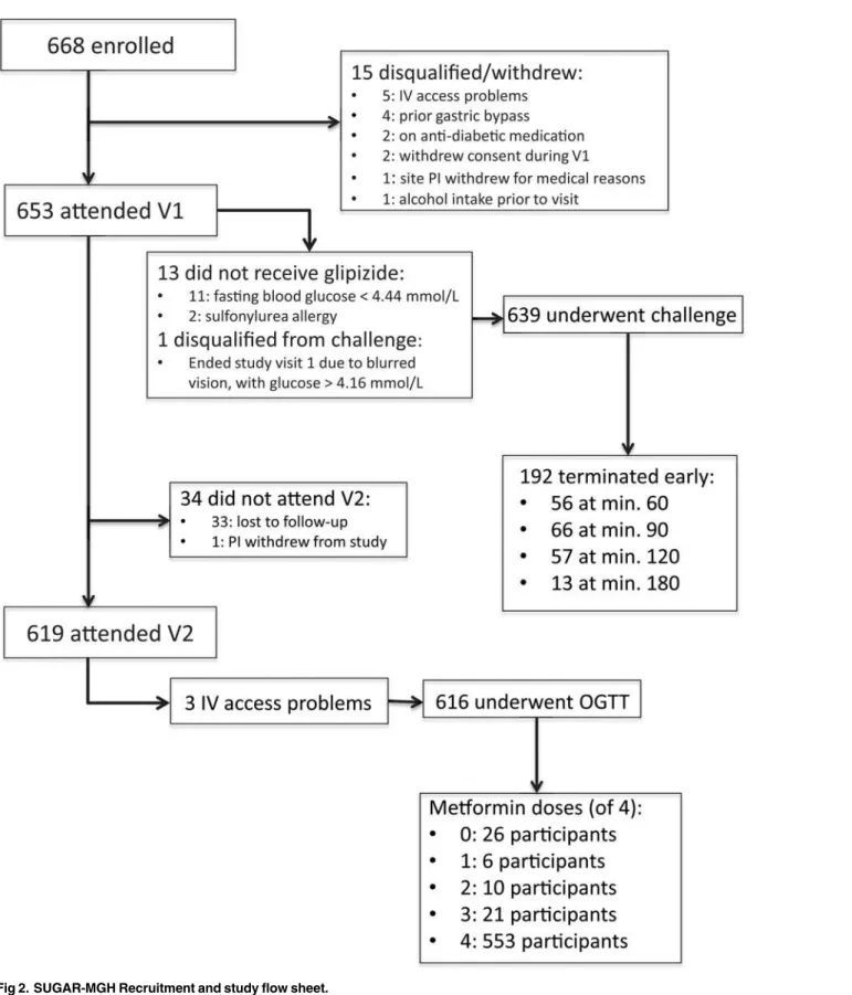
Among 1,797 individuals screened for SUGAR-MGH, 1,324 were found to be eligible, and 668 were enrolled as of March 2013. The participant disposition following enrollment is
de-picted inFig. 2. Of the 668 enrolled, 13 participants did not receive glipizide at the initial visit.
The most common reason for not administering glipizide at Visit 1 was a fasting capillary
blood glucose value<4.44 mmol/L. Six hundred and thirty-nine participants underwent the
glipizide challenge, and 192 (30.0%) terminated the observation period earlier than 240 min-utes due to reaching one of the pre-specified safety thresholds for hypoglycemia. No participant experienced a hypoglycemia-related serious adverse event, and no participant required intrave-nous glucose or glucagon to treat hypoglycemia. Six hundred and nineteen participants (94.8% of those who attended Visit 1) attended Visit 2. The most common reason for loss to follow-up in Visit 2 was failure to attend the visit by the study participant. There was no difference be-tween the participants who attended and who did not attend Visit 2 with respect to age, sex, BMI, fasting plasma glucose, fasting insulin, race or ethnicity, or any other measured baseline
characteristic (allP>0.05). The majority of Visit 2 participants took four doses of metformin
as specified in the protocol; approximately 10% took three or fewer doses.
Biochemical response to glipizide
Glipizide raised serum insulin and lowered blood glucose, as expected (Figs. 3A and 3B). Insu-lin peaked at 60 minutes, and blood glucose values nadired at 120 minutes. Glucagon levels during the glipizide challenge peaked at 180 minutes, a time point after mean blood glucose had reached its lowest value (Fig. 3C).
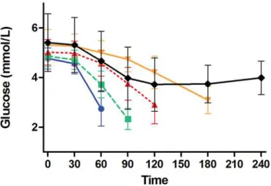
Approximately 30% of participants terminated the glipizide challenge early due to hypogly-cemia or hypoglyhypogly-cemia-related symptoms, with 56, 66, 57, and 13 participants terminating at 60, 90, 120, and 180 minutes, respectively; 447 subjects completed the entire challenge. Partici-pants who completed the entire glipizide challenge had higher fasting glucose values prior to receiving glipizide and higher trough glucose during the glipizide challenge than did partici-pants who terminated the glipizide challenge due to hypoglycemia or hypoglycemia-related symptoms (Fig. 4). Specifically, mean fasting blood glucose levels of participants who complet-ed 240 minutes of the glipizide challenge were significantly higher than those of participants
who terminated the challenge at 60 min (5.41 ± 1.17vs. 4.77 ± 0.43 mmol/L respectively; group
difference 0.64 [95% CI 0.33, 0.95];P<0.0001), at 90 min (4.86 ± 0.42 mmol/L; group
differ-ence 0.55 [95% CI 0.26, 0.83];P<0.0001) and at 120 min (5.02 ± 0.51 mmol/L; group difference
0.39 [95% CI 0.08, 0.70];P<0.0001), but were not different from those of participants who
ter-minated the challenge at 180 min (5.32 ± 0.63 mmol/L; group difference 0.09 [95% CI -0.54,
0.73];P= 0.61). Mean glucose trough values of participants who completed 240 minutes of the
glipizide challenge were significantly higher than those participants who terminated the
chal-lenge at 60 min (3.18 ± 0.74vs. 2.74 ± 0.69 mmol/L respectively; group difference 0.43 [95% CI
0.23, 0.64];P<0.0001), at 90 min (2.33 ± 0.43 mmol/L; group difference 0.85 [95% CI 0.66,
1.03];P<0.0001) or at 120 min (2.85 ± 0.72 mmol/L; group difference 0.33 [95% CI 0.12, 0.53];
P= 0.002), but were not different from those of participants who terminated the challenge at
180 min (3.06 ± 0.51 mmol/L; group difference 0.12 [95% CI -0.28, 0.53];P= 0.43).
Biochemical response to metformin
Biochemical measures at Visit 1 and Visit 2 are shown inTable 2. Among all participants, fasting
glucose, insulin, HOMA-IR, and proinsulin were all lower at Visit 2 than at Visit 1 (P= 0.0008,
P= 0.003,P= 0.0004, andP<0.0001, respectively,Table 2); fasting glucagon was not different
between the two visits (P= 0.32,Table 2). In a sub-analysis, the change in HOMA-IR was similar
Fig 2. SUGAR-MGH Recruitment and study flow sheet.
years were excluded, suggesting that the effect of metformin on insulin sensitivity was not strongly influenced by changes in menstrual cycle.
For participants who took any number of metformin doses (1, 2, 3, or 4; n = 583), fasting blood glucose, fasting insulin, and fasting HOMA-IR were lower at Visit 2 than at Visit 1
(P= 0.0002,P= 0.0006, andP<0.0001, respectively,Table 2). For participants who took no
doses of metformin (n = 26), fasting blood glucose, fasting insulin, and fasting HOMA-IR were
not different at Visit 1 and Visit 2 (P= 0.22,P= 0.07,P= 0.06, respectively,Table 2). As a
re-sult, fasting blood glucose, fasting insulin, and fasting HOMA-IR at Visit 2 were lower for par-ticipants who took any dose of metformin as compared with parpar-ticipants who took no dose of
metformin (5.04 ± 0.89vs. 5.85 ± 1.48 mmol/L; group difference -0.81 [95% CI -1.18, -0.45],
P<0.0001 for glucose; 29.37 [17.21, 56.48]vs. 51.87 [27.44, 86.62] pmol/L, group log difference
-0.43 units [95% CI -0.80, -0.07],P= 0.02 for insulin; and 6.31 [3.50, 12.70]vs. 13.19 [5.93,
27.88] mmol
pmol/L2, group log difference -0.57 units [95% CI -0.96, -0.18],P= 0.004 for
HOMA-IR). There was no difference in the magnitude of fasting glucose, insulin, or
Fig 3. Glucose and insulin changes during glipizide challenge.Shown are A) mean±standard deviation for blood glucose (mmol/L), B) median [IQR] for insulin (pmol/L), and C) median [IQR] for glucagon (ng/L) values prior to and during the glipizide challenge.
HOMA-IR change between Visit 1 and Visit 2 between participants who took 1, 2, 3, or 4 met-formin doses (n = 6, 10, 21, and 546, respectively).
During the OGTT in the presence of metformin, glucose and insulin both increased, as ex-pected (S1 Fig.). After adjustment for differences in fasting glucose, glucose AUC during the Fig 4. Glucose values during glipizide challenge stratified by time of challenge termination.Shown are mean±standard deviation for blood glucose (mmol/L) during the glipizide challenge stratified by the time of challenge termination: 60 minutes (blue solid line with blue circles), 90 minutes (green dashed line with green squares), 120 minutes (red dashed line with red triangles), 180 minutes (orange solid line with orange inverted triangles), and 240 minutes (black solid line with black diamonds).
doi:10.1371/journal.pone.0121553.g004
Table 2. Biochemical response to metformin.
Visit 1a Visit 2a Paired difference Mean (95% CI) P-valueb
Glucose (mmol/L) 5.27±1.06 5.08±0.94 -0.20 (-0.31, -0.08) 0.0008
Took any metformin 5.27±1.07 5.04±0.89 -0.23 (-0.34, -0.11) 0.0002
Took no metformin 5.42±0.08 5.85±1.48 0.44 (-0.28, 1.15) 0.22
Insulin (pmol/L) 36.12 [22.62, 61.46] 29.97 [17.36, 57.60] -0.16 (-0.26, -0.05) 0.003
Took any metformin 36.18 [22.76, 61.65] 29.37 [17.21, 56.48] -0.19 (-0.29, -0.08) 0.0006
Took no metformin 30.03 [17.22, 55.04] 51.87 [27.44, 86.62] 0.44 (-0.91, 0.03) 0.07
HOMA-IR (mmol*pmol/L2) 8.31 [4.99, 14.29] 6.49 [3.60, 13.22] -0.20 (-0.30, -0.09) 0.0004
Took any metformin 8.31 [4.99, 14.33] 6.31 [3.50, 12.70] -0.23 (-0.34, -0.12) <0.0001
Took no metformin 8.33 [4.12,12.24] 13.19 [5.93, 27.88] 0.50 (-0.02, 1.03) 0.06
Glucagon (ng/L) 26.00 [17.00, 37.00] 26.00 [17.00, 36.00] 0.02 (-0.06, 0.02) 0.32
Took any metformin 26.00 [17.00, 36.00] 26.00 [17.00, 36.00] -0.02 (0.06, 0.02) 0.35
Took no metformin 32.00 [22.00, 46.00] 24.00 [19.00, 42.00] -0.04 (-0.26, 0.17) 0.68
Proinsulin (mmol/L) 14.0 [8.70, 24.25] 12.25 [7.70, 22.00] -0.12 (-0.16, -0.08) <0.0001
Took any metformin 13.75 [8.45, 24.00] 12.00 [7.53, 21.00] -0.13 (-0.17, -0.09) <0.0001
Took no metformin 17.50 [13.38, 29.25] 18.00 [11.75, 30.75] 0.07 (-0.08, 0.21) 0.33
Values are shown at Visit 1 and Visit 2 for all participants and stratified by those participants who took no or any number of (1, 2, 3, or 4) metformin pills.
aThe mean±SD for glucose and median [interquartile range] for non-normally distributed measures (insulin, HOMA-IR, glucagon, and proinsulin) are
shown at Visit 1 and Visit 2. For statistical comparison, non-normally distributed data were log-transformed and paired difference for log-transformed measures is the mean of the log-transformed value at Visit 2 minus log-transformed value at Visit 1.
bP-value is for paired t-test for glucose and for log-transformed insulin, HOMA-IR, glucagon, and proinsulin.
SD: standard deviation. CI: confidence interval. HOMA-IR: homeostatic model assessment of insulin resistance.
OGTT was significantly higher in participants who took no metformin doses as compared with
participants who took any dose of metformin (424.0 ± 216.9vs. 267.1 ± 150.9 minmmol/L;
group difference 156.8 [95% CI 94.0, 219.7];P= 0.002,Fig. 5A). There was no statistically
sig-nificant difference in glucose AUC during the OGTT among participants who took one, two, three, or four metformin doses after adjustment for baseline glucose.
Fig 5. Glucose values during oral glucose tolerance test (OGTT) in the presence and absence of metformin.Shown are mean±standard deviation for A) blood glucose (mmol/L) prior to and during the SUGAR-MGH oral glucose tolerance test (OGTT) stratified by participants who took no metformin doses (blue dashed line with blue circles) and participants who took any dose of metformin (black solid line with black squares); B) blood glucose (mmol/L) prior to and during an oral glucose tolerance test in the subset of participants who underwent an OGTT as part of the CAMP MGH study before receiving metformin (red dashed line with red circles) and after receiving four doses of metformin as part of the SUGAR-MGH study (black solid line with black squares).
A subset of SUGAR-MGH participants had undergone fasting glucose and insulin mea-sures and had completed an OGTT in the absence of metformin or other anti-hyperglycemic agents as part of another study (CAMP MGH). Participants had completed the CAMP MGH study an average of 10 months (range 3.4 years to 2 days) before completing the SUGAR-MGH study. Among these participants, all of whom took four doses of metformin during SUGAR-MGH, fasting glucose, fasting insulin, and HOMA-IR were higher prior to
the metformin intervention than after the metformin intervention (5.47 ± 0.78vs. 4.89 ± 0.61
mmol/L; paired difference 0.58 [95% CI 0.44, 0.73],P<0.0001; 61.80 [26.34, 96.00] vs.32.04
[12.96, 57.09] pmol/L; paired log difference 0.57 units [95% CI 0.43, 0.72];P<0.0001; and
16.48 [5.74, 26.32]vs. 7.47 [2.60, 13.76] mmol
pmol/L2; paired log difference 0.68 units [95%
CI 0.53, 0.84];P<0.0001, respectively). After adjustment for fasting glucose, the glucose AUC
during the OGTT in the absence of metformin was the same as the glucose AUC during
OGTT in the presence of metformin (236.1 ± 161.0vs. 236.9 ± 126.2 minmmol/L; paired
dif-ference -0.85 [95% CI -43.8, 26.7];P= 0.63Fig. 5B). Insulin sensitivity, as estimated by the
Matsuda index [25], was higher in the presence of metformin than in the absence of metfor-min (20.14 ± 16.0 units in the absence of metformetfor-min; paired difference 20.3 [95% CI 12.5,
28.0];P<0.0001).
Novel phenotypes for acute pharmacological and physiological
responses
SUGAR-MGH is a resource to understand the acute effects of pharmacological perturbations. With adequate description of the biological responses to these interventions at two-thirds study enrollment, we here describe phenotypes of the human response to acute glipizide and metformin challenges. Upon full enrollment in SUGAR-MGH, these novel phenotypes can be employed in SUGAR-MGH to understand the influence of genetic variation on the human re-sponse to metformin and glipizide.
Phenotypes of glipizide response (Table 3A) were divided into those that are glucose-based (attempting to reflect a clinically relevant endpoint) and insulin-based (attempting to reflect a more proximal pharmacodynamic endpoint). We also considered several potential glucose-based measures to define the counter-regulatory response during the recovery from hypoglyce-mia. During the glipizide challenge, both glucose trough (S2 Fig.) and time to glucose trough differed across participants (S3 Fig.). Therefore, we considered phenotypes that captured both the magnitude and time component of the glipizide response: 1) glucose trough adjusted for baseline glucose (S4 Fig.), 2) time to glucose trough with adjustment for baseline glucose (S5 Fig.), and 3) insulin peak adjusted for baseline insulin (S6 Fig.). For the counter-regulatory recovery after hypoglycemia, we selected the slope in glucose between trough and the end of
the study Visit 1 (240 minutes,S7 Fig.), capturing both magnitude and time in one measure.
This measure of counter-regulatory response excludes participants who, due to hypoglycemia, received rescue carbohydrate and therefore reflects the endogenous response to hypoglycemia driven by counter-regulatory hormones alone.
Novel phenotypes of metformin response (Table 3B) were divided into those that were glu-cose-based, insulin-based, and HOMA-IR based. Fasting glucose at Visit 2, adjusted for fasting glucose at Visit 1 (S8 Fig.), was chosen as the most robust, clinically relevant phenotype.
The putative effect of ethnicity on all response phenotypes was tested. The glucose and
insu-lin changes following glipizide administration in each ethnic group are provided inS9 Fig. The
glucose and insulin changes following the 75-g oral glucose load in the presence of metformin
in each ethnic group are provided inS10 Fig. After adjusting for age, sex, and BMI, there were
metformin by ethnic group. These analyses may be underpowered and will need to be con-firmed in the completed cohort.
Genetic association preliminary results
While genetic analyses performed at two-thirds study enrollment may be underpowered, we tested whether known genetic associations with sufficiently large effects could be validated cur-rently in SUGAR-MGH.
Table 3. Proposed glipizide and metformin challenge endpoints.
Glipizide Challenge Metformin Challenge
Glucose-based Glucose-based
Glucose trough Δfasting glucose Visit 1 and Visit 2 Glucose trough adjusted for baseline
glucose*
Fasting glucose at Visit 2 adjusted for fasting glucose at Visit 1*
Δ(glucose 0 min to trough) Δfasting glucose Visit 1 and Visit 2 adjusted for BMI
Δ(glucose 0 to 90 min) Fasting glucose at Visit 2 adjusted for baseline glucose at Visit 1 and BMI
Glucose at 120 min adjusted for baseline glucose
Δ(glucose 0 to 120 min)
Area over the glucose curve from 0 to 120 min
Time to glucose trough*
Time to glucose trough adjusted for baseline glucose*
Δ(glucose 0 min to trough)/time to trough
Insulin-based Insulin-based
Insulin peak Δfasting insulin Visit 1 and Visit 2 Insulin peak adjusted for baseline
insulin*
Fasting insulin at Visit 2 adjusted for fasting insulin at Visit 1
Insulin at 60 min adjusted for baseline
insulin Δ
fasting insulin Visit 1 and Visit 2 adjusted for BMI
Δ(insulin 0 min to peak insulin) Fasting insulin at Visit 2 adjusted for fasting insulin at Visit 1 and BMI
Δ(insulin 0 to 60 min)
Area under the insulin curve from 0 to 240 min
Time to insulin peak
Δ(insulin 0 min to peak insulin) / time to peak insulin
Recovery period HOMA-IR based
Δ(glucose 120 to 240 min) ΔHOMA-IR Visit 1 and Visit 2
Δ(glucose trough to 240 min) HOMA-IR at visit 2 adjusted for HOMA-IR at Visit 1
Δ(glucose trough to 240 min) /time from trough to 240 min*
ΔHOMA-IR Visit 1 and Visit 2 adjusted for BMIHOMA-IR at Visit 2 adjusted for HOMA-IR at Visit 1 and BMI
Δ: change
*Selected endpoints
Δ: change
BMI: body mass index; HOMA-IR: homeostatic model assessment of insulin resistance.
First, we tested whether the T risk allele at the single nucleotide polymorphism (SNP)
rs7903146 in the type 2 diabetes-associated locusTCF7L2was associated with higher fasting
glucose in our cohort, as previously shown by the MAGIC investigators [26]. Consistent with
these published findings, which showed an effect of +0.023 ± 0.004 mmol/L
allele in a cohort
of>45,000 individuals, the same risk allele was associated with higher fasting glucose in 527
SUGAR-MGH participants with genotype information (β= +0.139 ± 0.05 mmol/L
allele,
P= 7×10⁻³).
Next we tested whether an aggregate genetic risk score (GRS) composed of SNPs confirmed
to be associated with glycemic traits in genome-wide association studies [26–28] are associated
with fasting glucose and insulin levels in SUGAR-MGH. The GRSs were constructed by sum-ming the number of risk alleles carried by each participant at loci previously associated with fasting glucose (for the fasting glucose GRS) or with fasting insulin (for the fasting insulin GRS). Thus, we combined 34 SNPs associated at genome-wide significance with higher fasting blood glucose levels and 14 SNPs associated with higher fasting insulin levels (S1 Table) to construct a
GRS for fasting blood glucose levels (range 0–68 on the basis of the number of risk alleles carried
per individual) and a separate GRS for fasting insulin levels (range 0–28). The median fasting
blood glucose GRS was 36 (range 24–47). Prior to adjustment for demographic characteristics,
the association between fasting blood glucose GRS and fasting blood glucose values at Visit 1
was not significant but trended in the expected direction of effect (β= +0.0113 mmol/Lallele,
P= 0.215). After adjustment for age, sex, and ethnicity, the relationship between the fasting
glu-cose GRS and fasting gluglu-cose became significant (β= +0.0256 mmol/Lallele,P= 0.006). The
in-crease in effect size was due to inclusion of the ethnicity co-variate in the model, and models
corrected for age and/or sex did not substantially change theβ-coefficient and remained
non-significant. The relationship between the fasting glucose GRS and fasting glucose was lower in
Non-Hispanic Black participants (β= +0.004 ± 0.01 mmol/Lallele,P= 0.81) than in other
race/ethnic groups (Non-Hispanic Whiteβ= +0.03 ± 0.01 mmol/L
allele,P= 0.01; Asian
β= +0.03 ± 0.02 mmol/Lallele,P= 0.23; Hispanicβ= +0.07 ± 0.03 mmol/Lallele,P= 0.02).
The median fasting insulin GRS was 15 (range 5–23). The direction of effect between the insulin
GRS and fasting insulin level trended in the expected direction but did not reach conventional
statistical significance (β= +0.078 pmol/L
allele,P= 0.08).
Discussion
It is highly important to translate the plethora of recent genetic findings for glycemic traits into
clinically relevant outcomes [5,29]. An initial step on this path involves the physiological
char-acterization of loci associated with type 2 diabetes or one of its related phenotypes, as a way to establish their likely anatomical site or mechanism of action. In addition, one of the paramount
promises of“precision medicine”is the potential use of genetic information to guide
therapeu-tic choices: for that promise to be realized, tests of specific hypotheses must be performed rigor-ously. This requires the construction of appropriate resources in the relevant model system (the human) where both tasks can be carried out.
In SUGAR-MGH, we have achieved a safe glipizide challenge in the fasting state, with no subject experiencing severe hypoglycemia. As sulfonylurea medications are not generally ad-ministered in the fasting state, this perturbation is unique and allows characterization of sulfo-nylurea-related biology and the counterregulatory response in the absence of exogenous glucose. In addition, we have shown a measureable glycemic effect of short-term metformin treatment, comparable to that seen in other short-term studies [30] and similar in magnitude to that observed after 1 year of metformin treatment in the Diabetes Prevention Program [14]. The effect of this intervention impacts both fasting glucose and fasting insulin measures. Among participants who underwent an OGTT in both the absence and presence of metformin, fasting glucose captures most of the effect of acute metformin treatment, as differences in glu-cose AUC during an OGTT before and after metformin treatment are explained by the differ-ences in the fasting measure. Interestingly, glucose AUC during an OGTT remains different between participants who took either 0 or any dose of metformin after adjustment for fasting glucose, suggesting that inter-individual variation, outside of the effect of metformin, contrib-utes to glucose excursions. Thus, additional opportunity exists, within the completed
SUGAR-MGH cohort, to examine genetic, environmental, and hormonal drivers of the hetero-geneous response to glipizide and metformin in the fasting and dynamic states.
We have selected a number of novel phenotypes for the acute response to glipizide and met-formin for potential pharmacogenetic experiments in this cohort. In addition, we have shown that there are no large ethnic influences on these phenotype outcomes, and that ethnic differ-ences in genetic analyses can be controlled by our statistical methods. These findings will be confirmed in the completed cohort. And finally, as a proof of concept, we have shown that in
this cohort the association of a polymorphism atTCF7L2and a GRS constructed on the basis
of previously known associations with fasting glucose are consistent with expectations. These are provided as evidence that genetic associations can be detected in SUGAR-MGH even prior to completion of the full cohort.
After full enrollment of the SUGAR-MGH resource, the ability to test genetic associations with pharmacological responses will be adequately powered based on our calculations. For ex-ample, the completed resource will be able to test relationships between known glycemic genet-ic variants with aspects of glipizide and metformin response. Similarly, SUGAR-MGH can be used to test directly the effect of novel variants, discovered in other cohorts and with unknown functional implications, on the human response to glipizide, metformin, and oral
glucose challenges.
Last, SUGAR-MGH could be employed to identify genetic variants that differentially influ-ence the responses to the two pharmacological challenges. Given the different time scales of the
interventions, this approach would require that a chosen glipizide phenotype (e.g. time to
trough glucose adjusted for baseline glucose) be normalized across all participants and
com-pared against a similarly normalized metformin phenotype (e.g, change in fasting glucose
ad-justed for baseline glucose). For this hypothetical example, there would be 85% power to detect an effect difference of 0.45 (medium to large effect) between the two endpoints for a variant with minor allele frequency of 5% in the SUGAR-MGH cohort. For a variant with a 20% minor allele frequency, there would be 85% power to detect an effect difference of 0.26 (small to medium effect). Given the large number of potential questions that can be tested using SUGAR-MGH, appropriate statistical thresholds, correcting for the number of hypotheses test-ed, must be used to limit false positive findings.
Future studies will evaluate the impact of specific genetic variants on the outcomes described here, and guide the design of prospective pharmacogenetic clinical trials.
Supporting Information
S1 Fig. Glucose and insulin response following 75-g oral glucose load in the presence of metformin.Shown are mean ± standard deviation for blood glucose (mmol/L, black solid line with black circles, left axis) and median [IQR] for insulin (pmol/L, blue dashed line with blue squares, right axis)
(TIF)
S2 Fig. Distribution of glucose trough following glipizide challenge.Shown is the number of participants at each trough glucose value (mmol/L) following administration of glipizide. (TIF)
S3 Fig. Distribution of time to glucose trough following glipizide challenge.Shown is the number of participants at each time point at which trough glucose (mmol/L) was reached fol-lowing administration of glipizide. Data were not collected at 150 minutes or 210 minutes and these categories are subsequently empty.
(TIF)
S4 Fig. Distribution of glucose trough following glipizide challenge adjusted for baseline glucose.Shown is the number of participants with residuals of the regression equation at each category in which glucose trough (mmol/L) was the dependent variable and baseline glucose (mmol/L) during the glipizide challenge was the covariate.
(TIF)
S5 Fig. Distribution of time to glucose trough following glipizide challenge adjusted for baseline glucose.Shown is the number of participants with the residuals of the regression equation at each category in which time to glucose trough (minutes) was the dependent vari-able and baseline glucose (mmol/L) during the glipizide challenge was the covariate.
(TIF)
S6 Fig. Distribution of insulin peak following glipizide challenge adjusted for baseline in-sulin.Shown is number of participants with residuals of the regression equation at each catego-ry in which insulin peak (pmol/L) was the dependent variable and baseline insulin (pmol/L) during the glipizide challenge was the covariate.
(TIF)
S7 Fig. Distribution of the slope in glucose between time of trough following glipizide ad-ministration and the end of study visit 1 (240 minutes).Shown is the number of participants at each data point for the relationship between the difference in glucose at trough and end of study visit (mmol/L) divided by difference in time of trough and 240 minutes (minutes). (TIF)
S8 Fig. Distribution of the fasting glucose at Visit 2 adjusted for fasting glucose at Visit 1.
Shown is the number of participants with the residual values of the regression equation at each category in which fasting glucose at second visit (mmol/L) was the dependent variable and fast-ing glucose at first visit (mmol/L) was the covariate.
(TIF)
inverted triangles), Black non-Hispanic (blue dashed line with blue squares), and Hispanic (green solid line with green triangles). In panel B are shown the median [IQR] for insulin (pmol/L) for the same groups.
(TIF)
S10 Fig. Glucose and insulin responses following 75-gram oral glucose load in the presence of metformin stratified by ethnic group.In panel A are shown the mean ± standard deviation for glucose (mmol/L) for White non-Hispanic (black solid line with black circles), Asian (red dashed line with red inverted triangles), Black non-Hispanic (blue dashed line with blue squares), and Hispanic (green solid line with green triangles). In panel B are the median [IQR] for insulin (pmol/L) for the same groups.
(TIF)
S1 File. Trial Consent Form.
(PDF)
S2 File. Trial Consent Form.
(PDF)
S3 File. Study data.
(XLSX)
S1 Protocol. Trial Protocol.
(DOCX)
S2 Protocol. Trial Protocol.
(DOCX)
S1 Table. Investigated genetic markers.Single nucleotide polymorphisms (SNPs) previously associated with fasting glucose were used to calculate the fasting glucose genetic risk score (GRS); SNPs previously associated with fasting insulin were used to calculated the fasting insulin GRS. For each SNP, an effect allele, which raises fasting glucose or fasting insulin, is provided. (DOCX)
S1 TREND Checklist. TREND Checklist.
(PDF)
Acknowledgments
The authors gratefully acknowledge the help and support of the Massachusetts General Hospi-tal Clinical Research Center and staff nurses at MGH, particularly Kathy Hall and Patricia
Moran; the Brigham and Women’s Hospital Center for Clinical Investigation (CCI) and CCI
nurses and staff; and the Joslin Diabetes Center CRC and CRC nurses and staff. We acknowl-edge Dr. Allan F. Moore, who was a significant contributor to the SUGAR-MGH project in its initial stages; Dr. Moore passed away in July 2008.
We thank Mary Lukowski for assistance with the MGH Cardiology and Metabolic Patient cohort database (CAMP MGH); Drs. James Meigs, Audrey Hendricks and Marco Dauriz for helpful discussions related to statistical analysis; the Partners Human Research Committee; and Dr. Enrico Cagliero for his role as safety officer.
Author Contributions
RRF AL CB RJA SQK RB LG ESS AFM CH VK MH AT LC AKM PH DW RMM JL MKT MSH JCF AKM. Analyzed the data: GAW NC JNT LKB MF. Contributed reagents/materials/ analysis tools: MH MKT RWG. Wrote the paper: GAW NC JNT MF JCF.
References
1. Altshuler D, Daly MJ, Lander ES. Genetic mapping in human disease. Science. 2008; 322(5903):881–8. doi:10.1126/science.1156409PMID:18988837
2. Manolio TA. Genomewide association studies and assessment of the risk of disease. N Engl J Med. 2010; 363(2):166–76. doi:10.1056/NEJMra0905980PMID:20647212
3. McCarthy MI. Genomics, type 2 diabetes, and obesity. N Engl J Med. 2010; 363(24):2339–50. doi:10. 1056/NEJMra0906948PMID:21142536
4. Mohlke KL, Scott LJ. What will diabetes genomes tell us? Curr Diab Rep. 2012; 12(6):643–50. doi:10. 1007/s11892-012-0321-4PMID:22983892
5. Billings LK, Florez JC. The genetics of type 2 diabetes: what have we learned from GWAS? Ann N Y Acad Sci. 2010; 1212(1):59–77. doi:10.1111/j.1749-6632.2010.05805.xPMID:21039589
6. Meigs JB, Shrader P, Sullivan LM, McAteer JB, Fox CS, Dupuis J, et al. Genotype score in addition to common risk factors for prediction of type 2 diabetes. N Engl J Med. 2008; 359(21):2208–19. doi:10. 1056/NEJMoa0804742PMID:19020323
7. de Miguel-Yanes JM, Shrader P, Pencina MJ, Fox CS, Manning AK, Grant RW, et al. Genetic risk re-classification for type 2 diabetes by age below or above 50 years using 40 type 2 diabetes risk single nucleotide polymorphisms. Diabetes Care. 2011; 34(1):121–5. doi:10.2337/dc10-1265PMID: 20889853
8. Hivert MF, Jablonski KA, Perreault L, Saxena R, McAteer JB, Franks PW, et al. Updated genetic score based on 34 confirmed type 2 diabetes Loci is associated with diabetes incidence and regression to normoglycemia in the diabetes prevention program. Diabetes. 2011; 60(4):1340–8. doi: 10.2337/db10-1119PMID:21378175
9. Nathan DM, Buse JB, Davidson MB, Ferrannini E, Holman RR, Sherwin R, et al. Medical management of hyperglycaemia in type 2 diabetes mellitus: a consensus algorithm for the initiation and adjustment of therapy: a consensus statement from the American Diabetes Association and the European Associa-tion for the Study of Diabetes. Diabetologia. 2009; 52(1):17–30. doi:10.1007/s00125-008-1157-y PMID:18941734
10. Inzucchi SE, Bergenstal RM, Buse JB, Diamant M, Ferrannini E, Nauck M, et al. Management of hyper-glycaemia in type 2 diabetes: a patient-centered approach. Position statement of the American Diabe-tes Association (ADA) and the European Association for the Study of DiabeDiabe-tes (EASD). Diabetologia. 2012; 55(6):1577–96. doi:10.1007/s00125-012-2534-0PMID:22526604
11. Florez JC. Pharmacogenetic perturbations in humans as a tool to generate mechanistic insight. Diabe-tes. 2013; 62(9): 3019–3021. doi:10.2337/db13-0871PMID:23970522
12. Inzucchi SE, Bergenstal RM, Buse JB, Diamant M, Ferrannini E, Nauck M, et al. Management of hyper-glycemia in type 2 diabetes: a patient-centered approach: position statement of the American Diabetes Association (ADA) and the European Association for the Study of Diabetes (EASD). Diabetes Care. 2012; 35(6):1364–79. doi:10.2337/dc12-0413PMID:22517736
13. Rodbard HW, Jellinger PS, Davidson JA, Einhorn D, Garber AJ, Grunberger G, et al. Statement by an American Association of Clinical Endocrinologists/American College of Endocrinology consensus panel on type 2 diabetes mellitus: an algorithm for glycemic control. Endocr Pract. 2009; 15(6):540–59. PMID:19858063
14. The Diabetes Prevention Program Research Group. Reduction in the incidence of type 2 diabetes with lifestyle intervention or metformin. N Engl J Med. 2002; 346(6):393–403. PMID:11832527
15. Ramachandran A, Snehalatha C, Mary S, Mukesh B, Bhaskar AD, Vijay V. The Indian Diabetes Pre-vention Programme shows that lifestyle modification and metformin prevent type 2 diabetes in Asian In-dian subjects with impaired glucose tolerance (IDPP-1). Diabetologia. 2006; 49(2):289–97. PMID: 16391903
16. Kahn SE, Haffner SM, Heise MA, Herman WH, Holman RR, Jones NP, et al. Glycemic durability of rosi-glitazone, metformin, or glyburide monotherapy. N Engl J Med. 2006; 355(23):2427–43. PMID: 17145742
17. Zeitler P, Hirst K, Pyle L, Linder B, Copeland K, Arslanian S, et al. A clinical trial to maintain glycemic control in youth with type 2 diabetes. N Engl J Med. 2012; 366(24):2247–56. doi:10.1056/
18. Tang K, Fu DJ, Julien D, Braun A, Cantor CR, Koster H. Chip-based genotyping by mass spectrometry. Proc Natl Acad Sci U S A. 1999; 96(18):10016–20. PMID:10468554
19. Matthews DR, Hosker JP, Rudenski AS, Naylor BA, Treacher DF, Turner RC. Homeostasis model as-sessment: insulin resistance and beta-cell function from fasting plasma glucose and insulin concentra-tions in man. Diabetologia. 1985; 28(7):412–9. PMID:3899825
20. Lewis GD, Wei R, Liu E, Yang E, Shi X, Martinovic M, et al. Metabolite profiling of blood from individuals undergoing planned myocardial infarction reveals early markers of myocardial injury. J Clin Invest. 2008; 118(10):3503–12. doi:10.1172/JCI35111PMID:18769631
21. Rhee EP, Souza A, Farrell L, Pollak MR, Lewis GD, Steele DJ, et al. Metabolite profiling identifies mark-ers of uremia. J Am Soc Nephrol. 21(6):1041–51. doi:10.1681/ASN.2009111132PMID:20378825
22. Wang TJ, Larson MG, Vasan RS, Cheng S, Rhee EJ, McCabe E, et al. Metabolite profiles and the risk of developing diabetes. Nature Medicine. 2011; 17(3).
23. Walford GA, Davis J, Warner AS, Ackerman RJ, Billings LK, Chamarthi B, et al. Branched chain and ar-omatic amino acids change acutely following two medical therapies for type 2 diabetes mellitus. Metab-olism. 2013; 62 (12): 1772–1778. doi:10.1016/j.metabol.2013.07.003PMID:23953891
24. Harris PA, Taylor R, Thielke R, Payne J, Gonzalez N, Conde JG. Research electronic data capture (REDCap)—A metadata-driven methodology and workflow process for providing translational research informatics support. J Biomed Inform. 2009; 42(2):377–81. doi:10.1016/j.jbi.2008.08.010PMID: 18929686
25. Matsuda M, DeFronzo RA. Insulin sensitivity indices obtained from oral glucose tolerance testing: com-parison with the euglycemic insulin clamp. Diabetes Care. 1999; 22(9):1462–70. PMID:10480510
26. Dupuis J, Langenberg C, Prokopenko I, Saxena R, Soranzo N, Jackson AU, et al. New genetic loci implicated in fasting glucose homeostasis and their impact on type 2 diabetes risk. Nat Genet. 2010; 42(2):105–16. doi:10.1038/ng.520PMID:20081858
27. Manning AK, Hivert M-F, Scott RA, Grimsby JL, Bouatia-Naji N, Chen H, et al. A genome-wide ap-proach accounting for body mass index identifies genetic variants influencing fasting glycemic traits and insulin resistance. Nat Genet. 2012; 44(6):659–69. doi:10.1038/ng.2274PMID:22581228
28. Scott RA, Lagou V, Welch RP, Wheeler E, Montasser ME, Luan Ja, et al. Large-scale association anal-yses identify new loci influencing glycemic traits and provide insight into the underlying biological path-ways. Nat Genet. 2012; 44(9):991–1005. doi:10.1038/ng.2385PMID:22885924
29. Manolio TA. Bringing genome-wide association findings into clinical use. Nat Rev Genet. 2013; 14:549–58. doi:10.1038/nrg3523PMID:23835440


![Fig 3. Glucose and insulin changes during glipizide challenge. Shown are A) mean ± standard deviation for blood glucose (mmol/L), B) median [IQR] for insulin (pmol/L), and C) median [IQR] for glucagon (ng/L) values prior to and during the glipizide challen](https://thumb-eu.123doks.com/thumbv2/123dok_br/16440697.196748/10.918.66.445.112.677/glucose-glipizide-challenge-standard-deviation-glucagon-glipizide-challen.webp)