CpG-ODN Class C Mediated
Immunostimulation in Rabbit Model of
Trypanosoma evansi
Infection
Parveen Kumar1,2, Rakesh Kumar2, Balvinder Kumar Manuja1, Harisankar Singha1, Anshu Sharma2, Nitin Virmani1, Suresh Chandra Yadav1, Anju Manuja1*
1National Research Centre on Equines, Sirsa road, Hisar-125001, Haryana, India,2Lala Lajpat Rai University of Veterinary and Animal Sciences, Hisar-125001, Haryana, India
Abstract
CpG oligodeoxynucleotides (CpG-ODN) stimulate immune cells from a wide spectrum of mammalian species. Class C CpG-ODN is relatively stable and has the combined immune effects of both A and B classes of CpG-ODN.Trypanosoma evansiproduces the state of immuno-suppression in the infected hosts. The current chemotherapeutic agents against this parasite are limited in number and usually associated with severe side effects. The present work aimed to determine the immunostimulatory effects of CpG-ODN class C in
T.evansiinfected rabbits. Rabbits inoculated with CpG C and challenged withT.evansi re-sulted in delayed onset of clinical signs with reduced severity in comparison to that ofT.
evansiinfected rabbits. The treatment also enhanced humoral immune responses. Histo-pathological findings in liver and spleen revealed enhancement of mononuclear cell infiltra-tion and secondary B cell follicles. These results demonstrate that CpG-ODN class C, has immunostimulatory properties in rabbit model of trypanosomosis. The use of booster doses or sustained delivery of CpG-ODN will further elucidate the prolonged CpG-ODN generated immune responses.
Introduction
Non-methylated cytosine-phosphate-guanosine (CpG) motifs present in viral and bacterial DNA are one of the pathogen associated molecular patterns (PAMPs) recognized by mammali-an innate immune system [1]. These unmethylated CpG motifs (CpG-ODN) are recognized as a danger signal by the innate immune system of the vertebrates. Synthetic CpG oligodeoxynu-cleotides (CpG-ODN) mimic bacterial DNA and are shown to have potent immunostimula-tory activity in vertebrates [2].
Three distinct classes of synthetic CpG-ODNs that differ in structure and function have been described [3,4].The class A ODN consists of phosphorothioate poly G sequences on both the 3’and 5’ends of a phosphodiester core containing CpG motifs [3]. This class of ODN is typically characterized by its ability to induce betterin vitronatural killer (NK) activity and
OPEN ACCESS
Citation:Kumar P, Kumar R, Manuja BK, Singha H, Sharma A, Virmani N, et al. (2015) CpG-ODN Class C Mediated Immunostimulation in Rabbit Model of
Trypanosoma evansiInfection. PLoS ONE 10(6): e0127437. doi:10.1371/journal.pone.0127437
Academic Editor:Selvakumar Subbian, Public Health Research Institute at RBHS, UNITED STATES
Received:January 5, 2015
Accepted:April 15, 2015
Published:June 3, 2015
Copyright:© 2015 Kumar et al. This is an open access article distributed under the terms of the
Creative Commons Attribution License, which permits unrestricted use, distribution, and reproduction in any medium, provided the original author and source are credited.
Data Availability Statement:All relevant data are within the paper and its Supporting Information files.
Funding:Funding was received from the Indian Council of Agricultural Research, Ministry of Agriculture, India (IXX00276). The funders had no role in study design, data collection and analysis, decision to publish, or preparation of the manuscript.
higher interferon-alpha (IFN-α) secretion by murine leukocytes than class B ODN [5,6]. The
Class A ODNs are rapidly degradedin vivowith a half-life of nearly 5 min [7] and therefore, are rarely used forin vivostudies. In contrast, class B ODNs having a nuclease-resistant phos-phorothioate backbone, are much more stable than class A ODNs and induce marked B cell proliferationin vitrobut are poor at NK cell activation [8,9]. CpG-ODN class C contains a phosphorothioate backbone, and is relatively stable [10]. These ODNs have the combined im-mune effects of both classes A and B ODNs.
Trypanosma evansiis the causative agent of surra, one of the most common and widespread trypanosomal disease of domestic and wild animals. The parasite is transmitted mechanically by biting flies such asTabanusandStomoxys[11]. Though this trypanosome can infect most of the mammals, the horses and camelsare the principal hosts and represent the most significant sources of economic loss. Surra is endemic in Africa, Asia and South America, where many ani-mals die during disease outbreaks each year. Trypanosomes are unusual among protozoan par-asites with regard to their unique property of possessing the thick immunogenic surface coat which is known as variant surface glycoprotein (VSG) [12].These parasites modify their VSG constantly leading to antigenic variation and thus evade the immune system of the host [13]. The resistance against this parasite was thought to be largely conferred by the adaptive im-munity that comprises VSG-specific B and T lymphocyte responses [14]. The current chemo-therapeutic agents are limited in number, usually associated with severe side effects and far from ideal. Antigenic variation, difficulties in large scale fly control, severe side effects of trypanocidal drugs, relapse of infection after treatment are the major hurdles in control of trypanosomosis.
Trypanosomes produce the state of immuno-suppression in the infected host which renders it more susceptible to secondary infections and results in poor immune response to bacterial and viral vaccines [15]. Therefore, considerable interest has been generated in finding ways to stimulate the innate immune system. Hemmi and his associates suggested the use of molecules that interact with pattern recognition receptors on immune cells to achieve this goal [16].
CpG-ODNs have been used to enhance innate immunity and for protection of experimental animal models from infections likeListeria monocytogenes[17] andFrancisella tularensis[18]. CpG ODNs have also been shown to confer protection againstTrypanosoma cruziand Trypa-nosoma bruceiinfection [19–22] and malaria [23] in susceptible BALB/c mice. Considering the plethora of literature available on use of CpG-ODN as immuno-modulator and ther-apeutic, it was thought pertinent to study the efficacy of CpG-ODN to counter the immuno-suppression caused byT.evansiinfection. We have observed previously that co-culturing ofT.
evansiantigen in horse PBMC’s with CpG A and CpG C resulted in synergistic effect in elicit-ing the immune response [24].The present work aimed to determine the immuno-modulatory effects of CpG-ODN class C in rabbit model ofT.evansi.
Materials and Methods
Animals and experimental design
Frozen stabilates of cryopreservedT.evansimaintained at Parasitology laboratory, NRCE, Hisar were expandedin mice for infection of rabbits. Briefly, Swiss albino mice were injected with 1x10⁴trypanosomes intra-peritoneally (I/P). The peripheral blood from tail of mice was examined daily for scoring degree of parasitemia.Table 1summarizes the treatments given to five groups of rabbits. First group of rabbits were injected intra-muscularly (I/M) with the mice blood in phosphate buffered saline with glucose (PBSG) containing 1×10⁵trypanosomes on day 3 of experiment and kept as positive control. Synthetic ODNs containing unmethylated CpG-ODN 2395 (CpG C) having sequence 5’-tcgtcgttttcggcgcgcgccg-3’(Sigma Genosys, India) was reconstituted using endotoxin free water. Two groups of rabbits (II and III) were given CpG C prepared in 10% oil-in-water emulsion (Sigma-Aldrich, St Louis, USA) I/M at the dose rate of 20μg/kg body weight (b.wt). Second group of rabbits were challenged with 1×10⁵
try-panosomes on day 3 of experiment by injectingT.evansimice blood I/M. The rabbits in group IV were injected with CpG-ODN class C alone at the dose rate of 20μg/kg b.wt. I/M.The
rab-bits of group V (negative control) were injected with endotoxin-free PBS.
Clinical, haematological and biochemical observations
In vivoexperiments performed, complied with the regulations set out by CPCSEA. Animals were monitored daily for seven weeks. Prolonged hyperthermia/hypothermia, (more than 72 hrs) and/or more than 15% pre-infection weight loss was considered as humane endpoints. The rectal temperature and clinical signs of all the animals were recorded daily. The change in the body weight of the rabbits of different groups was determined weekly. The blood samples were collected in tubes containing EDTA on days 0, 3, 7, 14, 21, 28, 35, 42 and 49. Plasma was separated by centrifugation and stored at -20°C until analysis. The blood haemoglobin was measured using Sahli’s acid haematin method. The blood glucose was measured on the spot using fresh drop of blood by blood glucometer. For demonstration ofT.evansiparasites, wet smears were examined daily. The thin blood smears were also prepared, dried, stained with Gi-emsa stain and examined for detection of parasites and morphological changes in the
blood cells.
Estimation of immunoglobulin-G (IgG) concentration
The quantitative turbidimetric assay was used for the measurement of IgG in test samples [25]. The anti-rabbit IgG antibodies formed insoluble complexes when mixed with samples contain-ing purified rabbit IgG (Invitrogen, Life technologies, Carlsbad, CA). The scattercontain-ing of light by the immune-complexes to determine the IgG concentration in the sample was quantified by
Table 1. Details of experimental groups and treatments.
Group Experimental design
I Inoculated with 1x10⁵ T. evansi (Horse strain) parasites/animal in phosphate buffered saline with glucose (PBSG) by i/m route on day 3 of experiment (Positive control).
II Inoculated with CpG C (20μg/kg body wt.) formulated with 10% oil-in-water emulsion on day
0 of experiment and challenged with 1x10⁵ T. evansi (Horse strain) parasites/animal in PBSG by i/m route on day 3 of experiment.
III Inoculated with CpG C (20μg/kg body wt.) formulated with 10% oil-in-water emulsion on day
0 of experiment by i/m route.
IV Inoculated with CpG C (20μg/kg body wt.) dissolved in PBS on day 0 of experiment by i/m
route.
V Inoculated endotoxin-free PBS (100μl/kg body wt.) on day 0 of experiment by i/m route
(Negative control).
comparing with a calibrator of known IgG concentration (5 mg/ml to 0.625 mg/ml). The 50μl
volume of each dilution was dispensed in duplicate in 96 well flat bottom microtitre plate (Greiner bio-one, Cellstar) and 50μl of anti-rabbit IgG prepared in tris buffer was added to
these dilutions. Absorbance of each well was measured at 540 nm by ELISA plate reader (Bio-tek instruments, powerwave X2, USA) and plotted between absorbance and IgG concentration of each dilution. The IgG standard curve was used to determine the concentration of IgG in test samples.
Estimation of
T
.
evansi
specific immunoglobulin-G
The whole cell lysate (WCL)T.evansiantigen was prepared using purified trypanosomes [26]. Parasite-specific antibody responses were measured by ELISA, as previously described with some modifications [27,28]. Briefly, 96-well plates were coated with 50μl of 500 ng WCLT.
evansiantigen in 0.1 M carbonate-bicarbonate buffer (pH 9.6) and incubated overnight at 4°C. After six washings with PBS containing 0.05% tween-20 (PBST), the wells were blocked with 5% skimmed milk in PBST for 1 h at 37°C. Subsequent to further six washings in PBST, 50μl
of 1:100 diluted plasma samples in blocking solution were added to the corresponding wells and incubated for 1 h at 37°C. After washing, equal quantity of 1:5000 dilution of goat anti-rab-bit IgG-horseradish peroxidase conjugate (Sigma) was added to each well and incubated for 1 h at 37°C. After six washings and subsequent addition of 1:20 tetramethylbenzidene (TMB/ H2O2) substrate solution, the reaction was stopped with 1 M sulphuric acid after 10-min
incu-bation at room temperature. The absorbance was read at 450 nm by ELISA reader (Bio Tek, USA) and results were expressed as mean OD 450 of duplicate samples. The relative percent positivity (RPP) of the samples was determined using formula [(mean OD of test sample-mean OD of negative sample)/(mean OD of positive sample- mean OD of negative sample)]100.
Histopathological evaluation
The IgG levels were monitored for a period of seven weeks. At the end of the experiment, either animals were euthanized for histopathological studies or treated with trypanocidal drug quina-pyramine sulfate. Animals were euthanized by injecting sodium thiopentone I/V.The proce-dure of euthanasia was quick and painless and in an atmosphere free from fear or anxiety. Liver and spleen of rabbits were dissected and fixed in 10% neutral buffered formalin. The tis-sues were washed thoroughly with water, processed by automatic tissue processor and then embedded in paraffin wax. The tissue pieces were sliced by microtome to about 3–4μm
thick-ness. The slices were fixed on glass slides and stained with hematoxylin and eosin stain. All the stained slides were examined microscopically to observe pathological changes due toT.evansi
infection and/or CpG treatment.
Statistical analysis
Data were analyzed using the statistical software programSigmaplotversion 12.5. The differ-ences in various clinical parameters within the groups and across the groups were investigated using one-way analysis of variance (ANOVA) applying non-parametric test. The means of the treatment groups were further compared using Holm-Sidak multiple comparison test. The data were analyzed statistically and p values<0.05 were considered significant.
Results and Discussion
severity of clinical signs of the diseases. CpG ODN treatment inrhesus macaques significantly reduced the severity of the lesions caused by a challenge withLeishmaniainfection [29]. We have observed synergistic effects ofT.evansiantigen and CpGs co-culture on proliferation of horse PBMC [24]. In the present study, the rabbits were inoculated with CpG-ODN C with the aim to boost the immune response and then challenged withT.evansito record the immuno-modulatory effects of CpG-ODN againstT.evansi. Rabbits were taken as experimental model due to the chronic nature of theT.evansiinfection in rabbits, mimicking naturally
infected animals.
Clinical signs
Acute clinical signsviz. rise in rectal temperature, loss of appetite, drop in feed consumption and dullness appeared within days 3 to 5 post-infection in all rabbits of control group (Group I) whereas these signs appeared after day 5 post-infection in rabbits of CpG treated andT.
evansichallenged group (Group II). Condition of none of the animal deteriorated to reach hu-mane endpoint. Since the rabbits were in growing stage, we observed increase in the body weight in all the groups irrespective ofT.evansiinfection (Table A inS1 File). None of the rab-bit showed hypothermia, prolonged hyperthermia (more than 72 hrs) or reduction in pre-in-fection body weight more than 15%. The clinical signs were rough hair coat, dullness,
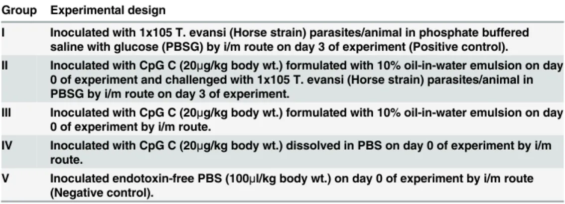
fluctuating rectal temperature, pale mucous membrane, swellings of external genitalia, incoor-dination of movement, lacrimation, deposition of white plaques in eyes and corneal opacity in some animals (Fig 1). Eyes were washed with 2% boric acid solution to provide relief to the rab-bits from lacrimation.The clinical signs ofT.evansiinfection with reduced severity appeared within days 26 to 30 post-infection in positive control group of rabbits (group I) and within days 34 to 38 post-infection in CpG treated group (group II).Abdul-majeedet al. (2007) ob-served the clinical signs in rabbits including rise in temperature during the first three days after infection, loss of appetite, progressive emaciation, and refusal to walk due to recumbency, de-pression, conjunctivitis, corneal opacity, and anemia in most of the infected rabbits [30].
Statistically significant changes in rectal temperature were reported in rabbits of two groups, which were experimentally infected withT.evansi(p<0.001). Slight increase in body tempera-ture was observed on day 3 of experiment in all CpG treated groups. In positive control group, two animals reflected significant increase in rectal temperature on day 3 post-infection (PI) i.e. day 6 of experiment. On days 8 and 9, rectal temperatures in all the six animals were on peak (p<0.001). The second peak of temperature was reported at an interval of 8–11 days. Whereas, in CpG treated andT.evansichallenged rabbits (group II), duration and rise in temperature on the first two peaks was lower than that observed in the positive control group. The remarked delay in the third peak of fever was also observed. Effects of CpG-ODN treatment and/orT.
evansiinfection on rectal temperature (°F) of animals from each of five groups of rabbits is shown inFig 2A. Clinical symptoms and other parameters ofTrypanosoma evansiinfected rab-bits (Group I) andTrypanosoma evansiinfected and challenged with CpG C rabbits (Group II) are summarized (Table B inS1 File).
Haematological and parasitological observations
on day 7 of experiment in these rabbits. The fluctuating pyrexia coincided with inconsistent parasitemia. Recurrent episodes of parasitemia occur during the course of the disease (Table C inS1 File).Thenumber of rabbits showing parasitemia in both the infected groups were deter-mined and expressed in percent. Overall, 58.94% rabbits of positive control group, showed parasitemic peaks during 42 days in comparison to 36.71% of rabbits in group II.Fig 2Bdepicts the percent of rabbitsof groups I and II showing parasitemia on different days.Fig 2Cshows the parasitemic scores shown by the rabbits of the two groups. The parasitemic scores were de-termined for eachT.evansiinfected group on different days, considering all the rabbits show-ing parasitemia in wet blood film (Table C inS1 File). The difference in the parasitemic scores between both the groups is statistically significant (p<0.012) for 28 days as determined by un-paired student’s-t test. After 28 days, the insignificant statistical difference in parasitemia sug-gests the need of booster dose of CpG. In a recent study in experimentally infected donkey mare, the parasite was not observed in wet blood film on microscopic examination, however in serum, a significant level of antitrypanosmal IgG antibodies in ELISA were present [31].
Fig 1. Photographs showing typical clinical signs inTrypanosoma evansiinfection in rabbits. A. LacrimationB. Corneal opacityC. Swelling of external genitalia. a.T.evansiinfected rabbit b. CpG C treated rabbit challenged withT.evansi
The mean haemoglobin level decreased from 12.63±0.45 g/dl to 8.97±0.29 g/dl inT.evansi
infected rabbits (group I) and from 12.60±0.33 g/dl to 9.57±0.45 g/dl in CpG treated andT.
evansichallenged rabbits (group II). The mean haemoglobin levels decreased by 28.9% inT.
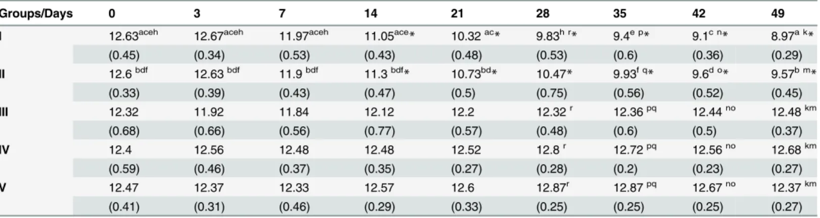
evansiinfected rabbits (group I), whereas the significant decrease in Hb levels in CpG treated andT.evansichallenged rabbits (group II) is 23.9% (Fig 2D). The difference in the decrease of mean haemoglobin values between both the groups is statistically significant (p<0.0001) as de-termined by student’s-t test. The values decreased significantly from day 14 in both the groups I and II with respect to day 0 observations (Table 2). These decreased haemoglobin values were found to be statistically significant by one way ANOVA using pair wise multiple comparison (Holm-Sidak method). There was no significant alteration in haemoglobin values in group III, IV and V throughout the observation period of 49 days (Table 2).
Fig 2. Effects of CpG-ODN C treatment inTrypanosoma evansiinfected and uninfected rabbits.The rabbits of group I were infected with 1x10⁵T.
evansiparasites/animal. The group II rabbits were treated with CpG C formulated with 10% oil-in-water emulsion and then challenged with 1x10⁵T.evansi
parasites/animal. The rabbits of group III, IV and V were inoculated with formulated CpG C, CpG C alone and PBS as negative control, respectively. Effects of CpG-ODN treatment and/orT.evansiinfection.A. Rectal temperature (°F).B. The number of rabbits showing parasitemia in wet blood film during the course of the disease in both the infected groups were determined and expressed in percentage. Figure B depicts the percent of rabbits of groups I and II showing parasitemia on different days.C. Parasitemic scores on different days in groups I and II. D. Percent decrease in haemoglobin level in groups infected withT.evansi.
Biochemical studies
The mean values of blood glucose (g/dl) along with their respective standard deviations of dif-ferent groups of rabbits are shown inTable 3.The significant fall in mean blood glucose values was reported on days 7 and 35 in positive control group (group I) at p<0.001, whereas mean blood glucose values did not show any significant alteration in CpG treated andT.evansi chal-lenged (group II) rabbits (p<0.05).
Hypoglycemia inT.evansiinfected animals has already been reported [32]. It might be due to the high metabolic rate caused by fever, hepatocyte degeneration or glucose consumption by the trypanosomes [33].
Immunoglobulin-G concentration
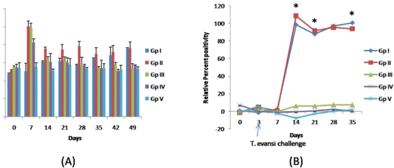
The IgG levels were the highest on day 7 of experiment in groups II, III and IV i.e. groups treat-ed with formulattreat-ed CpGs (group II and III) and CpGs alone (group IV) (Fig 3A). Furthermore,
Table 2. Effects of CpG ODN inoculation and/ orTrypanosoma evansiinfection on mean haemoglobin values (g/dl) in different groups of rabbits.
Groups/Days 0 3 7 14 21 28 35 42 49
I 12.63aceh 12.67aceh 11.97aceh 11.05ace* 10.32ac* 9.83h r* 9.4e p* 9.1c n* 8.97a k*
(0.45) (0.34) (0.53) (0.43) (0.48) (0.53) (0.6) (0.36) (0.29)
II 12.6bdf 12.63bdf 11.9bdf 11.3bdf
* 10.73bd
* 10.47* 9.93f q
* 9.6d o
* 9.57b m *
(0.33) (0.39) (0.43) (0.47) (0.5) (0.75) (0.56) (0.52) (0.45)
III 12.32 11.92 11.84 12.12 12.2 12.32r 12.36pq 12.44no 12.48km
(0.68) (0.66) (0.56) (0.77) (0.57) (0.48) (0.6) (0.5) (0.37)
IV 12.4 12.56 12.48 12.48 12.52 12.8r 12.72pq 12.56no 12.68km
(0.59) (0.46) (0.37) (0.35) (0.27) (0.28) (0.2) (0.23) (0.27)
V 12.47 12.37 12.33 12.57 12.6 12.87r 12.87pq 12.67no 12.37km
(0.41) (0.31) (0.46) (0.29) (0.33) (0.25) (0.25) (0.25) (0.27)
*Significant decrease in values with respect to day 0 within groups (p<0.05)
a c e h b d f.The values are signi
ficantly different within group across the days (p<0.050)
p q r k m n o.The values are signi
ficantly different across the groups (p<0.050).
doi:10.1371/journal.pone.0127437.t002
Table 3. Effects of CpG ODN inoculation and/ orTrypanosoma evansiinfection on mean blood glucose values (g/dl) in different groups of rabbits.
Groups/Days 0 3 7 14 21 28 35 42
I 129.67a 131.33a 93a,b
* 106.5 105.5 104.17 98.5a
* 111
(5.88) (6.18) (8.91) (16.14) (27.82) (19.85) (8.66) (10.50)
II 124.8 126.6 106.4 115.2 116.2 109.4 118.6 115.8
(6.52) (5.43) (7) (7.96) (15.16) (11.11) (11.48) (9.66)
III 125 125 137.5b 129 126.5 121 130 120.5
(8) (4) (7.5) (1) (5.5) (3) (1) (2.5)
IV 129.67 125.33 131.67b 132.33 131 130 129.33 125.67
(2.49) (8.26) (2.05) (4.03) (5.72) (1.63) (7.59) (3.68)
V 123.5 122 124.5 137.5 134.5 124 131.5 120.5
(2.5) (2) (7.5) (0.5) (0.5) (2) (3.5) (1.5)
*Means with superscripts vary significantly with respect to days 0 and 3 (p<0.05) a. The values are significantly different within group across the days (p<0.050) b. The values are significantly different across the groups (p<0.050).
the levels of IgG were higher in groups II and III than group IV. The values of IgG decreased in all these three groups during next observations and decrease in IgG values of group II was less-er in comparison to that of group III and IV. On day 35 onwards the regular elevation in the IgG values of group I was observed.
The highest IgG concentration was observed after CpG inoculation in (T.evansiinfected and uninfected) rabbits treated with formulated CpGs and CpGs alone. CpG-ODN provokes humor-al immune responses with less toxicity, superior to those induced by humor-alum [34,35]. The values of IgG in rabbits receiving CpG-ODN decreased later which might be indicative of degradation of CpG-ODN, suggesting requirement of booster doses. After day 35 of experiment the regular ele-vation in the IgG values ofT.evansiinfected rabbits was observed which supported the earlier observation of increased plasma globulin levels inT.evansiinfected donkeys [36]. Similar obser-vations have been documented in other mammalian hosts infected withT.evansi[37,38].
Trypanosoma evansi
specific immunoglobulin G
The detectable antibody levels againstT.evansiwere observed 11th dpi (or day 14 post CpG treatment) onwards in all theT.evansiinfected rabbits in both the groups (Fig 3B). The plasma samples of five rabbits showed increasing trend of antibody titres from day 11 (98.49%) on-wards and peak antibody titres were observed on 35thdpi in Group I (positive control). In Group II peak antibody titres were observed on day 11 dpi (108.78%). The levels ofT.evansi
specific IgG levels were significantly higher than the rabbits in group I on day’s upto 21 days post CpG treatment or 18 dpi, which might be due to degradation of CpG-ODN. The regular elevation in the IgG values ofT.evansiinfected rabbits was because of cumulative effect and in-creasing number of blood parasites. Thereafter, antibody titres decreased as compared to rab-bits of Group I. Similar observations for both the groups of rabrab-bits were observed in IgG concentrations measured by quantitative turbidity test. Determination ofT.evansispecific IgG levels can be compared between two groups only, which are receivingT.evansiparasite, but
Fig 3. Effects of CpG ODN inoculation and/ orT.evansiinfection. A. Immunoglobulin G values (g/l) in different groups of rabbits. The anti-rabbit IgG antibodies formed insoluble complexes when mixed with samples containing purified rabbit IgG. IgG concentration in the sample was quantified by comparing with a calibrator of known IgG concentration.B. Relative percent positivity forT.evansispecific Immunoglobulin G.T.evansispecific IgG levels were determined by ELISA and expressed as relative percent positivity with respect to knownT.evansipositive serum sample.
estimation of IgG levels using quantitative turbidity test provide comparison amongst all the groups irrespective ofT.evansiinfection.
Histopathological evaluation
At necropsy, rabbits of positive control group (group I) and rabbits of CpG treated andT.
evansichallenged group (group II) showed splenomegaly and multifocal areas of necrosis in spleen. Similar observations were reported inT.evansiinfected donkeys and rabbits [36,39]. Histopathological changes in liver ofT.evansiinfected (group I) rabbits revealed hydropic de-generation of hepatocytes, progressive destruction of hepatic parenchyma, dilated sinusoids, heamorrhages in hepatic parenchyma and the inflammatory reaction extended from the portal tract to the parenchyma causing extensive hepatic necrosis and loss of normal hepatic architec-ture (Fig 4A). Hepatomegaly, congestion, necrotic foci and destruction of hepatocytes with in-filtration of inflammatory cells were observed in the liver ofT.evansiinfected rats, buffaloes and goats also [40–43]. In rabbits treated with CpG C and challenged withT.evansi(group II), there was mild to moderate hydropic degeneration, mononuclear cell infiltrations and de-creased severity of necrosis (Fig 4B) whereas in group III (treated with formulated CpG), the areas of mononuclear cell infiltration in portal triad along with mild hydropic degeneration of hepatocytes were observed (Fig 4C). Histopathological changes in spleen ofT.evansiinfected (group I) rabbits revealed formation of secondary follicles, moderate lymphocytic necrosis, haemorrhages and edematous fluid (Fig 4D) whereas in spleen of group II (CpG C treated and challenged withT.evansi) secondary follicles were comparatively more in number as compared to group I rabbits (Fig 4E). Depletion of lymphocytes and necrosis was also observed. Diseases caused by trypanosomes induce the formation of high levels of systemic antigen-antibody im-mune complexes and their consequent deposition in the heart, liver, brain and kidneys may possibly play a role in tissue damage [44]. However, some reports indicated that trypanosomes can cause tissue inflammation directly as a result of the infection [42,45]. The observed spleen lesions with follicular hyperplasia might be indicative of an immunological response by the in-fected rabbits. Following CpG-ODN stimulations, splenomegaly accompanied by proliferation of splenic B cells was also reported in mice [46].
This study was performed to observe the impact of single dose of CpG-ODN class C onT.
evansiinfected animals. The impact of booster doses of CpG-ODN can also be explored to pro-long the CpG-ODN generated immune responses. Recently, multifunctional magnetic nanopar-ticles loaded with CpG- ODNs were used to show the impact of immune activation on human head and neck squamous cell carcinoma cells [47,48]. Earlier, we reported that the nano-delivery of quinapyramine sulfate-loaded nanoparticles provided sustained release of quinapyramine sul-fate and is highly effective inT.evansiinfected mice [49]. Thus, either nano delivery of CpGs in form of sustained release formulations or booster doses can be tried in this context.
Conclusion
We observed delayed onset of clinical signs with reduced severity in CpG treated andT.evansi
Future Perspective
In endemic areas, the disease increases significantly during the rainy season due to highbiting fly populations. The property of trypanosomes to rapidly change their surface glycoproteins to avoid the immune is the major obstacle to provide any vaccine against the parasite. Approaches to en-hance the innate immune response in the animals either by CpG-ODN can be exploited in endem-ic areas with low infection or with the antitrypanosomal treatment to avoid relapse of infection.
The combined CpG ODN C along with conventional chemotherapy againstT.evansimay provide new alternatives to control the disease with reduced frequency and doses of the treat-ment with trypanocidal drugs. Novel approaches for trypanocidal therapy along with these po-tential new molecules to enhance their therapeutic value need further elucidation.
Supporting Information
S1 File. Supporting information.Table A. Effects of CpG ODN inoculation and/ or Trypano-soma evansiinfection on body weight (percent increase) in different groups of rabbits. Table B. Clinical symptoms, hematological and biochemistry parameters ofTrypanosoma evansi in-fected rabbits (Group I) andTrypanosoma evansiinfected and challenged rabbits with CpG C (Group II). Table C. Parasitemia shown byTrypanosoma evansiinfected rabbits (Group I) and
Trypanosoma evansiinfected and challenged with CpG C rabbits (Group II).T.evansi
Fig 4. Histopathological changes in liver and spleen ofTrypanosoma evansiinfected and uninfected rabbits in response to CpG-ODN treatment. A. Liver tissue ofT.evansiinfected (positive control) group I, extensive necrosis and loss of normal hepatic architecture is shown by double arrow (H&E 400X). B. Liver tissue of group II (CpG C treated and challenged withT.evansi), decreased severity of necrosis (black colored arrow) (H&E 400X).C. Liver tissue of group III (treated with formulated CpG alone),mononuclear cell infiltration in portal triad (black colored arrow) (H&E 400X).D. SpleenT.evansi
infected (positive control) group I, secondary follicles (white block arrows), haemorrhages and edematous fluid(H&E 100X). E. Spleen of group II CpG C treated and challenged withT.evansi. Secondary follicles comparatively more in number (white block arrows) (H&E 100X)
Infection was given on day 3. (DOCX)
Acknowledgments
Authors thank Indian Council of Agricultural Research, Ministry of Agriculture, Government of India for providing financial support for the institutional project (IXX00276). The funding agency had no role in study design, data collection and analysis, decision to publish, or prepa-ration of the manuscript. The support of Sh. Om Prakash, for assistance in handling of animals is acknowledged.
Author Contributions
Conceived and designed the experiments: AM BKM RK. Performed the experiments: PK AM BKM SCY. Analyzed the data: AM BKM HS AS NV. Contributed reagents/materials/analysis tools: AM BKM SCY AS. Wrote the paper: AM BKM.
References
1. Weiner GJ, Liu HM, Wooldridge JE, Dahle CE, Krieg AM. Immunostimulatory oligodeoxynucleotides containing the CpG motif are effective as immune adjuvants in tumor antigen immunization. Proc. Natl Acad Sci USA 1997; 94 (20). 10833–37 PMID:9380720
2. Krieg AM. CpG motifs in bacterial DNA and their immune effects. Annu Rev Immunol. 2002; 20: 709–
60. PMID:11861616
3. Klinman DM. Immunotherapeutic uses of CpG oligodeoxynucleotides. Nat Rev Immunol. 2004; 4: 249–58. PMID:15057783
4. Krieg AM. Therapeutic potential of Toll-like receptor 9 activation. Nat Rev Drug Discov. 2006; 5: 471–
84. PMID:16763660
5. Ballas ZK, Rasmussen WL, Krieg AM. Induction of NK activity in murine and human cells by CpG motifs in oligodeoxynucleotides and bacterial DNA. J Immunol. 1996; 157(5): 1840–1845. PMID:8757300
6. Sands H, Gorey-Feret LJ, Cocuzza AJ, Hobbs FW, Chidester D. Trainor G. Bio-distribution and metab-olism of internally 3H-labeled oligonucleotides I. Comparison of a phosphodiester and a phosphorothio-ate. Mol Pharmacol. 1994; 45: 932–43. PMID:8190109
7. Krug A, Rothenfusser S, Hornung V, Jahrsdorfer B, Blackwell S, Ballas ZK et al. Identification of CpG oligonucleotide sequences with high induction of IFN-alpha/beta in plasmacytoid dendritic cells. Eur J Immunol. 2001; 31: 2154–63. PMID:11449369
8. Vollmer J, Weeratna R, Payette P, Jurk M, Schetter C Laucht M et al.. Characterization of three CpG oli-godeoxynucleotide classes with distinct immunostimulatory activities. Eur J Immunol. 2004; 34: 251–
62. PMID:14971051
9. Zhao Q, Yu D, Agrawal S. Site of chemical modifications in CpG containing phosphorothioate oligo-deoxynucleotide modulates its immunostimulatory activity. Bioorg Med Chem Lett. 1999; 9: 3453–58.
PMID:10617090
10. Jurk M, Schulte B, Kritzler A, Noll B, Uhlmann E, Wader T, et al.. C-Class CpG ODN. Sequence require-ments and characterization of immunostimulatory activities on mRNA level. Immunobiology, 2004; 209: 141–54. PMID:15481148
11. Barret MP. The Trypanosomoses. Lancet, 2004; 362: 1469–80 ().
12. Cross GA. Cellular and genetic aspects of antigenic variation in trypanosomes. Annu rev immunol. 1990; 8: 83–110. PMID:2188680
13. Borst P, Rudenko G. Antigenic variation in African trypanosomes. Science, 1994; 264: 1872–73.
PMID:7516579
14. Baral TN. Immunobiology of African trypanosomes: need of alternative interventions. J Biomed Bio-technol. 2010; 2010:389153 doi:10.1155/2010/389153Epub 2010 Feb 23. PMID:20182644 15. Juyal PD. Newer perspective in the diagnosis and control of Trypanosomosis (Surra) in domestic
live-stock in India. Tropmed—Internationale Wissenschaftliche Publikationen. 2011;1–13
16. Hemmi H, Takeuchi O, Kawai T. A toll-like receptor recognizes bacterial DNA. Nature, 2000; 408: 740–
17. Rad M, Ahmadi M, Farid Hosseini R, Ashkar AA, Nejati M, Tabaraie B, et al. Protection againstListeria monocytogenesby ODN containing CpG motifs in BALB/c and C57BL/6 mice. J Appl Anim Res. 2006; 29: 145–147.
18. Elkins KL, Rhinehart-Jones TR, Stibitz S, Conover JS, Klinman DM. Bacterial DNA containing CpG mo-tifs stimulates lymphocyte-dependent protection of mice against lethal infection with intracellularbac-teria. J Immunol. 1999; 162: 2291–98. PMID:9973506
19. Corral RS, Petray PB. CpG DNA as a Th1-promoting adjuvant in immunization againstTrypanosoma cruzi. Vaccine, 2001; 19(2/3): 234–242.
20. Frank FM, Petray PB, Cazorla SI, Munoz MC, Corral RS, Malchiodi EL. Use of a purified Trypanosoma-cruziantigen and CpG oligodeoxynucleotides for immunoprotection against a lethal challenge with try-panomastigotes. Vaccine, 2003; 22 (1): 77–86. PMID:14604574
21. Harris TH, Mansfield JM, Paulnock DM. CpG oligodeoxynucleotide treatment enhances innate resis-tance and acquired immunity to African trypanosomes. Infect Immun. 2007; 75(5): 2366–73. PMID:
17339353
22. Manuja A, Manuja BK, Kaushik J, Singha H, Singh RK. Immunotherapeutic potential of CpG oligodeox-ynucleotide in veterinary species. Immunopharmacol Immunotoxicol. 2013; 35(5): 535–44. doi:10.
3109/08923973.2013.828743PMID:23981003
23. Gramzinski RA, Doolan DL, Sedegah M, Davis HL, Krieg AM, Hoffman SL. Interleukin-12 and gamma interferon-dependent protectionagainst malaria conferred by CpG oligodeoxynucleotide in mice. Infect Immun. 2001; 69: 1643–49. PMID:11179339
24. Manuja A, Kumar P, Kumar R, Manuja BK, Singha H, Sharma RKet al. CpG-ODN class C-mediated immunostimulation and its potential againstTrypanosoma evansiin equines. Int Immunopharmacol. 2014; 22, 366–370. doi:10.1016/j.intimp.2014.07.016PMID:25066759
25. Tietz Textbook of Clinical Chemistry, 3rd Ed. Burtis CA, Ashwood ER. WB Saunders Co., (1999). 26. Yadav SC, Kumar R, Manuja A, Goyal L, Gupta AK. Early detection ofTrypanosoma evansiinfection
and monitoring of antibody levels by ELISA following treatment. J Parasit Dis. 2014; 38(1): 124–7. doi:
10.1007/s12639-012-0204-2PMID:24505190
27. Yadav SC, Kumar R, Kumar S, Tatu U, Singh RK, Gupta AK. Identification and characterization of Try-panosoma evansicysteine proteinases for immunodiagnosis. Parasitol Res. 2011; 109, 559–565. doi:
10.1007/s00436-011-2284-9PMID:21350794
28. Kumar R, Kumar S, Khurana SK, Yadav SC. Development of an antibody-ELISA for seroprevalence of
Trypanosoma evansiin equids of North and North-western regions of India. Vet Parasitol. 2013; 196 (3): 251–257. doi:10.1016/j.vetpar.2013.04.018PMID:23664710
29. Flynn B, Wang V, Sacks DL, Seder R A, Verthelyi D. Prevention and treatment of cutaneous leishmani-asis in primates by using synthetic type D/A oligodeoxynucleotides expressing CpG motifs. Infect and immune. 2005; 73 (8): 4948–4954. PMID:16041009
30. Abdul-majeed HA, Abbas FH, Abbood HF. Blood picture and hepatic changes in rabbits experimentally infected withTrypanosoma evansiIraqi strain. Med J Baby, 2007; 4 (3&4): 358–64.
31. Kumar R, Kumar S, Virmani N, Yadav SC. Transplacental transmission ofTrypanosoma evansifrom experimentally infected donkey mare to neonatal foal. J Equine Vet Sci. 2015; 4: 337–341.
32. Silva RA, Ramirez L, Souza SS, Ortiz AG, Pereira SR, Davila AM. Hematology of natural bovine trypa-nosomsis in the Brazilian Pantanal and Bolivian Wetlands. Vet Parasitol. 1999; 85(1): 87–93. PMID:
10447196
33. Von Brand T. Biochemistry of parasites 2ed. New York: Academic Press (1973).
34. Davis HL, Weeratna R, Waldschmidt TJ, Tygrett L, Schorr J, Krieg AM. CpG DNA is a potent enhancer of specific immunity in mice immunized with recombinant hepatitis B surface antigen. J Immunol. 1998; 160: 870–76. PMID:9551923
35. Weeratna R, McCluskie MJ, Yu X, Davis HL. CpG DNA induces stronger immune responses with less toxicity than other adjuvants. Vaccine, 2000; 18: 1755–62. PMID:10699323
36. Cadioli FA, Marques LC, Machado RZ, Alessi AC, Aquino LP, Barnabe PA. ExperimentalTrypanosoma evansiinfection in donkeys: hematological, biochemical and histopathological changes. Arq Bras Med Vet Zootec. 2006; 58(5): 749–756.
37. Monzon CM, Villavicencio V. Serum proteins in guinea pigs and horses infected withTrypanosoma evansi(Steel, 1885). Vet. Parasitol. 1990; 36: 295–15. PMID:2399649
38. Soodan JS, Sood NK, Khahra SS,Singh KB, Juyal PD. Clinico-pathological studies in donkeys experi-mentally infected withTrypanosoma evansi. Indian J Anim Sci. 1996; 66: 443–48.
40. Biswas D, Choudhury A, Misra KK. Histopathology ofTrypanosoma,Trypanozoon evansiinfection in bandicoot rat. I. Visceral organs. Exp Parasitol. 2001; 99(3):148–59. PMID:11846525
41. Dargantes AP, Reid SA, Copman DB. ExperimentalTrypanosoma evansiinfection in the goat. I. Clinical signs and clinical pathology. J Comp Pathol. 2005; 133(4): 261–66. PMID:16213515
42. Damayanti R, Graydon RJ, Ladds PW. The pathology of experimentalTrypanosoma evansiinfection in the Indonesian buffalo (Bubalus bubalis). J Comp Pathol. 1994; 110(3): 237–52. PMID:8040389
43. Tizard IR.Imunologia veterinária: uma introducao. 5ed São Paulo: Roca: 331–332 (1998).
44. Sudarto MW, Tabel H, Haines DM. Immunohistochemical demonstration ofTrypanosoma evansiin tis-sues of experimentally infected rats and a naturally infected water buffalo (Bubalus bubalis). J Parasitol. 1990; 76: 162–67. PMID:2319414
45. Ngeranwa JJ, Gathumbi PK, Mutiga ER, Agumbah GJ. Pathogenesis ofTrypanosoma (brucei) evansi
in small east African goats. Res Vet Sci. 1993; 54 (3): 283–89. PMID:8337477
46. Zhao Q, Temsamani J, Zhou RZ, Aggarwal S. Pattern and kinetics of cytokine production following ad-ministration of phosphorothioate oligonucleotides in mice. Antisense Nucleic Acid Drug Dev. 1997; 7 (5): 495–02. PMID:9361908
47. Shukoor MI, Natalio F, Tahir MN, Barz M, Weber S, Brochhausen C, et al. CpG-DNA loaded multifunc-tional MnO nanoshuttles for TLR9-specific cellular cargo delivery, selective immune-activation and MRI. J Mater Chem 2012; 22: 8826–34.
48. Manuja A, Kumar B, Singh RK. Nanotechnology developments: Nanotechnology developments: oppor-tunities for animal health and production. Nanotechnology Dev.: 2012; doi:10.4081/nd.2012.e4 49. Manuja A, Kumar S, Dilbaghi N, Bhanjana G, Chopra M, Kaur H, et al.. Quinapyramine sulphate-loaded