Introduction
Tuberculosis (TB) is a major global health
issue and one of the leading causes of death
mediated by infectious agents. Despite
enormous efforts to control the disease,
estimations show that there are about 9
million new cases of TB infection with
1.5 million deaths per year.
1Although
TB affects several organs of the body,
it majorly and specifically affects the
lungs. In most circumstances, infections
with
Mycobacterium tuberculosis do
not demonstrate symptoms and clinical
manifestations, which is known as latent
tuberculosis (LTBI). However, almost
10% of cases with latent infections further
develop to active disease. The most
common clinical symptoms of active TB
include a prolonged cough combined with
blood-containing sputum, sweating, fever,
and weight loss.
2Initiation of TB infection occurs once
M. tuberculosis reaches the pulmonary
alveoli, where the bacterium invades and
then replicates within the endosomes
of alveolar-resident macrophages. The
behavior of immune response determines
the fate of infection by modifying the rate
of actively replicating M. tuberculosis in
patients with concomitant alterations in
TB disease risks. During latent and early
activated phases of TB, the infection is
mainly intracellular and, consequently,
T-cell responses play important roles for
doi 10.15171/ijbsm.2016.10
Cross-Talk Between the Immune System and
Tuberculosis Pathogenesis; A Review With Emphasis
on the Immune Based Treatment
Mohammad Reza Javan
1, Ahmad Ali Jalali Nezhad
2, Sarieh Shahraki
2, Amin Safa
1, Halimeh Aali
3*,
Zohre Kiani
41Department of Immunology, Faculty of Medicine, Zabol University of Medical Sciences, Zabol, Iran 2Department of Physiology, Faculty of Medicine, Zabol University of Medical Sciences, Zabol, Iran 3Department of Internal Medicine, Faculty of Medicine, Zabol University of Medical Sciences, Zabol, Iran 4Student Research Committee, Kerman University of Medical Sciences, Kerman, Iran
Abstract
As a globally major health problem, tuberculosis (TB) causes almost two million cases of death annually. Epidemiological studies demonstrate that a third of the world’s individuals is infected with Mycobacterium tuberculosis. Approximately 10% of infected patients with M. tuberculosis
develop chronic manifestation as TB. Due to HIV coinfection and emerging the drug-resistant TB, the disease has been increasing and its control has been frustrated in several parts of the world. Current diagnostic techniques and therapeutic tools for TB are not satisfactory. Consequently, it is urgently essential to establish new therapies concerning vaccines, immunotherapeutic agents to provide prosperous attempts for TB controlling. To achieve this goal, it is required to be armed with comprehensive understanding of immunobiology and immunopathogenesis of TB. This would be beneficial in designing new immune-based protections, drug discoveries, personalized medicine by choosing highly-effective immunotherapeutic interventions, identification and development of novel drug candidates. Hopefully, immunotherapies could be advantageous in modulating the immune system in patients with TB, providing efficient control of M. tuberculosis
infection perpetuation and, therefore, its pathogenesis. This review herein attempts to describe the function of immune system in response to TB that is of the therapeutical and clinical importance. Moreover, new insights based on therapeutics to resolve TB with immunological orientation will be discussed.
Keywords: Mycobacterium tuberculosis, Tuberculosis, Therapeutic agents, Immune system. *Correspondence to
Halimeh Aali, Department of Internal Medicine, Faculty of Medicine, Zabol University of Medical Sciences, Zabol, Iran.
Email: ali@zbmu.ac.ir
Received July 10, 2016
Revised September 7, 2016
Accepted November 9, 2016
Published online November 21, 2016
http://ijbsm.zbmu.ac.ir/
Copyright © 2016 The Author(s); Published by Zabol University of Medical Sciences. This is an open-access article distributed under the terms of the Creative Commons Attribution License (http://creativecommons.org/licenses/by/4.0), which permits unrestricted use,
the protective immunity. In this immune response, CD8
+T cells as well as CD4
+helper 1 T cells (Th1) through
producing cytokines including tumor necrosis factor
(TNF)-α, interferon (IFN)-γ, and interleukin (IL)-2
participate in controlling the M. tuberculosis replication.
3-5Development of TB is likely due to dysregulated immune
response as well as impaired immune regulation. In
addition, a bulk of the lung damage related to TB is due
to host-mediated immunopathology instead of direct
virulence factors derived from M. tuberculosis. The
modulation of immune regulation through divergence
of a protective response by Th1 and CD8
+T cells, with
Th2-related cytokines, TGF-β, regulatory T cells (Treg),
and other immunosuppressive mediators is a key player
in this regard.
6-8Thus, host-directed therapy by applying
immunomodulators might be a promising therapeutic
approach to control TB. This paper aimed to focus on the
immunology of TB and the immune-based therapies to
control this disease.
Methods
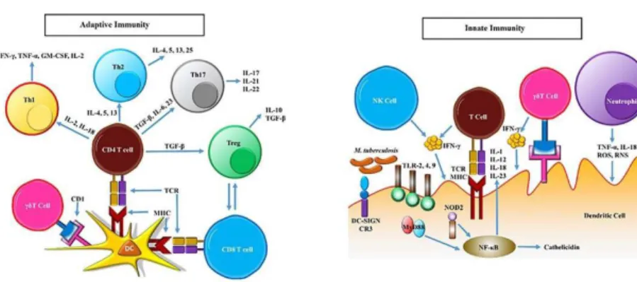
Mechanisms of the Innate Immunity to
M. tuberculosis
Following
M. tuberculosis entrance into the host lungs,
the surface antigens interact with several receptors like
pattern recognition receptors (PRRs) including toll-like
receptors (TLRs),
9mannose receptor,
10complement
receptor 3 (CR3),
11scavenger receptor,
10and dendritic
cell (DC)-specific
intercellular-adhesion-molecule-3-grabbing non-integrin (DC-SIGN), which are commonly
located on the surface of macrophages and DCs. The
antigens of M. tuberculosis which are recognized with these
receptors include CpG-containing DNA, lipoprotein,
phosphatidylinositol mannoside, and mannose-capped
lipoarabinomannan. Furthermore, M. tuberculosis surface
lipoarabinomannan is recognized by the pulmonary
surfactant protein D (SP-D), resulting in limitation of
the intracellular growth of M. tuberculosis by means
of promoted infusion of phagosome and lysosome.
12Moreover, there is a cooperation among TLR-2, C-type
lectin dectin-1 that interacts with M. tuberculosis,
and cytosolic nucleotide binding and oligomerization
domain-like receptors like NOD2 that binds to muramyl
dipeptide, which eventuates in activation of the nuclear
factor-kappa B (NF-κB) signaling pathway, which in turn
facilitates the production of pro-inflammatory cytokine.
13The cytokines which are produced and secreted upon
the activation of NF-κB include IL-1, IL-12, IL-18,
TNF-α, and chemokines. These chemokines mediate the
recruitment of immune cells such as natural killer (NK)
cells, neutrophils, T cells, DC, and macrophages to the
infected tissue.
14,15M. tuberculosis secrets protein
ESAT-6, which suppresses the activation of NF-kB signaling
pathway by inhibiting the interaction of MyD88 with the
downstream molecule, namely, interleukin-1
receptor-associated kinase 4(IRAK4).
16Activation of TLR signaling
causes up-modulation of expression of both the vitamin D
receptor (VDR) and the vitamin D-1-hydroxylase genes,
which mediate the conversion of pro-vitamin D into the
active form of vitamin D, 1,25(OH)2D3. Overexpression
of these genes ultimately eventuates in production
of the antimicrobial peptides such as β-defensin and
cathelicidin to kill the intracellular pathogen.
17-20On the
other side, activation of DC-SIGN signaling causes the
production of immunosuppressive cytokines like IL-10
and transforming growth factor (TGF)-β.
14Macrophages are heterogeneous and play different roles
during immune response toward M. tuberculosis infection.
The type 1 IL-23-producing macrophages promote a
protective Th1 mediated immunity against infection, and
type 2 IL-10-producing macrophages play a role in the
suppression of immune response to M. tuberculosis.
21In
addition, type 2 macrophages participate in the induction
of CD4
+T cells to be converted to CD25
+FoxP3
+mTGFβ-1
+Tregs, which further suppress the immune system.
22Activated T cells and NK cells both secret IFN-γ which
plays a role in the activation of macrophages to kill
bacteria by promoting the phagosomal maturation and
production of reactive oxygen species (ROS) and reactive
nitrogen intermediates.
23,24While IFN-γ contributes to
the fusion of phagosome and lysosome by cell signaling
pathway IRGm1 (LRG-47)
25,26and PI3K,
27both IL-4
and IL-13 secreted by Th2 inhibit autophagy-associated
killing of bacteria by Akt signaling pathway.
28TNF-αhas
also a role in killing the intracellular M. tuberculosis by
activating the reactive nitrogen species (RNS), thereby
participating in the development of granuloma.
29Neutrophils are the first immune cells attracted to the
infected sites that express many surface receptors as
well as antimicrobial agents.
14In vitro experiments on
neutrophils demonstrate that these cells are activated
during incubation with M. tuberculosis and are capable
of limiting the bacterial growth.
29. Neutrophils produce
the cathelicidin LL-37, human neutrophil peptides
1-3, and lipocalin 2, which can either kill or limit the
growth of M. tuberculosis.
30On the other hand, apoptotic
neutrophils have a role in the activation of macrophages
by producing the heat shock protein 72,
31and granule
proteins.
32Interestingly, some studies have suggested that
neutrophils have no beneficial roles in infection with M.
tuberculosis and show a pathological function rather than
a protective role in the control of active TB
30,33(Figure 1).
Mechanisms of the Adaptive Immunity to
M. tuberculosis
DCs and macrophages infected with M. tuberculosis
are common antigen presenting cells (APCs), which
present bacterial antigens to T and B cells of the adaptive
immunity. It has been shown that IL-12p40 plays an
important role in the activation of pulmonary DC during
pathogen induced stimulation.
34Furthermore, more
efficient antigen presentation could occur by released
apoptotic vesicles from the macrophage apoptosis
procedures that provide bacterial antigens to primary
mycobacteria uninfected DCs. Activation of CD8
+T cells
can be diminished by decreased antigen delivery through
blocking the macrophage apoptosis.
35during
M. tuberculosis infection is carried out by the
MHC restricted CD4
+and CD8
+T cells as well as γ-δ
T lymphocytes, which participate in IFN-γ production.
Thus, CD4
+Th1 cells are major players in TB protection
and demonstrate powerful IFN-γ response, compared
with CD8
+T cells in patients with mycobacterial
infection.
36,37Moreover, depletion of CD4
+T cells in M.
tuberculosis
infection culminates in the procrastinated
chemotaxis of activated CD8
+T cells from the lymph
nodes, resulting in the impaired immune protection.
38During
M. tuberculosis infection, CD4
+T cells can
differentiate into several subtypes of Th1, Th2, Th17,
and even regulatory T (Treg) cells. Cytokines produced
by Th1 cells, namelyIL-2, IFN-γ, TNF-α, TNF-β, and
granulocyte-monocyte colony-stimulating factor
(GM-CSF)cause further differentiation and activation of Th1
cells, cytotoxic T lymphocytes (CTL), macrophages,
and granulocytes. On the other hand, Th2 cytokines
including IL-4, IL-5, IL-6, IL-9, andIL-13 cause activation
and stimulation of B cells, providing antibody response.
Additionally, Th2 cytokines suppress the Th1 associated
immune response, that mediates a non-protective
immune response toward TB. The Th17 cells play a role
in the early phase of inflammatory response by producing
specific cytokines such as IL-17F, IL-21, and IL-22. These
cytokines are involved in the production of defensin as
well as recruitment of inflammatory cells like neutrophils
and monocytes to the site of infection.
39-41Tregs mainly belong to CD4
+CD25
+FoxP3
+T cells, which
suppress numerous mechanisms of immune response by
producing IL-10 and TGF-β.
42,43Moreover, CD8
+Treg
cells are involved in the inhibition of T cell proliferation
and function.
44Tregs play a role in the modulation of
CD4
+T cell differentiation to the Th1, Th2, or Th17
subsets. The natural FoxP3 expressing Tregs have been
observed to show greater expansion during M. tuberculosis
infection.
45These cells participate in the inhibition of
IFN-γ production from γ/δ memory T cells responding
to antigens from M. tuberculosis.
46Treg cells also mediate
their action by producing TGF-β, which prevents the
activation and proliferation of CD4
+T cell,
47facilitating
mycobacterial infection dissemination and therefore,
exacerbation of infection manifestations.
48,49Increased
numbers of Treg cells have been observed in patients with
active TB,
49and depletion of Treg cells has been observed
to improve the protective efficacy of vaccines toward
infection with M. tuberculosis.
50,51For many years, B cells have been thought to have little
impact in protection against TB. Animal studies have
revealed a beneficial role of B cells through interactions
with players of the cellular immunity as well as activation
of complements, which provide an optimal protection
in mice with M. tuberculosis infection.
52,53Activation of
the three complement alternative, classical, and lectin
pathways by Bacillus Calmette–Guérin (BCG) causes
the fixation of the main complement component of
C3b on the surface antigens of bacteria, contributing to
phagocytosis and killing of mycobacteria.
54Various profiles of secreted cytokines during M.
tuberculosis
infection determine the fate of CD
+T cell
differentiation and therefore play roles in the beneficial
or deleterious quality of immune response to TB. A
protective Th1 related response is mediated by IFN-γ,
IL-12, and IL-18, whereas Th2 development is carried
out by another cytokine set including IL-4, IL-5, and
IL-13. TGF-β in lower doses alongside with IL-6, IL-21,
and IL-23 are involved in Th17 development; however,
TGF-β at higher concentrations in combination with
IL-2 promote Treg differentiation.
42,43IL-6 is involved
in inflammatory responses by suppressing the TGF-β
induced Treg cell development as well as by inducing the
Th17 differentiation alongside with TGF-β
55(Figure 1).
Results
Immune System-Based Therapeutics of Tuberculosis
Other Uncommon Mycobacteria
Application of Mycobacterium vaccae as an
immunotherapeutic tool has been shown. Investigations
demonstrated that single injection of M. vaccae could
increase the conversion of sputum culture after 1 month
and cause a significant improvement, regarding the
radiographic manifestations, after 6 months.
56However,
a study could not show the beneficial effects of M.
vaccae
injection.
57On the other hand, a meta-analysis
concluded that intradermal injection of M. vaccae based
immunotherapy was beneficial, considering the sputum
conversion enhancement as well as amelioration of
radiographic outcomes.
58In addition, enhanced sputum
conversion in newly treated cases with TB was shown
after oral administration of M. vaccae.
59Promising
observations have been reported using other mycobacteria
like M. indicuspranii in the animal models of TB.
60Vaccines
DNA vaccines have been explored to treat the TB. Several
DNA vaccines, based on genes expressing M.
tubercu-losis
proteins such as ESAT-6, Hsp65, and Ag85A have
been indicated to have fruitful results in mice with M.
tuberculosis infection.
61-64A developed DNA vaccine with
cDNA3.1 plasmid as a vector that expresses IL-2 as well as
Hsp65 genes that integrate into the virus-free envelopes,
originated from the hemagglutinating virus of Japan. The
intramuscular administration of the above-mentioned
DNA vaccine that contained both Hsp65 and IL-12 genes
resulted in the promoted survival rate of mice, which were
infected with multidrug resistance (MDR) or extensively
drug-resistant (XDR) TB. Moreover, almost the same
beneficial effects were reported in another survey, which
demonstrated that the DNA vaccine containing Hsp65
and IL-12 genes improved the survival rate of primates
with M. tuberculosis infection.
65On the other side, vaccines containing other molecules
have been explored to see if there are potential benefits.
RUTI is a vaccine, which is built using the detoxified
cellular fragments of M. tuberculosis and liposomes as a
delivery approach.
66It has been reported that RUTI has
the potential to be used as a tool for the immunotherapy
and prophylaxis against M. tuberculosis infection in
animal models.
67The immunogenicity and safety of this
vaccine have been established in Phase I and II clinical
trials.
68,69Mesenchymal Stem Cells
Mesenchymal stem cells (MSCs) are multipotent stromal
cells that differentiate into several cell types.
70,71There
are MSCs in several tissues and organs of the body
like lungs,
72,73providing a potential in the repair of
damaged tissues.
74,75MSCs modify a tissue with chronic
inflammation to a condition with capability to stimulate
a strong pathogen-specific immune response. The
cell-to-cell contact as well as release of mediators like
prostaglandin E2 and TGF-β have been considered as
the functional pathways of MSCs.
76Administration of
MSCs during 4 weeks of TB treatment as a phase I clinical
trial demonstrated beneficial effects by measuring the
radiological changes.
71V5 Immunitor
V5 immunitor is derived from the blood of patients with
hepatitis Band C virus-positive and is inactivated by
chemical agents and heat. It was first developed to treat
patients with chronic hepatitis B and C infections.
77It has
been proposed that blood donors may have circulating
M. tuberculosis cell fragments and antigens, which drive
immune responsestoTB.
78Moreover, the circulating
cytokines and other immune mediators from the blood
of donors could increase the T-cell responses to M.
tuberculosis. However, there may be some other unknown
agents, providing adjuvant function. V5 immunitor oral
therapy, during a Phase I clinical trial, culminated in a
remarkable conversion of sputum culture after 1 month
of treatment.
79,80Cytokines and Inhibitors
Application of IFN-γ and IL-12 as adjuncts resulted
in beneficial outcomes in some cases with MDR TB.
81-83Moreover, IFN-γ administration in combination
with intranasal IgA in murine models of TB caused a
decreased load of M. tuberculosis in the lungs.
84There
are controversial observations in the advantageous effects
of IL-2to treat TB patients.
85,86However, intradermal
injection of IL-2 to patients with MDR TB resulted in
improved sputum conversion.
87Moreover, IL-2 caused an
increased activity of a pyrophosphate to promote γδ T cell
responses as well as a diminished load of M. tuberculosis in
the lungs of monkeys infected with the bacterium.
88Anti-TNF-α antibodies play a role in increasing the risk of TB
reactivation.
89However, anti-TNF-α therapy is beneficial
in the patients with active TB alongside with TB multidrug
therapy. The benefits of anti-TNF-α therapy may be
mediated by increased susceptibility of M. tuberculosis to
the bactericidal activity of other drugs.
90IL-4 and TGF-β
blockade can improve Th1 type immunity and contribute
to decline in the bacterial load of M. tuberculosis in the
lungs of mice infected with the bacterium.
91,92Antibodies
B-cell-deficiency causes a higher bacterial load and,
therefore, severe manifestations upon M. tuberculosis
infection.
93,94Use of monoclonal antibodies against M.
tuberculosis antigens has conflict
ing outcomes. This
issue may be due to differences in the strategies of
administration as well as the types of antibodies.
95-97Improved phagocytosis rate of mycobacteria has been
shown following the administration of sera from the
vaccinated patients with BCG. This intervention could
also enhance the ability of macrophages in killing the
intracellular M. tuberculosis.
98Conclusion
Many studies have been performed on the promising
immunotherapy of TB. The immunotherapy may
contribute to improve the management and control of
MDR/XDR TB cases. A third of population are infected
with
M. tuberculosis. Hence, immunotherapeutic
approaches which can help to eliminate the latent infection
with M. tuberculosis could have a marked impression on
TB control. The impacts of novel immunotherapeutics/
vaccines on the reactivation or the progression of latent
cases of TB in humans need to be further studied.
Ethical Approval
Competing Interests
Authors declare that they have no competing interests.
References
1. Glaziou P, Sismanidis C, Floyd K, Raviglione M. Global
epidemiology of tuberculosis. Cold Spring Harb Perspect Med. 2014;5(2):a017798. doi:10.1101/cshperspect. a017798.
2. Mert A, Bilir M, Tabak F, et al. Miliary tuberculosis:
clinical manifestations, diagnosis and outcome in 38 adults. Respirology. 2001;6(3):217-224.
3. Brighenti S, Andersson J. Induction and regulation of CD8+
cytolytic T cells in human tuberculosis and HIV infection. Biochem Biophys Res Commun. 2010;396(1):50-57. doi: 10.1016/j.bbrc.2010.02.141.
4. Seder RA, Darrah PA, Roederer M. T-cell quality in
memory and protection: implications for vaccine design. Nat Rev Immunol. 2008;8(4):247-258. doi: 10.1038/ nri2274.
5. Han Q, Bagheri N, Bradshaw EM, Hafler DA,
Lauffenburger DA, Love JC. Polyfunctional responses by human T cells result from sequential release of cytokines. Proc Natl Acad Sci U S A. 2012;109(5):1607-1612. doi: 10.1073/pnas.1117194109.
6. Rook GA, Lowrie DB, Hernández-Pando R.
Immunotherapeutics for tuberculosis in experimental animals: is there a common pathway activated by effective protocols? J Infect Dis. 2007;196(2):191-198.
7. Rook GA, Dheda K, Zumla A. Immune systems in
developed and developing countries; implications for the design of vaccines that will work where BCG does not. Tuberculosis. 2006;86(3):152-162.
8. Rook GA, Dheda K, Zumla A. Immune responses to
tuberculosis in developing countries: implications for new vaccines. Nat Rev Immunol. 2005;5(8):661-667.
9. Trinchieri G, Sher A. Cooperation of Toll-like receptor
signals in innate immune defence. Nat Rev Immunol. 2007;7(3):179-190.
10. Ernst JD. Macrophage receptors for Mycobacterium tuberculosis. Infect Immun. 1998;66(4):1277-1281. 11. Schlesinger LS, Bellinger-Kawahara CG, Payne
NR, Horwitz MA. Phagocytosis of Mycobacterium tuberculosis is mediated by human monocyte complement receptors and complement component C3. J Immunol. 1990;144(7):2771-2780.
12. Ferguson JS, Martin JL, Azad AK, et al. Surfactant protein D increases fusion of Mycobacterium tuberculosis-containing phagosomes with lysosomes in human macrophages. Infect Immun. 2006;74(12):7005-7009. 13. Divangahi M, Mostowy S, Coulombe F, et al.
NOD2-deficient mice have impaired resistance to Mycobacterium tuberculosis infection through defective innate and adaptive immunity. J Immunol. 2008;181(10):7157-7165. 14. Korbel DS, Schneider BE, Schaible UE. Innate immunity
in tuberculosis: myths and truth. Microbes Infect. 2008;10(9):995-1004.
15. Liu PT, Modlin RL. Human macrophage host defense against Mycobacterium tuberculosis. Curr Opin Immunol. 2008;20(4):371-376.
16. Pathak SK, Basu S, Basu KK, et al. Direct extracellular
interaction between the early secreted antigen ESAT-6 of Mycobacterium tuberculosis and TLR2 inhibits TLR signaling in macrophages. Nat Immunol. 2007;8(6):610-618.
17. Liu PT, Stenger S, Li H, et al. Toll-like receptor triggering of a vitamin D-mediated human antimicrobial response. Science. 2006;311(5768):1770-1773.
18. Chocano-Bedoya P, Ronnenberg AG. Vitamin D and tuberculosis. Nutr Rev. 2009;67(5):289-293.
19. Méndez-Samperio P. Role of antimicrobial peptides in host defense against mycobacterial infections. Peptides. 2008;29(10):1836-1841.
20. Liu PT, Stenger S, Tang DH, Modlin RL. Cutting edge: vitamin D-mediated human antimicrobial activity against Mycobacterium tuberculosis is dependent on the induction of cathelicidin. J Immunol. 2007;179(4):2060-2063.
21. Verreck FA, de Boer T, Langenberg DM, et al. Human 23-producing type 1 macrophages promote but IL-10-producing type 2 macrophages subvert immunity to (myco) bacteria. Proc Natl Acad Sci U S A. 2004;101(13):4560-4565.
22. Savage ND, de Boer T, Walburg KV, et al. Human anti-inflammatory macrophages induce Foxp3+ GITR+ CD25+ regulatory T cells, which suppress via membrane-bound TGFβ-1. J Immunol. 2008;181(3):2220-2226. 23. Kaufmann SH. How can immunology contribute to the
control of tuberculosis? Nat Rev Immunol. 2001;1(1):20-30.
24. MacMicking JD, North RJ, LaCourse R, Mudgett JS, Shah SK, Nathan CF. Identification of nitric oxide synthase as a protective locus against tuberculosis. Proc Natl Acad Sci U S A. 1997;94(10):5243-5248.
25. Singh SB, Davis AS, Taylor GA, Deretic V. Human IRGM induces autophagy to eliminate intracellular mycobacteria. Science. 2006;313(5792):1438-1441. 26. MacMicking JD, Taylor GA, McKinney JD. Immune
control of tuberculosis by IFN-γ-inducible LRG-47. Science. 2003;302(5645):654-659.
27. Gutierrez MG, Master SS, Singh SB, Taylor GA, Colombo MI, Deretic V. Autophagy is a defense mechanism inhibiting BCG and Mycobacterium tuberculosis survival in infected macrophages. Cell. 2004;119(6):753-766. 28. Harris J, De Haro SA, Master SS, et al. T helper 2 cytokines
inhibit autophagic control of intracellular Mycobacterium tuberculosis. Immunity. 2007;27(3):505-517.
29. Flynn JL, Chan J. Tuberculosis: latency and reactivation. Infect Immun. 2001;69(7):4195-4201.
30. Martineau AR, Newton SM, Wilkinson KA, et al. Neutrophil-mediated innate immune resistance to mycobacteria. J Clin Invest. 2007;117(7):1988-1994. 31. Persson YAZ, Blomgran-Julinder R, Rahman S, Zheng
L, Stendahl O. Mycobacterium tuberculosis-induced apoptotic neutrophils trigger a pro-inflammatory response in macrophages through release of heat shock protein 72, acting in synergy with the bacteria. Microbes Infect. 2008;10(3):233-240. doi: 10.1016/j.micinf.2007.11.007.. 32. Soehnlein O, Weber C, Lindbom L. Neutrophil granule
33. Cook JL. Pathogenesis of Human Pulmonary Tuberculosis: Insights from the Rabbit Model By Arthur M. Dannenberg, Jr. Washington, DC: ASM Press; 2006. 34. Khader SA, Partida-Sanchez S, Bell G, et al. Interleukin
12p40 is required for dendritic cell migration and T cell priming after Mycobacterium tuberculosis infection. J Exp Med. 2006;203(7):1805-1815.
35. Schaible UE, Winau F, Sieling PA, et al. Apoptosis facilitates antigen presentation to T lymphocytes through MHC-I and CD1 in tuberculosis. Nat Med. 2003;9(8):1039-1046. 36. Ngai P, McCormick S, Small C, et al. Gamma
interferon responses of CD4 and CD8 T-cell subsets are quantitatively different and independent of each other during pulmonary Mycobacterium bovis BCG infection. Infect Immun. 2007;75(5):2244-2252.
37. Kaufmann SH, McMichael AJ. Annulling a dangerous liaison: vaccination strategies against AIDS and tuberculosis. Nat Med. 2005;11:S33-S44.
38. Wang J, Santosuosso M, Ngai P, Zganiacz A, Xing Z. Activation of CD8 T cells by mycobacterial vaccination protects against pulmonary tuberculosis in the absence of CD4 T cells. J Immunol. 2004;173(7):4590-4597. 39. Flynn JL, Chan J, Triebold KJ, Dalton DK, Stewart TA,
Bloom BR. An essential role for interferon gamma in resistance to Mycobacterium tuberculosis infection. J Exp Med. 1993;178(6):2249-2254.
40. Surcel H, Troye-Blomberg M, Paulie S, et al. Th1/Th2 profiles in tuberculosis, based on the proliferation and cytokine response of blood lymphocytes to mycobacterial antigens. Immunology. 1994;81(2):171.
41. Zhang M, Lin Y, Iyer DV, Gong J, Abrams JS, Barnes PF. T-cell cytokine responses in human infection with Mycobacterium tuberculosis. Infect Immun. 1995;63(8):3231-3234.
42. Kaufmann SH, Parida SK. Tuberculosis in Africa: learning from pathogenesis for biomarker identification. Cell Host Microbe. 2008;4(3):219-228. doi: 10.1016/j. chom.2008.08.002.
43. Dorhoi A, Kaufmann SH. Fine-tuning of T cell responses during infection. Curr Opin Immunol. 2009;21(4):367-377. doi: 10.1016/j.coi.2009.07.004.
44. Joosten SA, Ottenhoff TH. Human CD4 and CD8 regulatory T cells in infectious diseases and vaccination. Hum Immunol. 2008;69(11):760-770. doi: 10.1016/j. humimm.2008.07.017.
45. Scott-Browne JP, Shafiani S, Ishida-Tsubota K, et al. Expansion and function of Foxp3-expressing T regulatory cells during tuberculosis. J Exp Med. 2007;204(9):2159-2169.
46. Li L, Wu C-Y. CD4+ CD25+ Treg cells inhibit human memory γδ T cells to produce IFN-γ in response to M tuberculosis antigen ESAT-6. Blood. 2008;111(12):5629-5636. doi: 10.1182/blood-2008-02-139899.
47. Kursar M, Koch M, Mittrücker H-W, et al. Cutting Edge: Regulatory T cells prevent efficient clearance of Mycobacterium tuberculosis. J Immunol. 2007;178(5):2661-2665.
48. Rahman S, Gudetta B, Fink J, et al. Compartmentalization of immune responses in human tuberculosis: few CD8+ effector T cells but elevated levels of FoxP3+ regulatory
t cells in the granulomatous lesions. Am J Pathol. 2009;174(6):2211-2224. doi: 10.2353/ajpath.2009.080941. 49. Chiacchio T, Casetti R, Butera O, et al. Characterization
of regulatory T cells identified as CD4+ CD25highCD39+ in patients with active tuberculosis. Clin Exp Immunol. 2009;156(3):463-470.
50. Jaron B, Maranghi E, Leclerc C, Majlessi L. Effect of attenuation of Treg during BCG immunization on anti-mycobacterial Th1 responses and protection against Mycobacterium tuberculosis. PloS One. 2008;3(7):e2833. doi: 10.1371/journal.pone.0002833.
51. Bayry J, Tchilian EZ, Davies MN, et al. In silico identified CCR4 antagonists target regulatory T cells and exert adjuvant activity in vaccination. Proc Natl Acad Sci U S A. 2008;105(29):10221-10226.
52. Abebe F, Bjune G. The protective role of antibody responses during Mycobacterium tuberculosis infection. Clin Exp Immunol. 2009;157(2):235-243.
53. Maglione PJ, Chan J. How B cells shape the immune response against Mycobacterium tuberculosis. Eur J Immunol. 2009;39(3):676-686.
54. Carroll MV, Lack N, Sim E, Krarup A, Sim RB. Multiple routes of complement activation by Mycobacterium bovis BCG. Mol Immunol. 2009;46(16):3367-3378. doi: 10.1016/j.molimm.2009.07.015.
55. Bettelli E, Carrier Y, Gao W, et al. Reciprocal developmental pathways for the generation of pathogenic effector TH17 and regulatory T cells. Nature. 2006;441(7090):235-238. 56. Johnson JL, Kamya RM, Okwera A, et al. Randomized
controlled trial of Mycobacterium vaccae immunotherapy in non-human immunodeficiency virus-infected Ugandan adults with newly diagnosed pulmonary tuberculosis. J Infect Dis. 2000;181(4):1304-1312.
57. Johnson J, Nunn A, Fourie P, et al. Effect of Mycobacterium vaccae (SRL172) immunotherapy on radiographic healing in tuberculosis. Int J Tuberc Lung Dis. 2004;8(11):1348-1354.
58. Yang X-Y, Chen Q-F, Li Y-P, Wu S-M. Mycobacterium vaccae as adjuvant therapy to anti-tuberculosis chemotherapy in never-treated tuberculosis patients: a meta-analysis. PloS One. 2011;6(9):e23826. doi: 10.1371/ journal.pone.0023826.
59. Butov DA, Efremenko YV, Prihoda ND, et al. Randomized, placebo-controlled Phase II trial of heat-killed Mycobacterium vaccae (Immodulon batch) formulated as an oral pill (V7). Immunotherapy. 2013;5(10):1047-1054. 60. Gupta A, Ahmad FJ, Ahmad F, et al. Efficacy of
Mycobacterium indicus pranii immunotherapy as an adjunct to chemotherapy for tuberculosis and underlying immune responses in the lung. PloS One. 2012;7(7):e39215. doi: 10.1371/journal.pone.0039215. 61. Lowrie DB, Silva CL. Enhancement of immunocompetence
in tuberculosis by DNA vaccination. Vaccine. 2000;18(16):1712-1716.
62. Okada M, Kita Y, Nakajima T, et al. Novel prophylactic vaccine using a prime-boost method and hemagglutinating virus of Japan-envelope against tuberculosis. Clin Dev Immunol. 2011;2011:549281. doi: 10.1155/2011/549281. 63. Tanghe A, D’Souza S, Rosseels V, et al. Improved
DNA vaccine encoding Ag85 by protein boosting. Infect Immun. 2001;69(5):3041-3047.
64. Derrick SC, Yang AL, Morris SL. A polyvalent DNA vaccine expressing an ESAT6–Ag85B fusion protein protects mice against a primary infection with Mycobacterium tuberculosis and boosts BCG-induced protective immunity. Vaccine. 2004;23(6):780-788. 65. Okada M, Kita Y, Nakajima T, et al. Novel therapeutic
vaccine: granulysin and new DNA vaccine against Tuberculosis. Hum Vaccin. 2011;7:60-67. doi: 10.4161/ hv.7.0.14563.
66. Cardona P-J, Amat I, Gordillo S, et al. Immunotherapy with fragmented Mycobacterium tuberculosis cells increases the effectiveness of chemotherapy against a chronical infection in a murine model of tuberculosis. Vaccine. 2005;23(11):1393-1398.
67. Vilaplana C, Gil O, Cáceres N, Pinto S, Díaz J, Cardona P-J. Prophylactic effect of a therapeutic vaccine against TB based on fragments of Mycobacterium tuberculosis. PloS One. 2011;6(5):e20404. doi: 10.1371/journal. pone.0020404.
68. Vilaplana C, Montané E, Pinto S, et al. Double-blind, randomized, placebo-controlled Phase I Clinical Trial
of the therapeutical antituberculous vaccine RUTI®.
Vaccine. 2010;28(4):1106-1116.
69. Nell AS, D’lom E, Bouic P, et al. Safety, tolerability, and immunogenicity of the novel antituberculous vaccine RUTI: randomized, placebo-controlled phase II clinical trial in patients with latent tuberculosis infection. PloS One. 2014;9(2):e89612. doi: 10.1371/journal. pone.0089612.
70. Herzog EL, Chai L, Krause DS. Plasticity of marrow-derived stem cells. Blood. 2003;102(10):3483-3493. 71. Skrahin A, Ahmed RK, Ferrara G, et al. Autologous
mesenchymal stromal cell infusion as adjunct treatment in patients with multidrug and extensively drug-resistant tuberculosis: an open-label phase 1 safety trial. Lancet Respir Med. 2014;2(2):108-122. doi: 10.1016/S2213-2600(13)70234-0.
72. Hogan BL, Yingling JM. Epithelial/mesenchymal interactions and branching morphogenesis of the lung. Curr Opin Genet Dev. 1998;8(4):481-486.
73. Sabatini F, Petecchia L, Tavian M, de Villeroché VJ, Rossi GA, Brouty-Boyé D. Human bronchial fibroblasts exhibit a mesenchymal stem cell phenotype and multilineage differentiating potentialities. Lab Invest. 2005;85(8):962-971. doi:10.1038/labinvest.3700300.
74. Tropea KA, Leder E, Aslam M, et al. Bronchioalveolar stem cells increase after mesenchymal stromal cell treatment in a mouse model of bronchopulmonary dysplasia. Am J Physiol Lung Cell Mol Physiol. 2012;302(9):L829-L837. doi: 10.1152/ajplung.00347.2011.
75. Sinclair K, Yerkovich ST, Chambers DC. Mesenchymal stem cells and the lung. Respirology. 2013;18(3):397-411. doi: 10.1111/resp.12050.
76. Joshi L, Chelluri LK, Gaddam S. Mesenchymal stromal cell therapy in MDR/XDR tuberculosis: a concise review. Arch Immunol Ther Exp (Warsz). 2015;63(6):427-433. doi: 10.1007/s00005-015-0347-9.
77. Batdelger D, Dandii D, Jirathitikal V, Bourinbaiar AS.
Open-label trial of therapeutic immunization with oral V-5 Immunitor (V5) vaccine in patients with chronic hepatitis C. Vaccine. 2008;26(22):2733-2737. doi: 10.1016/j.vaccine.2008.03.021.
78. Butov DA, Pashkov YN, Stepanenko AL, et al. Phase IIb randomized trial of adjunct immunotherapy in patients with first-diagnosed tuberculosis, relapsed and multi-drug-resistant (MDR) TB. J Immune Based Ther Vaccines. 2011;9(1):1. doi: 10.1186/1476-8518-9-3. 79. Arjanova OV, Prihoda ND, Yurchenko LV, et al.
Adjunct oral immunotherapy in patients with re-treated, multidrug-resistant or HIV-coinfected TB. Immunotherapy. 2011;3(2):181-191.
80. Butov DA, Efremenko YV, Prihoda ND, et al. Adjunct immune therapy of first-diagnosed TB, relapsed TB, treatment-failed TB, multidrug-resistant TB and TB/ HIV. Immunotherapy. 2012;4(7):687-695. doi: 10.2217/ imt.12.59.
81. Condos R, Rom WN, Schluger NW. Treatment of multidrug-resistant pulmonary tuberculosis with interferon-γ via aerosol. Lancet. 1997;349(9064):1513-1515.
82. Giosue S, Casarini M, Ameglio F, et al. Aerosolized interferon-alpha treatment in patients with multi-drug-resistant pulmonary tuberculosis. Eur Cytokine Netw. 2000;11(1):99-104.
83. Koh W-J, Kwon OJ, Suh GY, et al. Six-month therapy with aerosolized interferon-γ for refractory multidrug-resistant pulmonary tuberculosis. J Korean Med Sci. 2004;19(2):167-171.
84. Balu S, Reljic R, Lewis MJ, et al. A novel human IgA monoclonal antibody protects against tuberculosis. J Immunol. 2011;186(5):3113-3119. doi: 10.4049/ jimmunol.1003189.
85. Johnson B, Bekker L, Rickman R, et al. rhulL-2 adjunctive therapy in multidrug resistant tuberculosis: a comparison of two treatment regimens and placebo. Tuber Lung Dis. 1997;78(3):195-203.
86. Johnson JL, Ssekasanvu E, Okwera A, et al. Randomized trial of adjunctive interleukin-2 in adults with pulmonary tuberculosis. Am J Respir Crit Care Med. 2003;168(2):185-191.
87. Shen H, Min R, Tan Q, et al. The beneficial effects of adjunctive recombinant human interleukin-2 for multidrug resistant tuberculosis. Arch Med Sci. 2015;11(3):584-590. doi: 10.5114/aoms.2015.52362. 88. Chen CY, Yao S, Huang D, et al. Phosphoantigen/IL2
expansion and differentiation of Vγ2Vδ2 T cells increase resistance to tuberculosis in nonhuman primates. PLoS Pathog. 2013;9(8):e1003501. doi: 10.1371/journal. ppat.1003501.
89. Fallahi-Sichani M, Flynn JL, Linderman JJ, Kirschner DE. Differential risk of tuberculosis reactivation among anti-TNF therapies is due to drug binding kinetics and permeability. J Immunol. 2012;188(7):3169-3178. doi: 10.4049/jimmunol.1103298.
91. Hernández‐Pando R, Orozco‐Esteves H, Maldonado H, et al. A combination of a transforming growth factor‐β antagonist and an inhibitor of cyclooxygenase is an effective treatment for murine pulmonary tuberculosis. Clin Exp Immunol. 2006;144(2):264-272. doi: 10.1111/j.1365-2249.2006.03049.x.
92. Roy E, Brennan J, Jolles S, Lowrie DB. Beneficial effect of anti-interleukin-4 antibody when administered in a murine model of tuberculosis infection. Tuberculosis. 2008;88(3):197-202.
93. Vordermeier H, Venkataprasad N, Harris D, Ivanyi J. Increase of tuberculous infection in the organs of B cell‐ deficient mice. Clin Exp Immunol. 1996;106(2):312-316. 94. Maglione PJ, Xu J, Chan J. B cells moderate inflammatory
progression and enhance bacterial containment upon pulmonary challenge with Mycobacterium tuberculosis. J Immunol. 2007;178(11):7222-7234.
95. López Y, Yero D, Falero-Diaz G, et al. Induction of a
protective response with an IgA monoclonal antibody against Mycobacterium tuberculosis 16kDa protein in a model of progressive pulmonary infection. Int J Med Microbiol. 2009;299(6):447-452. doi: 10.1016/j. ijmm.2008.10.007.
96. Roy E, Stavropoulos E, Brennan J, et al. Therapeutic efficacy of high-dose intravenous immunoglobulin in Mycobacterium tuberculosis infection in mice. Infect Immun. 2005;73(9):6101-6109.
97. Hamasur B, Haile M, Pawlowski A, Schröder U, Källenius G, Svenson S. A mycobacterial lipoarabinomannan
specific monoclonal antibody and its F (ab′) 2 fragment
prolong survival of mice infected with Mycobacterium tuberculosis. Clin Exp Immunol. 2004;138(1):30-38. 98. De Valliere S, Abate G, Blazevic A, Heuertz R, Hoft D.