Expression profile of genes associated with mastitis in dairy cattle
Isabela Fonseca
2, Priscila Vendramini Silva
2, Carla Christine Lange
1, Marta F.M. Guimarães
1,
Mayara Morena Del Cambre Amaral Weller
2, Katiene Régia Silva Sousa
2, Paulo Sávio Lopes
2,
José Domingos Guimarães
3and Simone E.F. Guimarães
21
Laboratório de Genética Molecular, Embrapa Gado de Leite, Juiz de Fora, MG, Brazil.
2Departamento de Zootecnia, Universidade Federal de Viçosa, Viçosa, MG, Brazil.
3Departamento de Medicina Veterinária, Universidade Federal de Viçosa, Viçosa, MG, Brazil.
Abstract
In order to characterize the expression of genes associated with immune response mechanisms to mastitis, we quantified the relative expression of theIL-2, IL-4, IL-6, IL-8, IL-10, IFN-gandTNF-agenes in milk cells of healthy cows and cows with clinical mastitis. Total RNA was extracted from milk cells of six Black and White Holstein (BW) cows and six Gyr cows, including three animals with and three without mastitis per breed. Gene expression was ana-lyzed by real-time PCR.IL-10 gene expression was higher in the group of BW and Gyr cows with mastitis compared to animals free of infection from both breeds (p < 0.05). It was also higher in BW Holstein animals with clinical mastitis (p < 0.001), but it was not significant when Gyr cows with and without mastitis were compared (0.05 < p < 0.10). Among healthy cows, BW Holstein animals tended to present a higher expression of all genes studied, with a signifi-cant difference for theIL-2 and IFN-ggenes (p < 0.001). For animals with mastitis no significant difference in gene ex-pression was observed between the two breeds. These findings suggest that animals with mastitis develop a preferentially cell-mediated immune response. Further studies including larger samples are necessary to better characterize the gene expression profile in cows with mastitis.
Key words:cytokine, gene expression, immune response, real-time PCR.
Received: September 17, 2008; Accepted: July 8, 2009.
Introduction
Dairy cattle farming is one of the most important ac-tivities of the Brazilian agricultural industry; however, many obstacles still need to be overcome to ensure the sustainability and competitiveness of the sector. In regard to health problems, infectious-contagious diseases are the most important, being mastitis the main cause of economic losses. Mastitis is characterized by the presence of an in-flammatory response in the mammary gland caused by metabolic and physiological alterations, injuries or, more frequently, environmental or contagious pathogenic micro-organisms (Oviedo-Boyso et al., 2007). Studies indicate that world economic losses due to mastitis can reach 35 bil-lion dollars per year, with no actual statistics being avail-able for Brazil, but it is believed that these numbers are significant (Polititis et al., 1995; Giraudo et al., 1997; Pereiraet al., 2001).
Zebu breeds and their crosses play an important role in the composition of Brazilian cattle, corresponding to about 80% of the national effective cattle herd. The Gyr
breed plays a key role in this context and its importance has been increasing in Brazil and other countries in Latin America, as well as in Africa and Asia, because this breed is widely used in crossings, especially with Holstein animals, aiming to incorporate rusticity, productivity, docility, and its effectiveness in the production of milk at low cost (Ferreiraet al., 2007; Vercesi Filhoet al., 2007). In con-trast, Holstein cattle is the most widely reared European breed in Brazil (Costaet al., 2007) and it is known world-wide as the major milk producer among cattle breeds.
One promising approach to reduce problems caused by infectious-contagious diseases, in addition to sanitary care, is the selection of animals that are resistant to disease and the incorporation of this trait into the herds. In the case of mastitis, Detilleuxet al.(1994) stated that it will be diffi-cult to produce effective vaccines due to the wide variety of microorganisms causing the disease. Within this context, studies aiming for a better understanding of the biological processes involved in the determination of resistance to dis-eases are fundamental to overcome these problems and to develop technological solutions.
Resistance to mastitis is a complex trait and genes in-volved in the immune response have been indicated as strong candidates (Shusteret al., 1993; Ferenset al., 1998;
Send corresponding to Simone Eliza Facioni Guimarães. Departa-mento de Zootecnia, Universidade Federal de Viçosa, 36570-000 Viçosa, MG, Brazil. E-mail: [email protected].
Alluwaimiet al., 2003; Rambeaud et al., 2003; Oviedo-Boysoet al., 2007;). Therefore, the objective of the present study was to characterize the expression of genes associ-ated with immune response mechanisms to mastitis. For this purpose, the expression of the interleukin 2 (IL-2),
IL-4,IL-6,IL-8,IL-10, interferon gamma (IFN-g) and tu-mor necrosis factor alpha (TNF-a) genes was investigated in Black and White (BW) Holstein and Gyr cows with and without clinical signs of mastitis.
Material and Methods
Six BW Holstein cows (Bos taurus) and six Gyr cows (Bos indicus) raised by two different commercial farms in the State of Minas Gerais, Brazil, were used. The animals selected from each breed were divided into two groups: 1) free of infection (n = 3) and 2) with clinical mastitis (n = 3).
A 150-mL aliquot of milk was collected from one quarter of each cow into sterile tubes. All animals were sub-mitted to clinical examination of the udder before collec-tion of the samples. The milk samples from cows with mastitis were collected from the quarter with clinical masti-tis immediately after the onset of clinical signs and before drug treatment, thus excluding artificial infection of the cows. In addition, all samples were submitted to microbio-logical analysis.
Total RNA was extracted from the milk samples us-ing the RNeasy Mini kit (Qiagen, Valencia, CA, USA) ac-cording to manufacturer instructions. The extracted RNA was quantified spectrophotometrically and the OD260/OD280 was used for evaluation of quality.
First-strand cDNA was synthesized using the SuperScript III First-Strand Synthesis SuperMix kit (Invitrogen, Carlsbad, CA, USA) and the average concentration of cDNA in the
samples was estimated by spectrophotometry. The cDNA samples were then stored at -20 °C until use in real-time PCR.
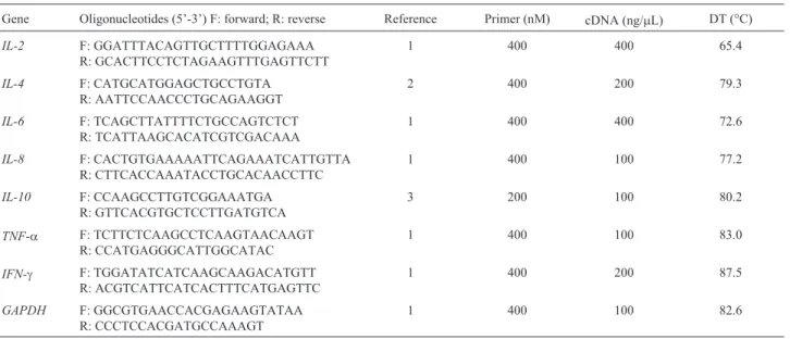
Real-time PCR was carried out using the SYBR Green® PCR Master Mix kit (BioRad, Hercules, CA, USA) according to manufacturer instructions. The primers used for the evaluation of gene expression were designed as described in the literature (Table 1). The glyceraldehyde-3-phosphate dehydrogenase (GAPDH) gene was used as endogenous reference (Leuteneggeret al., 2000). After 40 amplification cycles, all samples were submitted to analy-sis of the dissociation curve in order to confirm the absence of nonspecific products and primer dimers.
Before real-time quantification, the PCR was opti-mized for all genes. For this purpose, three cDNA concen-trations (10, 100 and 200 ng/mL) and three primer dilutions (100, 200 and 400 nM) were tested. After determination of the best PCR conditions, a standard curve was constructed for each gene in which the serial cDNA dilutions were plot-ted against their respective cycle thresholds (Ct). The effi-ciency of amplification of the target genes and endogenous control was similar, ranging from 0.61 to 0.74, and the dis-sociation curves showed no peaks corresponding to primer dimers or nonspecific products for any of the target genes or endogenous control. Table 1 shows the cDNA and primer concentrations optimized for each gene. For theIL-2
andIL-6genes, the best efficiency of the reactions was ob-tained when 400 ng cDNA/mL was used. The coefficient of variation in Ct obtained for each sample in duplicate reac-tions did not exceed 5%, with the widest variation being ob-served for theIL-6gene in one Gyr cow without mastitis (2.95%).
Each sample was analyzed in duplicate in 96-well op-tical reaction plates sealed with opop-tical adhesive film. The
Table 1- Primer pairs and optimized conditions used for determining bovine gene expression by quantitative real-time PCR
Gene Oligonucleotides (5’-3’) F: forward; R: reverse Reference Primer (nM) cDNA (ng/mL) DT (°C)
IL-2 F: GGATTTACAGTTGCTTTTGGAGAAA
R: GCACTTCCTCTAGAAGTTTGAGTTCTT
1 400 400 65.4
IL-4 F: CATGCATGGAGCTGCCTGTA
R: AATTCCAACCCTGCAGAAGGT
2 400 200 79.3
IL-6 F: TCAGCTTATTTTCTGCCAGTCTCT
R: TCATTAAGCACATCGTCGACAAA
1 400 400 72.6
IL-8 F: CACTGTGAAAAATTCAGAAATCATTGTTA
R: CTTCACCAAATACCTGCACAACCTTC
1 400 100 77.2
IL-10 F: CCAAGCCTTGTCGGAAATGA
R: GTTCACGTGCTCCTTGATGTCA
3 200 100 80.2
TNF-a F: TCTTCTCAAGCCTCAAGTAACAAGT R: CCATGAGGGCATTGGCATAC
1 400 100 83.0
IFN-g F: TGGATATCATCAAGCAAGACATGTT R: ACGTCATTCATCACTTTCATGAGTTC
1 400 200 87.5
GAPDH F: GGCGTGAACCACGAGAAGTATAA
R: CCCTCCACGATGCCAAAGT
1 400 100 82.6
samples were amplified separately using the ABI Prism 7300 Sequence Detection System (Applied Biosystem) by the amplification program (denaturation at 95 °C for 5 min, followed by 40 cycles with denaturation at 95 °C for 15 s, and annealing and extension at 60 °C for 1 min). The real-time PCR results were analyzed with the REST© pro-gram (Pfafflet al., 2002), which uses the Pair Wise Fixed Reallocation Randomisation Test©to compare differences in expression across treatments.
Results
Microbiological analysis indicated Streptococcus
spp. as the causative agent in the three BW Holstein cows with mastitis andKlebsiella pneumoniain one Gyr cow. Isolation in culture was not possible in the remaining cows due to the presence of multiple causative agents.
The expression of theIL-2,IL-4,IL-6,IL-8,IL-10,
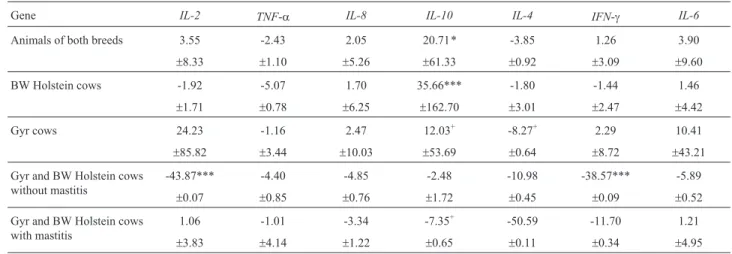
IFN-gandTNF-agenes were compared in: I) animals with-out mastitis vs. animals with mastitis of both breeds (Table 2); II) BW Holstein cows without mastitisvs.BW Holstein cows with mastitis (Table 2); III) Gyr cows with-out mastitisvs.Gyr cows with mastitis (Table 2); IV) BW Holstein cows without mastitisvs.Gyr cows without masti-tis (Table 2); V) BW Holstein cows with mastimasti-tisvs.Gyr cows with mastitis (Table 2).Considering the two breeds to-gether (Table 2),IL-10expression was 20.71 times higher in cows with mastitis compared to animals without mastitis (p < 0.05). For the other genes, no significant increase or decrease in expression across groups were observed (p > 0.05).
Similarly, when analyzing only BW Holstein cows (Table 2),IL-10expression was also higher (35.66 times) in cows with mastitis compared to animals free of infection (p < 0.001), whereas no difference in expression between animals with and without intramammary infection was ob-served for the other genes studied (p > 0.05). In the case of
Gyr cows (Table 2), no significant difference (p > 0.05) in
IL-10 gene expression was observed between cows with and without mastitis; however,IL-10expression showed a trend to be 12.03 times higher in cows with mastitis com-pared to healthy animals (p < 0.10).
Comparison of animals of the two breeds without mastitis (Table 2) showed a significantly lower expression of the IL-2 and IFN-g genes (p < 0.001) in Gyr cows, whereas no significant difference in expression between Gyr and BW Holstein cows was observed for the other genes (p > 0.05). For animals with mastitis (Table 2), no significant difference in the expression of any gene was ob-served between the two breeds (p > 0.05).
Discussion
In the present study, higher expression of theIL-10
gene was observed in BW Holstein and Gyr cows with mas-titis compared to healthy animals, whereas the other studied genes presented no significant difference in expression. IL-10 is an anti-inflammatory cytokine able to inhibit natu-ral killer cells (Barnatu-ral-Netoet al., 1995), as well as the pro-duction of IL-1 and TNF-aby macrophages (Fiorentinoet al., 1991), and also the production of IFN-g and IL-2 by Th1 lymphocytes (Mosmann and Moore, 1991). This fact suggests that high levels of IL-10 may have suppressed the expression ofIL-2, as well as the expression ofTNF-aand
IFN-g. The present results agree with the findings of Riollet
et al.(2001) who observed a higher expression ofIL-10in milk cells of animals with chronic mastitis caused by
Staphylococcus aureuscompared to healthy cows. In addi-tion, various studies have shown thatIL-10is expressed in milk cells of udders infected with different pathogens (Ban-nermanet al., 2004a, 2004b, 2005).
Similarly, separate analysis of BW Holstein cows also showed a higher expression of theIL-10gene in ani-mals with mastitis compared to aniani-mals free of infection.
Table 2- Relative gene expression in animals with mastitis compared to animals without mastitis and the respective standard errors of the mean.
Gene IL-2 TNF-a IL-8 IL-10 IL-4 IFN-g IL-6
Animals of both breeds 3.55 -2.43 2.05 20.71* -3.85 1.26 3.90
±8.33 ±1.10 ±5.26 ±61.33 ±0.92 ±3.09 ±9.60
BW Holstein cows -1.92 -5.07 1.70 35.66*** -1.80 -1.44 1.46
±1.71 ±0.78 ±6.25 ±162.70 ±3.01 ±2.47 ±4.42
Gyr cows 24.23 -1.16 2.47 12.03+ -8.27+ 2.29 10.41
±85.82 ±3.44 ±10.03 ±53.69 ±0.64 ±8.72 ±43.21
Gyr and BW Holstein cows without mastitis
-43.87*** -4.40 -4.85 -2.48 -10.98 -38.57*** -5.89
±0.07 ±0.85 ±0.76 ±1.72 ±0.45 ±0.09 ±0.52
Gyr and BW Holstein cows with mastitis
1.06 -1.01 -3.34 -7.35+ -50.59 -11.70 1.21
±3.83 ±4.14 ±1.22 ±0.65 ±0.11 ±0.34 ±4.95
However, analysis of Gyr cows revealed no significant dif-ference inIL-10gene expression between animals with and without mastitis, althoughIL-10expression showed a trend to be higher in cows with mastitis compared to healthy ani-mals (p < 0.10). These results suggest a higher expression of theIL-10gene in BW Holstein cows with mastitis when compared to Gyr cows with mastitis. In fact, as can be seen in Table 2, expression of this gene tended to be 7.35 times lower in Gyr cows with mastitis when compared to BW Holstein cows with mastitis, but this difference was not sig-nificant (p > 0.05). In addition, the sigsig-nificant difference in gene expression between animals of both breeds with and without mastitis (Table 2) was probably due to the large dif-ference in expression between healthy animals and BW Holstein cows with mastitis (Table 2).
For Gyr cows (Table 2), expression of theIL-4gene showed a trend to be lower in cows with mastitis compared to healthy animals, but this difference was not significant (p < 0.10). IL-4 exerts an antagonistic action to IFN-gand its main function is the regulation of IgE-mediated immune responses. In addition, IL-4 stimulates the differentiation of Th0 and Th2 lymphocytes,i.e., the humoral immune re-sponse. Also, no difference in expression between both groups of animals was observed for the other genes. These findings are not conclusive since a similar pattern of ex-pression ofIL-4andIL-10would be expected because these interleukins produced by Th2 lymphocytes responsible for the humoral immune response. In addition, an expression pattern of IL-10 opposite to that of the IL-2, TNF-aand
IFN-ggenes would be expected, but this was only observed in the case ofIFN-g.Despite the non-significant difference in expression and the divergent results obtained for some genes, these findings suggest that animals with mastitis de-velop a preferentially cell-mediated immune response. Further studies are necessary to better characterize and un-derstand the gene expression profiles in cows with mastitis, especially Gyr animals, since no studies of this type are yet available for Zebu cattle and, therefore, the results cannot be compared with other studies.
A significant difference in expression (p < 0.001) was observed when healthy cows of the two breeds were com-pared (Table 2), with the expression of theIL-2andIFN-g genes being higher in BW Holstein cows. IL-2 induces the proliferation of activated mononuclear cells as well as of some activated epithelial cells. IL-2 can be suppressed by the expression ofIL-10since an increase in IL-10 indirectly inhibits the differentiation of Th0 lymphocytes into Th1 lymphocytes. This result led to a reduction of IL-2 since this cytokine is mainly produced by Th1 lymphocytes (DeFrancoet al., 2007). IFN-gis associated with the con-version of Th0 into Th1 lymphocytes and activation of macrophages and neutrophils, in addition to potentiating the action of TNF-a(Janewayet al., 2002). Therefore, this cytokine is related to a Th1 immune response,i.e., a cellular
immune response. In addition, IFN-gdeficiency is associ-ated with increased susceptibility to infections caused by intracellular microorganisms. Despite the significant dif-ference inIFN-g andIL-2expression, further studies are necessary to evaluate the expression profile in different breeds under different conditions.
IL-8, together with IL-1 and TNF-a, has been indi-cated as an important mediator of neutrophil recruitment to sites of inflammation. Some studies have shown that TNF-aand IL-8 are present in large amounts in milk from udders infected with Gram-negative bacteria such as E. coli,K. pneumoniaeorPseudomonas aeruginosa, but are detected at lower concentrations or are absent in milk of cows whose mammary gland is infected withS. aureus. In the present study, no significant difference in the expres-sion ofTNF-aorIL-8was observed when animals of the two breeds with mastitis were compared; however, expres-sion of the two genes seemed to be higher in BW Holstein cows (Table 2), a fact that disagrees with the results ob-tained by others since in the present study all BW Holstein cows with mastitis were infected withStaphylococcusspp. andK. pneumoniaewas the causative agent of mastitis in one Gyr cow (Shuster et al., 1997; Riollet et al., 2000; Bannermanet al., 2004a, 2004b, 2005).
These divergent results might be explained by the fact that the immune response can differ according to bacterial strain and host, with the observation of wide individual variation. In agreement with the present findings, in anin vitrostudy Lahouassaet al.(2007) demonstrated that dif-ferentS. aureusstrains elicit different responses in epithe-lial cells of the mammary gland. In addition, in that study the intensity and level of expression of the genes analyzed (IL-8,GRO-a,GRO-b,TNF-a,IL-1b,TGF-b1andIL-10) varied according to the step of infection (3, 10 and 24 h af-ter the addition of bacaf-teria to the cell culture). According to those authors, these different responses imply in alternative routes of activation or at different signal transduction lev-els, reflecting what is observedin vivo. In the present study, the milk samples were collected immediately after the man-ifestation of clinical signs of mastitis,i.e., it was not possi-ble to determine when the animal was infected since two animals may manifest a disease on different days even when they were infected at the same date. In addition, there was no control of the type of strain that caused the infection and in some cases the causative agent of mastitis could not be identified (two Gyr cows infected with multiple agents). It should also be noted that the milk samples were collected from animals reared in commercial herds and, therefore, it was not possible to determine whether these animals were free of other infections or diseases, a fact that may have also influenced the present results.
regula-tion were not studied. Thus, it would be interesting to per-form proteomic studies to confirm these results. In addition, analysis of the structure of the genes that were dif-ferentially expressed in this study, such asIL-10, would be useful to identify marker SNPs for mastitis resistance and susceptibility. Mastitis is a multifactorial disease which is influenced by numerous genes. Therefore, further studies including a larger number of genes and animals in different steps of infection are necessary to better understand the im-mune response mechanism and to develop more efficient strategies for the control and eradication of this disease. In addition, studies using experimental populations, including the history of mastitis, somatic cell count of each animal, calving order and lactation stage, as well as studies inocu-lating specific bacterial strains and collecting samples at different times after inoculation, would greatly contribute to the understanding of the physiopathology of this disease.
It should be emphasized that theIL-4,IL-6,IL-8and
TNF-agenes were not differentially expressed in any of the conditions studied (p > 0.05), demonstrating that these genes are not good markers or indicators of mastitis under the analyzed conditions. Further experiments will be per-formed to investigate not only the genes studied here, but also other genes that might be involved in mastitis resis-tance, especially in Zebu animals, since most studies so far have been conducted on breeds of European origin. These studies may contribute to a better understanding of the im-mune responses that occur in mastitis, a disease that signifi-cantly affects milk quality and yield especially in Zebu breeds which represent the basis of the cattle-raising indus-try in Brazil.
Acknowledgments
The authors thank CAPES, CNPq, FAPEMIG and FINEP for the financial support of this work.
References
Alluwaimi AM, Leutenegger CM, Farver TB, Rossitto PV, Smith WL and Cullor JS (2003) The cytokine markers in Staphylo-coccus aureus mastitis of bovine mammary gland. J Vet Med 50:105-111.
Bannerman DD, Paape MJ, Lee JW, Zhao X, Hope JC and Rainard P (2004a) Escherichia coli and Staphylococcus aureuselicit differential innate immune responses following intramammary infection. Clin Diagn Lab Immunol 11:463-472.
Bannerman DD, Paape MJ, Hare WR and Hope JC (2004b) Char-acterization of the bovine innate immune response to intra-mammary infection withKlebsiella pneumoniae.J Dairy Sci 87:2420-2432.
Bannerman DD, Chockalingam A, Paape MJ and Hope JC (2005) The bovine innate immune response during experimen-tally-induced Pseudomonas aeruginosa mastitis. Vet Immunol Immunopathol 107:201-215.
Barral-Neto M, Barral A, Brodskyn C, Carvalho EM and Reed SB (1995) Cytotoxicity in human mucosal and cutaneous leishmaniasis. Parasite Immunol 17:21-28.
Costa CN, Freitas AF, Cobuci JA, Valloto AA, Ribas Neto PG, Horst JA, Barra RB, Campos LS and Marques A (2007) Sumário Nacional de Touros da Raça Holandesa. Embrapa Gado de Leite, Juiz de Fora, 64 pp.
DeFranco A, Locksley RM and Robertson M (2007) Immunity: The immune response in infectious and inflammatory dis-ease. New Science Press, London, 387 pp.
Detilleux JC, Koehler KJ, Freeman AE, Kehrli ME and Kelley DH (1994) Immunological parameters of periparturient Hol-stein cattle: Genetic variation. J Dairy Sci 77:2640-2650. Ferens WA, Goff WL, Davis WC, Fox LK, Deobald C, Hamilton
MJ and Bohach GA (1998) Induction of type-2 cytokines by a Staphylococcal enterotoxins superantigen. J Nat Toxins 7:193-213.
Ferreira MBD, Lopes BC, Ledic IL, Fernandes LO and Ribeiro SA (2007) Características reprodutivas de touros da raça Gir. Revista Gir Leiteiro 7:30-38.
Fiorentino DF, Zlotnik A and Vieira P (1991) IL-10 acts on the an-tigen-presenting cell to inhibit cytokine production by Th1 cells. J Immunol 146:3444-3451.
Giraudo JA, Calzolari A, Rampone H, Rampone A, Giraudo AT, Bogni C, Larriestra A and Nagel R (1997) Field trials of vac-cine against bovine mastitis. 1. Evaluation in heifers. J Dairy Sci 80:845-853.
Janeway CA, Tavers P, Walport M and Sholmchik MJ (2002) Imunologia: O Sistema Imune na Saúde e na Doença. 5th edition. Artmed, Porto Alegre, 776 pp.
Lahouassa H, Moussay E, Rainard P and Riollet C (2007) Differ-ential cytokine and chemokine responses of bovine mam-mary epithelial cells toStaphylococcus aureusand Esche-richia coli. Cytokine 38:12-21.
Leutenegger CM, Alluwaimi AM, Smith WL, Perani L and Cullor JS (2000) Quantitation of bovine cytokine mRNA in milk cells of healthy cattle by real-time TaqMan® polymerase chain reaction. Vet Immunol Immunopathol 77:275-287. Mosmann TR and Moore KW (1991) The role of IL-10 in
cros-sregulation of Th1 and Th2 responses. Immunol Today 12:A49-53.
Moussay E, Stamm I, Taubert A, Baljer G and Menge C (2006)
Escherichia coliShiga toxin 1 enhances Il-4 transcripts in bovine ileal intraepithelial lymphocytes. Vet Immunol Immunopathol 113:367-382.
Oviedo-Boyso J, Valdez-Alarcón JJ, Cajero-Juárez M, Ochoa-Zarzosa A, López-Meza JE, Bravo-Patiño A and Baizabal-Aguirre VM (2007) Innate immune response of bovine mammary gland to pathogenic bacteria responsible for mas-titis. J Infect 54:399-409.
Pereira AR, Machado PF and Sarríes GA (2001) Contagem de células somáticas e características produtivas de vacas da raça Holandesa em lactação. Sci Agric 58:649-654. Pfaffl MW, Horgan GW and Dempfle L (2002) Relative
Expres-sion Software Tool (REST©) for group wise comparison and statistical analysis of relative expression results in real-time PCR. Nucleic Acids Res 30:e36.
Rambeaud M, Almeida RA, Pighetti GM and Oliver SP (2003) Dynamics of leukocytes and cytokines during experimen-tally inducedStreptococcus uberismastitis. Vet Immunol Immunopathol 96:193-205.
Riollet C, Rainard P and Poutrel B (2000) Differential induction of complement fragment C5a and inflammatory cytokines during intramammary infections withEscherichia coliand
Staphylococcus aureusinfection. Clin Diagn Lab Immunol 7:161-167.
Riollet C, Rainard P and Poutrel B (2001) Cell subpopulations and cytokine expression in cow milk in response to chronic
Staphylococcus aureusinfection. J Dairy Sci 84:1077-1084. Shuster DE, Kehrli Jr ME and Stevens MG (1993) Cytokine
pro-duction during endotoxin-induced mastitis in lactating dairy cows. Am J Vet Res 54:80-85.
Shuster DE, Kehrli Jr ME, Rainard P and Paape M (1997) Com-plement fragment C5a and inflammatory cytokines in
neu-trophil recruitment during intramammary infection with
Escherichia coli. Infect Immun 65:3286-3292.
Vercesi Filho AE, Faro LE, Paz CCP, Cardoso VL, Silva DAL and Junqueira FS (2007) Fatores que influenciam o preço do sêmen de touros da raça Gir Leiteiro. Revista Gir Leiteiro 7:78-81.
Waldvogel AS, Hediger-Weithaler BM, Eicher R, Zakher A, Zarlenga DS, Gasbarre LC and Heussler VT (2000) Inter-feron-gama and Interleukin-4 mRNA expression by periph-eral blood mononuclear cells from pregnant and non-preg-nant cattle seropositive for bovine viral diarrhea virus. Vet Immunol Immunopathol 77:201-212.
Associate Editor: Luiz Lehmann Coutinho