D e te ctio n o f im m uno glo bulin G in the
lung and live r o f ham ste rs with visce ral
le ishm aniasis
1Laboratório de Soroepidemiologia e Imunobiologia Celular e Molecular,
Instituto de Medicina Tropical de São Paulo, and 2Departamento de Medicina
Preventiva, Faculdade de Medicina, Universidade de São Paulo, São Paulo, SP, Brasil
3Departamento de Clínica e Cirurgia Veterinária, Universidade Federal do Piauí,
Teresina, PI, Brasil R. Mathias1,
F.A.L. Costa1,3
and H. Goto1,2
Abstract
Several organs are affected in visceral leishmaniasis, not only those rich in mononuclear phagocytes. Hypergammaglobulinemia occurs during visceral leishmaniasis; anti-Leishmania antibodies are not primarily important for protection but might be involved in the patho-genesis of tissue lesions. The glomerulonephritis occurring in visceral leishmaniasis has been attributed to immune complex deposition but in other organs the mechanism has not been studied. In the current study we demonstrated the presence of IgG in the lung and liver of hamsters with visceral leishmaniasis. Hamsters were injected intra-peritoneally with 2 x 10⁷ amastigotes of Leishmania (Leishmania)
chagasi and the presence of IgG in the liver and lung was evaluated at 7, 15, 30, 45, 80 and 102 days postinfection (PI) by immunohis-tochemistry. The parasite burden in the spleen and liver increased progressively during infection. We observed a deposit of IgG from day 7 PI that increased progressively until it reached highest intensity around 30 and 45 days PI, declining at later times. The IgG deposits outlined the sinusoids. In the lung a deposit of IgG was observed in the capillary walls that was moderate at day 7 PI, but the intensity increased remarkably at day 30 PI and declined at later times of infection. No significant C3 deposits were observed in the lung or in the liver. We conclude that IgG may participate in the pathogenesis of the inflammatory process of the lung and liver occurring in experimen-tal visceral leishmaniasis and we discuss an alternative mechanism other than immune complex deposition.
Co rre spo nde nce
H. Goto
Laboratório de Soroepidemiologia e Imunobiologia Celular e Molecular IMTSP, USP
Av. Enéas C. Aguiar, 470 Prédio II, 4º andar 05403-000 São Paulo, SP Brasil
Fax: + 55-11-3062-3622 E-mail: hgoto@ usp.br
Research supported by FAPESP (Nos. 98/15414-3 and 98/12092-5), CNPq (No. 300866/98-4 to H. Goto) and LIM/38 (HC-FMUSP).
Received April 12, 2000 Accepted February 16, 2001
Ke y wo rds
·Leishm ania (Leishm ania) chagasi
·Visceral leishmaniasis
·Immunopathology
·Immunoglobulin G
·Liver
·Lung
Leishmaniasis is a disease caused by a protozoan of the genus Leishmania. Visceral leishmaniasis affects mononuclear phago-cytes mainly in the spleen and in the liver where hyperplasia and hypertrophy of these cells occur, but other organs such as lung and kidney are affected during progression of the disease (1). Interstitial inflammation is
cryosections of the organs using a polyclo-nal rabbit anti-hamster C3 antibody produced by us (8). Anti-Leishmania antibody titer was determined by immunofluorescence us-ing Leishmania (L.) amazonensis antigen. The parasite burden in the spleen and liver was calculated by counting the number of cells and parasites in each microscopic field of organ imprints up to 1000 cells or para-sites. Parasite burden was calculated by the formula: (number of parasites/number of cells) x weight of the organ (mg) x 2 x 10⁴.
Parasite burden in the spleen and liver and anti-Leishmania antibody titer in in-fected hamsters increased progressively dur-ing infection. Usdur-ing immunohistochemistry we observed some background staining in control animals but in the liver of infected animals we clearly observed a weak deposit of IgG at day 7 PI that increased progres-sively until it reached highest intensity around 30 and 45 days PI, declining at later times (Table 1). IgG deposits outlined the sinuso-ids (Figure 1A). In the lung a deposit of IgG was observed in the capillary walls (Figure 1B) which was of moderate intensity at day 7 PI, but increased remarkably at day 30 PI and declined at later times of infection (Table 1). No significant C3 deposits were observed either in the lung or in the liver. In two preliminary experiments with 3-4 infected visceral leishmaniasis. In the kidney,
im-mune complex deposition has been claimed as the mechanism of lesion (5-7). To study the immunopathogenesis of the lesions in the liver and lung, in the current study we evaluated the presence of IgG in these or-gans in hamsters with visceral leishmaniasis. Twenty-eight outbred 45-60-day-old male hamsters (Mesocricetus auratus) from the Animal Breeding Facility of the Medical School of the University of São Paulo were maintained in the Animal Facilities of the Institute of Tropical Medicine of São Paulo during the experiment. Seventeen hamsters were inoculated intraperitoneally with puri-fied 2 x 10⁷ amastigotes of Leishmania (
Leish-mania) chagasi (MHOM/BR/72/strain 46)
in RPMI 1640 medium (Gibco BRL, Gai-thersburg, MD, USA), and sacrificed at 7, 15, 30, 45, 80 and 102 days postinfection (PI). Eleven control animals were injected with RPMI 1640 medium and sacrificed at the same times. At the time of sacrifice, spleen, liver, kidney and lung were obtained and the presence of IgG in the liver, kidney and lung was evaluated by immunohis-tochemistry using biotinylated goat anti-ham-ster IgG antibody (20 µg/ml) (Rockland, Gilbertsville, PA, USA) in formalin-fixed and paraffin-embedded tissue sections. Complement fraction C3 was detected in
Table 1 - Intensity of IgG deposits detected by immunoperoxidase staining using biotinylated goat anti-hamster IgG antibody in the liver, lung and kidney of anti-hamsters infected w ith 2 x 10⁷ amastigotes of
Leishmania (L.) chagasi and non-infected control animals at different times of infection.
- = Negative. Intensity of positive reactions graded from + through ++++. Data are reported as the mean of 2-4 animals/group.
Days post- Liver Lung Kidney
infection
Control Infected Control Infected Control Infected
7 - + + ++ - +
15 + ++ + +++ - ++
30 + +++ + ++++ ++ +++
45 + +++ + +++ + ++
80 ++ ++ + +++ + ++
and control animals sacrificed at the same times, we observed similar results by immu-nofluorescence using polyclonal rabbit anti-hamster total immunoglobulin serum.
Since the absence of C3 deposits is not compatible with immune complex deposi-tion in the pathogenesis of the disease and since an immune complex-mediated mech-anism has been shown in the kidney in vis-ceral leishmaniasis (5-7), as a control we examined kidney tissue from the same ani-mals. In the kidney the deposit of IgG was observed outlining the capillary walls (Fig-ure 1C). The intensity of the deposit was highest at 30 and at 102 days PI and slightly lower at other time points (Table 1). C3 deposits of moderate intensity were detected in the kidney throughout the experiment, with a slight increase in intensity at day 102. Thus, we conclude that the conditions for the presumed formation of an immune complex were present in the experimental animals. Furthermore, one of the factors for immune complex deposition is the incapacity to clear immune complexes due to dysfunction of the reticuloendothelial system (9), which is likely to occur in visceral leishmaniasis due to Leishmania parasite proliferation in mono-nuclear phagocytes.
The absence of C3 deposits in the liver and lung still contradicts the mechanism of immune complex deposition. However, the presence of IgG deposits in the liver and lung might indicate its role in the pathogenesis. IgG deposits in the lung have been reported in some situations such as the presence of anti-basement membrane antibody in the
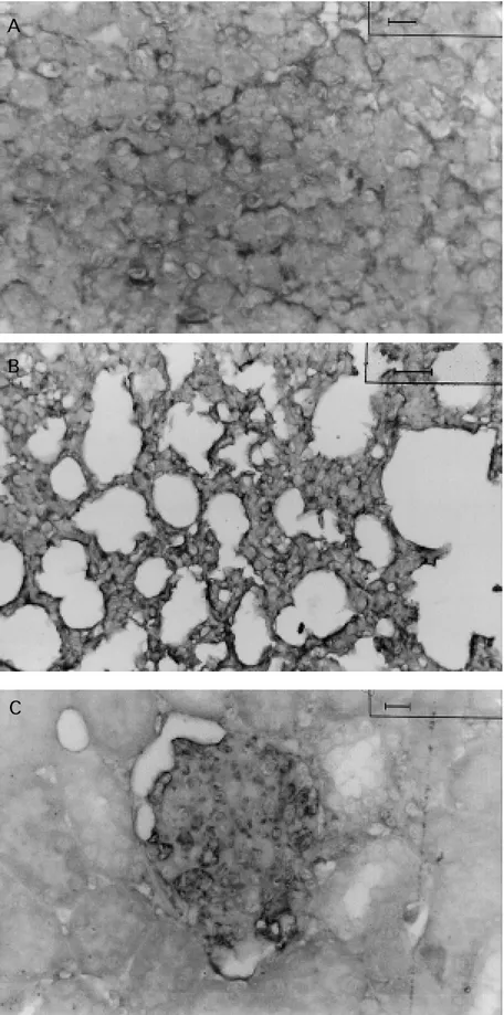
sep-Figure 1 - Detection of IgG deposits in the liver, lung and kidney of hamsters infected w ith 2 x 10⁷
amasti-gotes of Leishmania (L.) chagasi by immunoperoxidase staining using biotinylated goat hamster IgG anti-body. A, IgG deposits along the sinusoid are seen in the liver of infected hamsters at 45 days of infection. B, IgG deposits in the capillary w all of the lung septum from infected animals at 30 days of infection. C, IgG depos-its outlining the capillary w all in the glomeruli of in-fected hamsters at 45 days of infection. Bar: 25 µm.
A
B
tum in Goodpasture syndrome (10) and in acute and chronic immune complex-medi-ated disease models (11). In the present study, we observed the presence of IgG deposits in the lung mainly in the vessel wall, in agree-ment with findings for rabbits with chronic serum sickness, in which interstitial pneu-monitis develops (12). In addition, in vis-ceral leishmaniasis the presence of Leishma-nia antigen has been observed in inflamma-tory foci in the lung (13). In immune com-plex-mediated diseases and in animal mod-els of acute and chronic serum sickness, a known model of immune complex-mediated disease (11), no liver lesion has been de-scribed. Furthermore, in diseases involving an immune mechanism such as viral hepati-tis and primary biliary cirrhosis, the antibod-ies are directed at the hepatocytes and bile ducts, respectively (14,15). Therefore, to our knowledge, this is the first observation of IgG along the sinusoids.
To correlate the lesion with the IgG de-posits histopathological analysis was per-formed. In the liver, a progressive increase of diffuse hyperplasia and hypertrophy of Kupffer cells were observed from 7 to 80 days PI, when hyperplasia became less pro-nounced. Foci of mononuclear cells were observed from 7 days PI, initially in peripor-tal and centrolobular spaces, but later more disseminated with no preference for any par-ticular zone. In the lung, foci of septal thick-ening due to congestion, edema and mixed infiltrate of polymorphonuclear neutrophils and mononuclear cells were observed at 7 days PI. Inflammation increased progres-sively and the polymorphonuclear neutro-phils were gradually replaced by mono-nuclear cells in the infiltrate, and at 30 days PI only mononuclear cells were practically seen. In the kidney, hypercellularity and an increase in mesangial matrix were observed at 30-45 days PI followed by a decrease at 45 days, and at 102 days PI a deposit of an amorphous eosinophilic substance was seen. The lesions in different organs were
progres-sive, with noticeable changes in their fea-tures during evolution. In the liver, the mono-nuclear cell inflammatory infiltrate became more prominent than hyperplasia of Kupffer cells at later times and in the lung the cell population in the interstitial infiltrate changed during the course of infection. The changes in the characteristics of the lesion and the occurrence of more intense IgG deposits around 30-45 days PI in the lesion might suggest that participation of IgG in the patho-genesis occurs at a certain time during the evolution of the infection.
Concerning the mechanism of lesion, the presence of IgG deposits in the liver which has not been shown in immune complex-mediated diseases and the absence of C3 deposits both in the lung and in the liver led us to consider alternative mechanisms. We have some evidence that pathogenic mechan-isms other than immune complex-mediated ones participate in the lesions of visceral leishmaniasis. We recently observed the pres-ence of T cells in the kidney in canine vis-ceral leishmaniasis (16). In addition, in sys-temic lupus erythematosus it has been shown that immunoglobulins might participate in the pathogenesis of glomerular lesions by a mechanism distinct from immune complex deposition (17,18). We have been working with the hypothesis that internalization of immunoglobulins by endothelial cells might have some role in the pathogenesis of vis-ceral leishmaniasis. We observed in vitro
mi-gration of mononuclear cells including T cells to the lesion, as observed in the ne-phropathy of canine visceral leishmaniasis (16). Our findings in the liver and lung are compatible with this hypothesis and studies are in progress for a better understanding of the immunopathogenesis of visceral leish-maniasis.
Ackno wle dgm e nts
We thank the biologist Edite H.Y. Kana-shiro for technical support and the Histopa-thology Laboratory of the Faculdade de Me-dicina da Universidade de São Paulo for the histopathological preparations.
Re fe re nce s
1. Duarte M IS & Corbett CEP (1994). Patolo-gia das principais doenças tropicais no Brasil. Leishmaniose visceral. In: Brasilei-ro Filho G, Pitella JEH, Pereira FEL, Bambirra EA & Barbosa AJA (Editors), Bogliolo-Patologia. 5th edn. Guanabara-Koogan, Rio de Janeiro, RJ, Brazil. 2. Bunn-M oreno M M , M adeira ED, M iller K,
M enezes JA & Campos-Neto A (1985). Hypergammaglobulinemia in Leishmania donovani infected hamsters: possible as-sociation w ith a polyclonal activation of B cells and w ith suppression of T cell func-tion. Clinical and Experimental Immunol-ogy, 59: 427-434.
3. Galvão-Castro B, Sá-Ferreira JA, M arzochi KF, M arzochi M C, Coutinho SG & Lam-bert PH (1984). Polyclonal B cell activa-tion, circulating immune complexes and autoimmunity in human American visceral leishmaniasis. Clinical and Experimental Immunology, 56: 58-66.
4. Sacks DL, Louis JA & Wirth DF (1993). Leishmaniasis. In: Warren KS (Editor), Im-munology and M olecular Biology of Para-sitic Infections. Blackw ell Scientific Publi-cations, Boston, M A.
5. Brito T, Hoshino-Shimizu S, Amato Neto V, Duarte M IS & Penna DO (1975). Glo-merular involvement in human kala-azar: a light, immunofluorescent and electron mi-croscopic study based on kidney biopsies. American Journal of Tropical M edicine and Hygiene, 24: 9-18.
6. Weisinger JR, Pinto A, Velazquez GA, Bronstein I, Dessene JJ, Duque JF, M on-tenegro J, Tapanes F & Rousse AR (1978). Clinical and histological kidney involve-ment in human kala-azar. American Jour-nal of Tropical M edicine and Hygiene, 27:
357-359.
7. Sartori A, Roque-Barreira M C, Coe J & Campos-Neto A (1987). Immune complex glomerulonephritis in experimental kala-azar. Parasite Immunology, 9: 93-103. 8. Laurenti M D, Corbett CEP, Sotto M N,
Sinhorini IL & Goto H (1996). The role of complement on the acute inflammatory process in the skin and on host parasite interaction in hamsters inoculated by Leishmania (Leishmania) chagasi. Interna-tional Journal of Experimental Pathology, 77: 15-24.
9. Law ley TJ & Frank M (1980). Immune complexes and immune complex dis-eases. In: Parker CW (Editor), Clinical Im-munology. WB Saunders Company, Phila-delphia.
10. Sturgill BC & Westervelt FB (1965). Im-munofluorescence studies in a case of Goodpasture’s syndrome. Journal of the American M edical Association,194: 172-174.
11. Cochrane CG & Dixon FJ (1978). Immune complex injury. In: Samter M (Editor), Im-munological Diseases. Little, Brow n and Company, Boston.
12. Brentjens JR, O’Connell DW, Paw low ski IB, Hsu KC & Andres GA (1974). Experi-mental immune complex disease of the lung. Journal of Experimental M edicine, 140: 105-125.
13. Duarte M IS, M atta VLR, Corbett CEP, Laurenti M D, Chebabo R & Goto H (1989). Interstitial pneumonitis in human visceral leishmaniasis. Transactions of the Royal Society of Tropical M edicine and Hygiene, 83: 73-76.
14. Eddleston ALWF (1980). Immunology and the liver. In: Parker CW (Editor), Clinical
Immunology. WB Saunders Company, Philadelphia.
15. Krams SM , De Water JV, Coppel RL, Esquivel C, Robert s J, Ansari A & Gershw in M E (1990). Analysis of hepatic T lymphocyte and immunoglobulin depos-its in patients w ith primary biliary cirrho-sis. Hepatology, 12: 306-313.
16. Costa FAL, Guerra JL, Silva SM M S, Klein RP, M endonça IL & Goto H (2000). CD4+
T cells participate in the nephropathy of canine visceral leishmaniasis. Brazilian Journal of M edical and Biological Re-search, 33: 1455-1458.
17. Itoh J, Nose M , Takahashi S, Ono M , Terasaki S, Kondoh E & Kyogoku M (1993). Induction of different types of glo-merulonephritis by monoclonal antibod-ies derived from an M RL/lpr lupus mouse. Am erican Journal of Pat hology, 143: 1436-1443.
18. Ono M , Yamamoto T, Kyogoku M & Nose M (1995). Sequence analysis of the germ-line VH gene corresponding to a nephrito-genic antibody in M RL/lpr lupus mice. Clinical and Experimental Immunology, 100: 284-290.
19. Goto H & Nose M (1997). Ingestion of hamster and visceral leishmaniasis serum IgG by endothelial cells as pathogenetic mechanism of interstitial inflammation. XXII Congresso da Sociedade Brasileira de Imunologia, M angaratiba, RJ, Brazil, September 28-October 1, 1997, 11.17: 115.