Pre and post functional
endoscopic sinus surgery nasal
cavity volume assessment by
acoustic rhinometry
Summary
Rodrigo de Paula Santos1, Walter Habermann2,
Thiemo Hofmann3, Heinz Stammberger4
1 Master and Doctoral student, Head of the clinical rhinology department at the Sao Paulo Federal University - Paulista Medical School. 2 MD, Ph.D., Assistant physician of the Otorhinolaryngology Department at the Graz University, Austria.
3 MD, Assistant physician of the Otorhinolaryngology Department at the Graz University, Austria. 4 MD, Ph.D., Head of the Otorhinolaryngology Department at the Graz University, Austria.
Sao Paulo Federal University/ Paulista Medical School.
Address for correspondence: Al. Jaú 1767 1º andar Cerqueira Cesar Sao Paulo SP 01420-002.
Paper submitted to the ABORL-CCF SGP (Management Publications System) on February 28th, 2006 and accepted for publication on June 7th, 2006.
A
coustic rhinometry is an objective method to determine nasal cavity geometry. The technique is based on sound wave reflexion analysis in the nasal cavity, and determines crossectional areas as a function of distance as well as volume. Aim: The purpose of this study is to analyse nasal cavity volume changes caused by functional endoscopic sinus surgery (FESS) in adults with chronic rhinosinusitis by acoustic rhinometry, and to correlate these changes with improvements in the sensation of nasal obstruction. Material and Method: Forty patients aged from 18 to 73 years were prospectively evaluated between August and October 1999 at the Graz University Hospital, Austria. All patients were diagnosed with chronic rhinosinusitis, and undertook acoustic rhinometry before and after FESS. Scientific design: A clinical prospective study. Results: The nasal cavity total volume increased significantly after surgery. Nasal obstruction was improved in 88% of the patients, 20% with partial improvement and 68% with total improvement. There was no correlation between volume increase and improvement of the sensation of nasal obstruction. Conclusion: Total nasal cavity volume significantly increased after surgery; however, there was no correlation between volume increase and improvements of nasal obstruction. No significant pre or postoperative increase in total nasal cavity volume after decongestion were observed.Keywords: nasal polips, paranasal sinus, sinusitis. ORIGINAL ARTICLE
INTRODUCTION
Functional endoscopic sinus surgery (FESS) was developed based on the pathophysiology theory of chronic rhinosinusitis proposed by Messerklinger, published in the 50s and 60s. In his original studies, this author used cadaver heads, impregnating the paranasal sinus mucus with a variety of substances. Using microscopes and endoscopes, he described the physiological pathways of paranasal sinus secretions. Based on these observations and correlations with findings in surgery on changes in mucociliary transport, Messerklinger was able to establish that the maxillary and frontal sinuses depend on their prechambers in the ethmoid and lateral nasal wall. Ventila-tion and drainage of these cavities are essential for normal nasal sinus function1.
FESS involves the opening of paranasal sinus prechambers aiming to restore drainage and ventilation. Messerklinger2 observed that eradication of the primary
disease in the anterior ethmoid sinus through a limited endoscopic surgical procedure resulted in recovery of the mucosa of adjacent paranasal sinuses (frontal and maxil-lary) with no direct surgical manipulation of these areas. This is a conservative approach to surgical procedures on the nasal septum and turbinates.
Most papers published on this topic present subjec-tive symptom relief results following FESS3,4. The subjective
perception patients have of their improvement is probably the best parameter to assess surgical efficiency, but objec-tive methods are desirable to evaluate the impact of any therapeutic strategy.
There are few published papers studying objective assessment of the results of FESS. Most studies evaluate patients undergoing combined surgical procedures such as septoplasty and inferior turbinectomy.
Acoustic rhinometry (AR) is an objective method to measure the nasal cavity geometry5. The technique
analyses the reflection of sound waves within the nose and measures the cross-sectional area of the nasal cav-ity in relation to the nares and defines the volume. This method was developed based on Jackson et al.’s6 studies
on acoustic pulses to study the geometry of lower airways. This exam adds to the objective assessment of the nasal cavity, previously done with anterior rhinomanometry, which measures nasal air flow and resistance.
The aim of this paper is to study volume changes in the nasal cavity caused by FESS in adults with chronic rhinosinusitis with or without nasal polyposis using AR, and to correlate these alterations with clinical improve-ments and the sensation of nasal obstruction.
MATERIAL AND METHODS
There were 40 patients (80 nasal cavities) included in this study, 21 women and 19 men, none of which had
undergone nasal surgery. Patients were admitted consecu-tively at the Otorhinolaryngology ward of the Graz Uni-versity Hospital in Austria, between August and October 1999, to undergo FESS. Age varied from 18 to 73 years. Patients with nasal septum deviation with an indication for septoplasty and patients with lower turbinate hypertrophy were excluded from the study.
All patients had symptoms of chronic rhinosinusitis for at least three months and had been treated with an-tibiotics, topical corticosteroids and antihistaminic drugs when indicated, with no significant improvement.
When medical treatment was not successful, patients would undergo rigid nasal endoscopy and computed tomography of the paranasal sinuses. The indication for surgery was based on the clinical history and the results of these two exams.
Rigid nasal endoscopy followed Messerklingler’s2
methodology, assessing signs such as changes in the mu-cosa of the ethmoid infundibulum, the presence of pus in the rhinopharynx originated from the superior or middle meatus, and anatomical variants that led to narrowing or obstruction of the ostium-meatal complex, all of which are signs of chronic sinus disease.
Computed tomography was done to assess the degree of sinus involvement and as a guiding tool for surgery by revealing the anatomical relationship between the paranasal sinuses, the optic nerve, and the internal carotid artery7.
General anesthesia was used in all cases based on Stammberger’s8 technique. Disease extension defined the
type of surgical procedure, which included uncinectomy, anterior ethmoidectomy and perforation of the basal lamella of the middle turbinate in all cases. Posterior eth-moidectomy, sphenoidectomy, widening of the frontal re-cess and the maxillary sinus ostium were done as needed. No patient underwent surgery of the nasal septum or the lower turbinates.
AR was used as an objective evaluation method to separately measure nasal cavity volumes before and ten minutes after the use of a topical vasoconstrictor (two 0.5 mg/ml jets of oxymetazoline chloridrate nasal spray in each nare) pre and postoperatively. The vasoconstrictor was used to annul or minimize the influence of the physiologi-cal nasal cycle upon nasal cavity volume. Measurements were obtained one day before surgery and between four and eight weeks postoperatively, using the Rhinoklack - RK 1000tm (Stimotron Co., Wendelstein, Germany) device as internationally standardized9.
Out of 40 patients, 25 returned for a second evalua-tion. Before the second measurement, patients underwent postoperative endoscopy to clean the nasal cavities, remov-ing clots that might interfere in exam results.
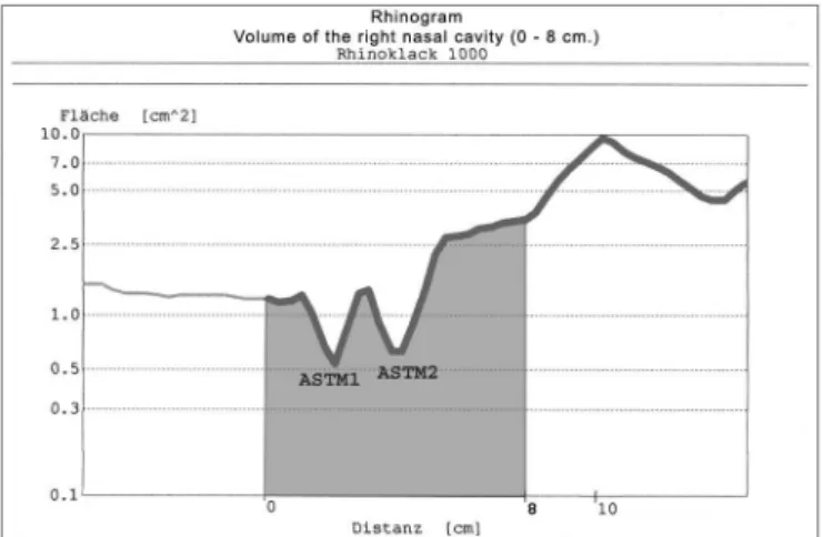
(Figure 1). On this chart the y-axis is the cross-sectional area (cm2) and the x-axis is the distance from the nasal adaptor into the nasal cavity (cm). The rhinogram of nor-mal adults has two “dips”. They are known as the mini-mal cross-sectional areas 1 and 2. The volume between any two points in the nasal cavity is computer-calculated after these points are charted. We measured the volume between 0 and 8 cm distance from the nare10,11. The total
nasal volume was calculated as the sum of the right and left nasal cavities (Figure 1).
Data were analyzed statistically (Wilcoxon nonpara-metric test) comparing pre and post-vasoconstrictor total nasal cavity volumes before surgery, pre and post-vasocon-strictor total nasal cavity volumes after surgery and in the pre and postoperative periods following vasoconstrictor use to minimize the influence of the physiological nasal cycle.
Patients were inquired about changes in the sen-sation of nasal obstruction postoperatively compared to the preoperative period. We used a visual analog scale in which patients marked one of the following options: (-1) worsening of the sensation of nasal obstruction; (0) unchanged; (1) partial improvement; (2) full improvement. Nasal cavity volume changes produced by surgery were compared with the sensation of improvement from nasal obstruction. We used the Kruskal-Wallis nonparametric statistical test.
Figure 1. Rhinogram of the right nasal cavity, showing minimal cross-sectional areas 1 and 2 and volume (0 - 8 cm).
RESULTS
Average nasal cavity volumes (right + left) before and after FESS and pre and post topical vasoconstrictor use are shown on Table 1. Nasal cavity volume increase following surgery was statistically significant.
Results on the subjective complaint of nasal
ob-struction based on the visual analog scale are shown on Chart 1.
We used the Kruskal-Wallis test to study the differ-ent groups in the subjective analysis of the sensation of nasal obstruction groups (unchanged, partial improvement, full improvement), noting that the difference in nasal cavity volumes produced by surgery was not significantly differ-ent between the three groups (p = 0.311).
DISCUSSION
FESS is currently the treatment of choice for chronic sinusitis that do not respond to medical treatment. Many studies with variable follow-up periods have focused on the subjective improvement of patients1,4,12-15. AR,
intro-duced by Hilberg et al.5, is an objective method to analyze
the nasal cavity geometry by the use of sound waves. It is a simple non-invasive method requiring minimal patient cooperation. AR has been used in the pre and
postopera-Table 1. Average pre and post-vasoconstrictor nasal cavity volumes, before and after FESS, in cm3.
Average preopera-tive volume of the nasal cavity (in cm3)
Average postope-rative volume of the nasal cavity (in cm3) Pre-vasoconstrictor 38,91 45,96 Post-vasoconstrictor 39,69 45,16
tive evaluation of rhinoplasties16, turbinoplasties and/or
turbinectomies17, polypectomies18, adenoidectomies19, in
assessing snorers and patients with obstructive sleep apnea syndrome20, choanal atresia21, subglottic stenosis22, and in
children with chronic rhinitis23.
This study was made to obtain measurable data on FESS, due to the lack of objective evaluation data in literature on the result of nasal surgery. Results from vari-ous centers may thus be compared, and a possible relation
between objective parameters and clinical improvement may be reached concerning nasal obstruction.
The total nasal cavity volume, obtained as the sum of the right and left nasal cavities, with an anterior-posterior distance of 0 to 8 cm, had an average value of 38.91cm3.
This number is higher than Roithmann et al.’s10 and Lund
& Scadding’s24 values. This variation may be due to
differ-ent inclusion criteria, ethnic differences, the type of device and the exam technique.
There was no significant difference between pre and post vasoconstrictor total nasal volumes both pre and postoperatively. There was an increase in nasal volume in normal individuals following the use of a vasoconstrictor, as reported in literature25. This effect was not observed in
our study, possibly because our patients had chronic rhino-sinusitis, where the mucosa does not behave normally.
The total nasal cavity volume was significantly increased following surgery (post-vasoconstriction), from 39.69cm3 to 45.16cm3 (p = 0.006), a 5.4cm3 average
increase. This is supported by Lund & Scadding’s24 and
Hofmann et al.’s26 findings, who found average volume
increases of 4.3cm3 and 4.1cm3 respectively. The nasal
cavity volume measured after surgery is influenced by the maxillary and ethmoid sinuses. All patients in this analysis underwent anterior ethmoidectomy and, when necessary, posterior ethmoidectomy and amplification of the maxillary sinus ostium. The resulting volume increase is strongly correlated with these procedures.
Symptom improvement on the visual scale showed that no patient reported subjectively worse results for nasal obstruction, which was present in 100% of patients preop-eratively. Three patients (12%) reported no change in nasal obstruction. There was improvement of nasal obstruction in 22 patients (88%), of which five patients (20%) reported partial improvement and 17 patients (68%) reported full improvement. These results are in agreement with nu-merous papers that assessed subjective improvements in patients following FESS, which reported improvement rates between 80% and 98%1,4,12-15. Clinical improvement rates
support the assumption that FESS operates on pathophysi-ological factors in chronic sinus disease2,3.
In the various groups of nasal obstruction changes (no improvement, partial improvement and full improve-ment), we noted that there was no significant difference between nasal cavity volumes as a result of surgery in the three groups. Possibly this may be due to the small number of patients in each group, which reduced the efficacy of the statistical test (Kruskal-Wallis) to recognize any differ-ence. Furthermore, the sensation of nasal permeability is more closely related to the minimal cross-sectional area than to the nasal cavity volume.
Various authors, however, have reported a low correlation between objective parameters obtained by acoustic rhinometry and rhinomanometry, and the
subjec-tive assessment of nasal obstruction11,26-29.
CONCLUSION
Total nasal cavity measurement was increased post-operatively following FESS (5.4cm3 on average), which was
statistically significant.
Acoustic rhinometry is useful to assess the FESS-related improvement of nasal obstruction, although there was not a linear relation between increased volume and the subjective improvement of nasal obstruction.
There was no significant difference between total nasal cavity volumes pre and post vasoconstrictor use both pre and postoperatively.
REFERENCES
1. Stammberger H, Posawetz W. Functional endoscopic sinus surgery: concepts, indications and results of the Messerklinger technique. Eur Arch Otorrhinolaryngol 1990;240:63-76.
2. Messerklinger W. Endoscopy of the nose. Baltimore: Urban & Schwar-zenberg; 1978. p. 1-18.
3. Stammberger H. Functional endoscopic sinus surgery. The Messerk-linger technique. Philadelphia: B.C. Decker; 1991. p. 1-278. 4. Kennedy DW. Prognostic factors, outcomes and staging in ethmoid
sinus surgery. Laryngoscope 1992;102(57):1-18.
5. Hilberg O, Jackson AC, Swift DL, Pedersen OF. Acoustic rhinometry: evaluation of nasal cavity geometry by acoustic reflections. J Appl Physiol 1989;66:295-303.
6. Jackson AC, Butler JB, Millet EJ, Hoppin FG, Dawson SV. Airway geometry by analysis of acoustic pulse response measurements. J Appl Physiol 1977;43(3):523-36.
7. Zinreich SJ, Kennedy DW, Rosembaum AE, Gayler BW, Kumar AJ, Stammberger H. CT of nasal cavity and paranasal sinuses: imag-ing requirements for functional endoscopic sinus surgery. J Radiol 1987;163:769-75.
8. Stammberger H. Endoscopic endonasal surgery - new concepts in the treatment of recurring sinusitis. Part. II: surgical technique. Oto-laryngol Head Neck Surg 1985;94:143-7.
9. Committee On Standardization Of Acoustic Rhinometry. Recom-mendations for technical specifications and standard operating pro-cedures. European Rhinologic Society/International Symposium of Infection and Allergy of the Nose, Meeting. 28 July-1 August, 1998, Vienna, Áustria.
10. Roithmann R, Cole P, Chapnik J, Shpirer I, Hoffstein V, Zamel N. Acoustic rhinometry in the evaluation of nasal obstruction. Laryngo-scope 1995;105:275-81.
11. Szücs E, Clement PAR. Acoustic rhinometry and rhinomanometry in the evaluation of nasal patency of patients with nasal septal devia-tion. Am J Rhinol 1998;12:345-52.
12. Lazar RH, Younes RT, Long TE. Functional endonasal sinus surgery in adults and children. Laryngoscope 1993;103:1-5.
13. Levine HL. Functional endoscopic sinus surgery: evaluation, surgery and follow-up of 250 patients. Laryngoscope 1990;100:79-84. 14. Lund VJ, Mackay IS. Outcome assessment of endoscopic sinus surgery.
Proceedings of Royal Society of Medicine 1994;87:70-2.
15. Rice DH. Endoscopic sinus surgery: results at two-years follow-up. Otolaryngol Head Neck Surg 1989;101:476-9.
16. Grymer LF. Reduction rhinoplasty and nasal patency: change in the cross-sectional area of the nose evaluated by acoustic rhinometry. Laryngoscope 1995;105(4pt1):429-31.
18. Lildholdt T. Surgical versus medical treatment of nasal polyps. Rhinol Suppl 1989;8:31-3.
19. Elbrond O, Hilberg O, Felding JU, Pederson OF, Andersen OB. Acoustic rhinometry, used as a method to demonstrate changes in the volume of the nasopharynx after adenoidectomy. Clin Otolaryngol 1991;16: 84-6.
20. Lenders H, Schaeffer J, Pirsig W. Turbinate hypertrophy in habitual snorers and patients with obstructive sleep apnea: findings of acoustic rhinometry. Laryngoscope 1991;101:614-8.
21. Djupesland PG, Kaastad E, Franzén G. Acoustic rhinometry in the evaluation of congenital choanal malformations. Int J Pediatr Oto-rhinolaryngol 1997;41:319-37.
22. Czaja JM, Mccaffrey TV. Acoustic measurement of subglottic stenosis. Ann Otol Laryngol 1996;105:504-9.
23. Carlini D. Rinometria acústica na avaliação de pacientes entre 7 e 13 anos de idade com obstrução nasal por rinite crônica hipertrófica não infecciosa. [tese]. São Paulo: Universidade Federal de São Paulo - Escola Paulista de Medicina; 1999.
24. Lund VJ, Scadding GK. Objective assessment of endoscopic sinus surgery in the management of chronic rhinosinusitis: an update. J Laryngol Otol 1994;108(9):749-53.
25. Grymer LF, Hilberg O, Pedersen OF, Rasmussen TR. Acoustic rhi-nometry: values from adults with subjective normal nasal patency. Rhinology 1991;29:35-47.
26. Hofmann T, Wolf G, Luxenberger W, Loidolt DL, Berghold A. Der stellenwert objektiver messverfahren der nasenatmung bei rhino-chirurgischen eingriffen. Trabalho apresentado no “27th International Scientific Meeting / 1st Combined Graz - Mayo Clinic Workshop on Functional Endoscopic Sinus Techniques”; September 7th to 9th, 2000, Graz, Áustria.
27. Reber M, Rahm F, Monnier PH. The role of acoustic rhinometry in pre- and postoperative evaluation of surgery for nasal obstruction. Rhinology 1998;36:184-7.
28. Schmäl F, Deitmer TH. Untersuchungen zur beurteilbarkeit der nas-endurchgängigkeit. Laryngo-Rhino-Otol 1993;72:611-3.