A Mechanism of Male Germ Cell Apoptosis
Induced by Bisphenol-A and Nonylphenol
Involving ADAM17 and p38 MAPK
Activation
Paulina Urriola-Mun˜oz., Rau´l Lagos-Cabre´.¤, Ricardo D. Moreno*
Departamento de Fisiologı´a, Facultad de Ciencias Biolo´gicas, Pontificia Universidad Cato´lica de Chile, Santiago, Chile
.These authors contributed equally to this work.
¤ Current address: Laboratorio de Comunicaciones Celulares, Centro de Estudios Moleculares de la Ce´lula (CEMC), Instituto de Neurosciencia Biome´dica (BNI), Facultad de Medicina, Universidad de Chile, Santiago, Chile
Abstract
Germ cell apoptosis regulation is pivotal in order to maintain proper daily sperm production. Several reports have shown that endocrine disruptors such as
Bisphenol-A (BPA) and Nonylphenol (NP) induce germ cell apoptosis along with a decrease in sperm production. Given their ubiquitous distribution in plastic products used by humans it is important to clarify their mechanism of action. TACE/ADAM17 is a widely distributed extracellular metalloprotease and participates in the
physiological apoptosis of germ cells during spermatogenesis. The aims of this work were: 1) to determine whether BPA and NP induce ADAM17 activation; and 2) to study whether ADAM17 and/or ADAM10 are involved in germ cell apoptosis induced by BPA and NP in the pubertal rat testis. A single dose of BPA or NP (50 mg/kg) induces germ cell apoptosis in 21-day-old male rats, which was prevented by a pharmacological inhibitor of ADAM17, but not by an inhibitor of
ADAM10.In vitro, we showed that BPA and NP, at similar concentrations to those
found in human samples, induce the shedding of exogenous and endogenous (TNF-a) ADAM17 substrates in primary ratSertolicell cultures and TM4 cell line. In
addition, pharmacological inhibitors of metalloproteases and genetic silencing of
ADAM17 prevent the shedding inducedin vitroby BPA and NP. Finally, we showed
thatin vivo BPA and NP induced early activation (phosphorylation) of p38 MAPK
and translocation of ADAM17 to the cell surface. Interestingly, the inhibition of p38 MAPK prevents germ cell apoptosis and translocation of ADAM17 to the cell surface. These results show for the first time that xenoestrogens can induce
OPEN ACCESS
Citation:Urriola-Mun˜oz P, Lagos-Cabre´ R, Moreno RD (2014) A Mechanism of Male Germ Cell Apoptosis Induced by Bisphenol-A and Nonylphenol Involving ADAM17 and p38 MAPK Activation. PLoS ONE 9(12): e113793. doi:10.1371/journal.pone.0113793
Editor:Yong Jiang, Southern Medical University, China
Received:July 4, 2014
Accepted:October 30, 2014
Published:December 4, 2014
Copyright:ß2014 Urriola-Mun˜oz et al. This is an open-access article distributed under the terms of theCreative Commons Attribution License, which permits unrestricted use, distribution, and repro-duction in any medium, provided the original author and source are credited.
Funding:This work was funded by a grant from the Chilean research council (FONDECYT) Grant number 1110778 to RDM (http://www.conicyt.cl/ fondecyt/). RLC and PUM are PhD fellows from CONICYT. The funders had no role in study design, data collection and analysis, decision to publish, or preparation of the manuscript.
activation of ADAM17 at concentrations similar to those found in human samples, suggesting a mechanism by which they could imbalance para/juxtacrine cell-to-cell-communication and induce germ cell apoptosis.
Introduction
Apoptosis is a regulated form of cell death and plays an important role in the events leading to germ cell differentiation during mammalian spermatogenesis. Several intrinsic and extrinsic factors induce an up-regulation of apoptosis, which leads to decreased sperm production that has been related to human male infertility [1–3].
It is believed that the function of apoptosis during spermatogenesis is to balance the number of germ cells to Sertoli cells in order sustain proper proliferation and differentiation during spermatogenesis. We have previously shown that the induction of germ cell apoptosis in rats can be regulated by activation of the transmembrane metalloprotease ADAM17 (A-Disintegrin and Metalloprotease-17) [4–6]. ADAM17 belongs to a family of metalloproteases that are structurally consisted of an N-terminal signal peptide, followed by a
prodomain, a metalloprotease domain, a disintegrin domain, a cysteine-rich region, an EGF-like domain, a transmembrane region and a cytoplasmic domain. Depending of their tissue expression pattern and function, some of the ADAM members may lack the metalloprotease domain (e.g. ADAM1) or have specific point mutations that render them inactive [7]. In the case of ADAM17, it is involved in the shedding of many protein ectodomains from the cell surface, including TNF-a, c-kit, FasL, Notch, APP and TrkA, among others, indicating strong participation in autocrine, paracrine and juxta/paracrine signaling [8,9]. One of the most interesting topics in ADAM protein biology is their regulation in different cellular contexts. Most models have shown basal (constitutive) and inducible shedding activity in different cell types [18]. In this sense, it has been reported that ADAM17 shedding activity may be regulated by p38 MAPK kinase and by phorbol ester (PMA), suggesting the involvement of protein kinase C (PKC) [10,11]. Some reports have shown that phosphorylation of the
intracellular domain at Thr735 by p38MAKP and trafficking to the cell surface are important steps in the shedding of substrates like TGF-a and TNF-a [12,13]. In addition, it seems that ancillary proteins such as Annexins, CD9 and irhom1/2 regulate the activity and substrate selectivity of ADAM17 [14–16].
Physiological and PMA-induced germ cell apoptosis could be prevented by using GW280264X, a pharmacological inhibitor of ADAM17 [6]. On the other hand, treatment with etoposide, which induces DNA fragmentation, promotes germ cell apoptosis, and up-regulation of ADAM17 protein and mRNA levelsin vivoandin vitro [5,17,18]. In addition, etoposide-induced germ cell apoptosis could be prevented by using pharmacological inhibitors of ADAM17 and the related isoform ADAM10 [5,17]. Interestingly, heat stress, which also induces germ cell apoptosis, does not induce activation of ADAM17 or ADAM10, suggesting that these enzymes are selectively activated by specific stimuli.
In recent years, several countries have experienced increases in the incidence of cryptorchisms and hypospadias [19,20], which are the most frequent congenital malformations in young boys, along with a trend in sperm count decline. It has been proposed that modern lifestyle and daily exposure to environmental toxicants (endocrine disruptors) could promote these reproductive disorders [21].
Endocrine disruptor chemical (EDC) is the common name given to a wide variety of molecules that are capable of inducing estrogenic and/or
anti-androgenic responses in adult and infant animals, including humans. In industry, Bisphenol A [2,2-bis(4-hydroxyphenyl)propane] (BPA) is used to harden
polycarbonate plastics in a wide variety of products, such as baby bottles, lunch boxes, toys and water pipes [22]. On the other hand, alkylphenolic compounds, such as 4-nonylphenol (NP), and their polyethoxylates are used as nonionic surfactants for the enhancement of products, or in processes where foaming, emulsification, solubilization, or dispersion are important, such as in the
Materials and Methods
Animals
Male Sprague–Dawley rats of 17- and 20-days old were acquired from the Animal Facility in Faculty of Biological Science, Pontifical Catholic University of Chile. The rats were housed under a 12L:12D cycle with water and rat chow ad libitum
and were euthanized by cervical dislocation.
Ethical statement
All the experiments in this work were conducted in accordance with the rules laid down by the Consortium for Developing a Guide for the Care and Use of Agricultural Animals in Agricultural Research and Teaching and by the National Research Council. All experimental protocols perform in this work were reviewed and approved by the Chilean National Fund of Science and Technology
(FONDECYT), and the ‘‘Bioethical and Biosecurity Committee’’ of the Faculty of Biological Sciences (permit number CBB-167/2010). Ricardo Moreno served on the Bioethics and Biosafety Committee at Pontificia Catholic University during the time when the animal research protocol was submitted for review and approval. However, he recused himself from the review and approval of his research group’s protocol.
Chemicals and antibodies
Bisphenol A [2,2-bis(4-hydroxyphenyl)propane] (BPA) and 4-nonylphenol (NP) were obtained from Sigma (St Louis, MO, USA). The p38 MAPK inhibitor, PD169316 (513030), and a general metalloprotease inhibitor, BB-94 (Batimastat), were obtained from Calbiochem (San Diego, CA, USA). Inhibitors GI254023X and GW280264X were kindly donated by Dr. Andreas Ludwig (Christian-Albrechts-University, Kiel, Germany). LipofectAMINE 2000 and p-nitrophenyl phosphate (p-NNP), the substrate of alkaline phosphatase, were obtained from Invitrogen (Carlsbad, CA, USA). TUNEL assay, Reverse Transcriptase and PCR Master Mix were obtained from Promega (Madison, WI, USA). TRIzol-Reagent, rat TNF-a ELISA kit and Propidium Iodide (P1304MP) were obtained from Invitrogen (Carlsbad, CA, USA).
Anti-rabbit UltraVision Detection Systems, EZ-Link Sulfo-NHS-SS-Biotin and NeutrAvidin Agarose Resins were obtained from Thermo Scientific (Fremont, CA, USA). Western Lightning Chemiluminescense Reagent Plus kit was obtained from PerkinElmer Inc. (Waltham, MA, USA).
Experimental procedure
Individual male rats (20-day-old) were weighed and randomly located to
experimental or control (vehicle) groups. After treatment all animals were left in indivual cages under a 12L:12D cycle with water and rat chow ad libitum. Usually this procedure was performed between 9–10 A.M. and involved the treatment of experimental and vehicle animals in parallel. Even after 24 h of treate no animal showed any sign of distress, infection of any anomalous behavior. This
experimental protocol was design to evaluate in vivo the effect of BPA and NP upon germ cell apoptosis and the role of ADAM17 in this process.
Xenoestrogens injections
Individual male rats (20-day-old) were weighed and then intraperitoneally injected (ip) with a single dose of BPA or NP (stock solutions were dissolved in ethanol) dissolved in olive oil. The injected volume never exceeded 20 ml. Rats were left for different periods of time under the same conditions described above and then sacrificed by cervical dislocation.
Intra-testicular injections
Twenty-day-old male rats were anesthetized with an intra-muscular (i.m.) injection of a mixture of ketamine:xylazine (1 mg/kg:750 mg/kg). The testes were exteriorized through a low midline incision. Ten microliters of a solution containing 10 mM of GW280264X, GI254023X and or 5mM of PD 169316 was infused via a 30G needle inserted through the tunica albuginae with the tip resting in the testicular interstitium [6,37]. Following drug delivery, the testes were returned to the peritoneum and the incision was closed. As a control, ethanol (at the same dilution than drugs) diluted in sterile PBS was injected into the testes. To ameliorate surgical pain, 5 mg/kg Ketoprofen was administrated prior to recovery. Water and food ingestion was monitored following surgery until the end of the experiment (up to 24 h). At least three different rats were used for each
experiment (n53).
Protein extraction and Western blotting
hydrochloride, 14 mME-64, 1 mM EDTA, and 1 mM leupeptin hemisulfatein, and then centrifuging for 10 min at 10,0006g at 4
˚
C. The samples were run on a 10%polyacrylamide gel (SDS–PAGE) under reducing and denaturing conditions, and then transferred to nitrocellulose at 400 mA for 2 h. Nitrocellulose was blocked with 3% (w/v) non-fat milk, 0.1% Tween in PBS or TBS, pH 7.4, and then incubated overnight at 4
˚
C with one of the following antibodies: anti-p38 MAPK (pThr180/Tyr182) phosphospecific (0.125 mg/ml), total p38 MAPK, anti-ADAM17, anti-SCP-3, anti-clusterin (0.2 mg/ml) or anti-b-actin (0.3 mg/ml), as a loading control. Membranes were then incubated with a secondary antibody conjugated with horseradish peroxidase-secondary antibodies (KPL,Gaithersburg, MD) diluted 1:3,000 in blocking solution for 1 h at room
temperature. Protein bands were revealed using the electrochemiluminescence kit (PerkinElmer Inc., Waltham, USA).
Testes histology
Rat testes were fixed in Bouin solution (picric acid-aqueous solution:formalde-hyde:glacial acetic acid, 15:5:1). Tissues were fixed at least overnight at room temperature. Then, they were embedded in paraffin, sectioned into 5 mm thick slices and mounted on xylanized slides. Histological sections were deparaffinized through a xylol series, hydrated through an alcohol series and then washed with water. Slides were stained with periodic acid-Schiff (PAS) and hematoxylin, according to standard procedures. Slides were observed under an Olympus BH-2 microscope (Olympus, Tokyo, Japan). Images were acquired by a digital Nikon photo camera model CoolPix 4500 (Nikon, Tokyo, Japan). The apoptotic index was calculated as the average number of apoptotic (pyknotic) cells per
seminiferous tubule cross-section. Three testicular histological sections per rat were used (n53, a total of nine sections), and 100 randomly selected tubules was counted in each tissue section (a total of 900 tubules were recorded per
treatment).The data represent the mean (SD) ¡ SEM.
Immunohistochemistry
Active caspase-3 was detected in paraffin-embedded cross-sections of rat testes fixed in Bouin’s solution and treated with sodium citrate 0.01 M, pH 6, to expose the antigens. The samples were first treated with 3% H2O2 for 10 min, then, to
Fremont, CA). Afterwards, slides were washed three times for 5 min in a Tris–HCl buffer, pH 7.6, with 0.3 M NaCl and 0.1% Tween-20. Finally, DAB
(3,3-diaminobenzidine tetrahydrochloride) Plus Substrate and DAB Plus Chromogen (Thermo Scientific, Fremont, CA) were applied for 1 min and washed in distilled water. Samples were stained with hematoxylin and observed under a phase contrast microscope (Optiphot-2, Nikon, Tokyo, Japan) and photographed with a digital camera (CoolPix 4500, Nikon, Tokyo, Japan).
Sertoli cells isolation and culture
Sertoli cells were obtained from the testes of 17-day-old male Sprague-Dawley rats that had been killed by cervical dislocation. Testes were removed, decapsulated and placed in PBS containing 0.1 mg/ml collagenase (Sigma, St Louis, MO). Then, the tubules were washed three times in PBS. Tubule cell isolation was performed by mechanical disaggregation in the presence of 0.1 mg/ml DNase (Sigma, St Louis, MO) using a 21G needle from different segments of the seminiferous tubules that were previously isolated in PBS. Then, the solutions were filtered through a mesh with a pore diameter of 200 mm and another with a pore diameter of 70 mm. Cells were resuspended in a solution containing PBS and distilled water (1:9) to produce hypotonic shock, which destroys germ cells but not Sertoli cells. Then, the cells were filtered again, and the filtered solutions were centrifuged for 3 min at 8006 g and resuspended in DMEM-F12 medium
without serum, and containing only 10% antibiotic and anti-mycotic (Gibco, Invitrogen, Carlsbad, CA). Following this, 16106 cells were cultured in
DMEM-F12 medium supplemented with 10% antibiotic and anti-mycotic, pH 7.2, at 37
˚
C and in 5% CO2. After 24 h, the cells were washed and cultivated with freshmedium, in the same conditions as above for 4 days, after which, every germ cell had been phagocytosed by Sertoli cells. The culture obtained had at least 90% purity; the other 10% was composed of peritubular cells.
Immunofluorescence
were incubated for 5 min at room temperature, before being washed as above. Finally, the slices were mounted with fluoromount (Sigma, St Louis, MO) and observed under a microscope (Zeiss LSM-510, Germany).
TUNEL analysis
Apoptotic fragmentation of DNA was evaluated by TUNEL analysis in deparaffinized sections of rat testes. The DeadEndTM Fluorometric TUNEL System Kit was used according to the manufacturer’s instructions (Promega, Madison, WI, USA). Samples were observed under phase contrast (Nikon model Optiphot-2, Tokyo, Japan) and micrographs were taken with a digital camera (Nikon model CoolPix 4500, Tokyo, Japan). TUNEL-positive germ cells, visualized as green cells, were quantified in each tissue section by counting the number of TUNEL-positive cells per seminiferous tubule cross-section in random areas. Three testicular histological sections were taken per rat (three rats total, n53), and 100 randomly selected tubules were counted in each tissue section (a total of 900 tubules per treatment). The data represent the mean (SD) ¡ SEM.
Surface ADAM17 detection by flow cytometry
Isolated intra-tubular cells (i.e. germ and Sertoli cells) were obtained from 21-day-old rats previously treated with BPA or NP (50 mg/kg), with or without inhibitors GW280264X, GI254023X and/or PD169316. Cells were blocked with DMEM-F12 supplemented with 3% BSA for 1 hour. Anti-ADAM17 antibody was diluted 1:100 in the same blocking buffer and incubated at 4
˚
C overnight. Then, cells were washed 3 times in PBS and incubated with anti-rabbit secondary antibody FITC conjugated for 1 hour. Next, cells were washed three times with PBS and the final pellet dissolved in PBS. The samples were analyzed by a flow cytometer(FAScanto) and 10,000 gated events were acquired in each sample. One sample was used as autofluorescence, while another was incubated with only primary antibody and the third sample with only secondary antibody. All data were analyzed using the FCS express V4.0 software (De Novo Software, Los Angeles).
Cell cycle analysis by flow cytometry
propidium iodide and 50 mg/ml RNase A (Invitrogen, Carlsbad, CA, USA) dissolved in distilled water. The samples were analyzed within 10 min of buffer addition on a flow cytometer (FAScanto) and 10,000 gated events were acquired for each sample. All data were analyzed with the software FCS express V4.0 (De Novo Software, Los Angeles).
Quantification of soluble TNF-
a
Soluble TNF-awas measured using theTNF-aRat ELISA Kit(Invitrogen, Carlsbad,
CA, USA). The lowest concentration detected by this assay is 4 pg/ml of rat TNF-a. Briefly, primary culture Sertoli cells were treated with 10mM BPA or NP with or without 10 mM BB-94 for 6 hours, before the supernatant was collected and centrifuged at 1,0006g for 3 min to eliminate cellular debris. The culture medium
was diluted in a standard buffer (included in the kit) and incubated in a 96-well plate coated with antibody against rat TNF-a. Then, the sample was washed and a secondary antibody coupled to biotin was added to the wells and incubated for 90 min at room temperature. Finally, the mixture was incubated with streptavidin coupled to peroxidase for 30 min at room temperature. The reaction was measured at 450 nm in an ELISA reader ELx800TM (BioTek, USA).
TM4 cell line transfection
TM4 cell lines were transfected with the plasmid (AP)-NRGb1, which was kindly donated by Dr. Carl Blobel (Hospital for Special Surgery, New York, USA), or the mouse ADAM17shRNA (pG4-T shRNA ADAM17 y pG4-T scramble), which was kindly donated by Dr. Yan (Tongji Medical College, Wuhan, China) [38]. Briefly, TM4 cells at 80% confluence cultivated for 12 h in DMEM-F12 medium with 10% FBS and 10% antibiotic and anti-mycotic. Then, the cells were washed and cultured in DMEM-F12 medium deprived of serum, antibiotic and anti-mycotic for 1 h. Later, cells were cultured for 6 h with a complex DNA-LipofectAMINE 2000, and cultured for another 24 or 48 h in DMEM-F12 medium with 10% FBS and 10% antibiotic and anti-mycotic. Transfection was evaluated by the presence of neuregulin a1 or the absence of ADAM17 by RT-PCR.
Alkaline Phosphatase activity measurement
Alkaline Phosphatase activity was measured from the culture medium of TM4 cells treated with BPA or NP. The medium was collected and incubated for 1 h with p-NPP (p-nitrophenyl phosphate) (4:1), which is a colorimetric and soluble alkaline phosphatase substrate, and then measured in a spectrophotometer at 450 nm (ELx800TM, BioTek, USA).
RNA extraction and RT-PCR
quantified, and after confirmation of its integrity, cDNA was generated from 1 mg RNA using random primers and Reverse Transcriptase (Promega, Madison, WI). The cDNA obtained was amplified by polymerase chain reaction (PCR) in 30 cycles using the PCR Master Mix (Promega, Madison, WI). Several primer sets were used to obtain the PCR products: ADAM17 forward 59 -GTTGGTGAG-CCTGACTCTA-39and reverse 59-CCTCTTGTGGAGACTTGA-39; Neuregulinb1 forward 59-ATCCACGACTGGGACCAG-39and reverse 59 -AAGCTTCTGCC-GCTGTTTC-39; and GAPDH forward 59-TCCACCACCCTGTTGCTGTA-39and reverse 59-ACCACAGTCCATGCCATCAC-39; these were run using the same conditions as previously described [18]. Aliquots of the PCR products were run on a 1% agarose gel and then stained with SYBR Green (Invitrogen, Carlsbad, CA, USA). Bands obtained were analyzed measuring the pixels with AdobeR
Photoshop 7.0 (Adobe System Incorporated, USA), and normalized to GAPDH mRNA levels.
Statistical analysis
For mean comparisons, we used analysis of variance (ANOVA). When the ANOVA test showed statistical differences, the Tukey post hoc test was used to discriminate between groups. Statistical significance was defined as p,0.05. Statistical analyses were performed using GraphPad Prism version 5.0 for Windows (GraphPad Software, San Diego California USA, www.graphpad.com).
Results
BPA and NP induce germ cell apoptosis in pubertal male rats
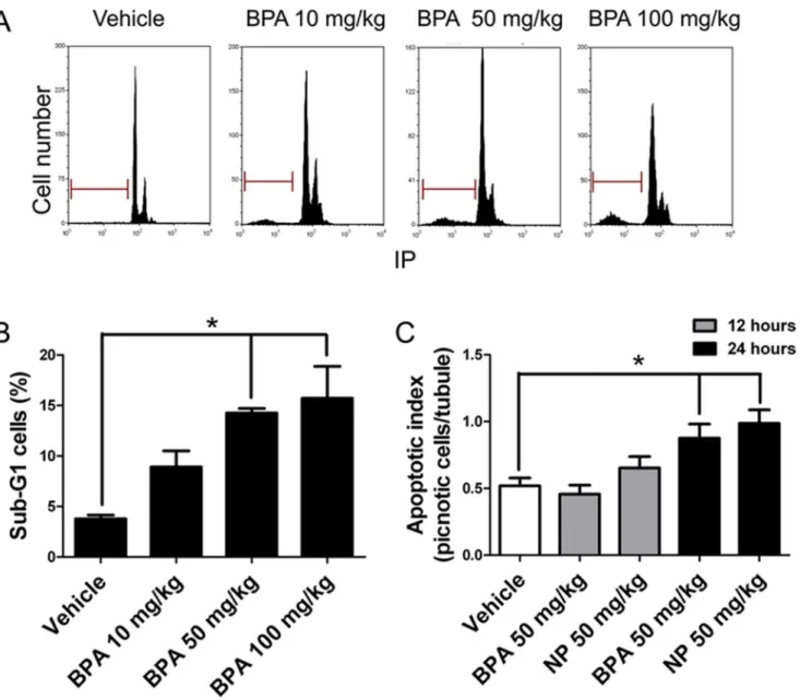
First, we aimed to determine the doses at which BPA or NP induces apoptosis in pubertal rat testes in vivo. To this end, 21-day-old male rats were subcutaneously injected with different doses of BPA and germ cell death was assayed 24 h later. We chose this experimental protocol because previous investigations stated that orally administered BPA and NP are rapidly absorbed and transformed to glucuronide derivatives during passage through the gut wall, and the liver [39,40]. In addition, this protocol has previously been successfully used for determination of the effect of EDCs in rodent models [41]. We chose to work with 21-day-old rats because it has been previously described that newborn and pubertal animals are more susceptible to EDCs than adult animals [42]. A dose-response curve using BPA showed that the percentage of sub-G1 cell population, which represents cells with fragmented and condensed DNA, increased significantly from
concentra-tion in the subsequent experiments, since it was the smallest concentraconcentra-tion that induced an increase in DNA fragmentation in rat testis cells in vivo. In addition, this dose has been previously used in similar experiments, and is within the range of the No Observed Adverse Effective Levels (NOAEL) [43–45]. Germ cells undergoing apoptosis are identified by counterstaining with periodic acid-Schiff and hematoxylin histological testes sections as having a round and pyknotic appearance. We have previously shown that these pyknotic cells express apoptotic markers such as active caspase-3 and stain positively for TUNEL [46]. The average
Figure 1. BPA and NP induce apoptosis in rat testes.A) Cell cycle analysis by flow cytometry of seminiferous tubule cells isolated from 21 day-old rats after 24 h of a single dose of 10, 50 or 100 mg/kg BPA. The horizontal bar indicates the sub-G1 cell population, which is a parameter of apoptosis. B) There is a significant increase in the percentage of sub-G1 cell population at 50 and 100 mg/kg. C) Quantification of pyknotic cells after the injection of 50 mg/kg of BPA or NP. * p,0.05, n53.
number of cells with apoptotic morphology (pyknotic) within a seminiferous slide (apoptotic index) increased significantly from 0.52¡0.06 to 0.88¡0.10 in the case of BPA and to 0.99¡0.10 in the case of NP (Fig. 1C) 24 h after treatment. There was no difference 12 h after injection. We also found that BPA or NP treatment induced an increase in active caspase-3 in germ cells but not Sertoli cells, which can be seen in histological sections (Figure 2A). Interestingly, we found that only spermatocytes (cells in meiosis) were intensely labeled in BPA-treated animals (Figure 2A, arrows), whereas spermatogonia (Figure 2A, arrowheads) and few spermatocytes were labeled in NP-treated animals. We found that the number of caspase-3-positive cells significantly increased 24 h after BPA or NP treatment (Fig. 2A). The number of active caspase-3-positive cells increased from 5.04¡0.56 per seminiferous tubule in vehicle-treated testes to 22.75¡1.98 in BPA- and to 22.54¡1.72 in NP-treated rats (Fig. 2A). We also evaluated the fragmentation of DNA in histological sections of 21-day-old rats treated with 50 mg/Kg of BPA or NP using the TUNEL assay. The results showed that BPA- and NP-treated animals had 1.78¡0.20 and 2.19¡0.18 TUNEL-positive cells per seminiferous tubule, respectively, which was significantly higher than that reported for vehicle-treated animals (0.48¡0.08 positive cells) (Fig. 2B). These results clearly indicate that BPA and NP induce apoptosis in rat germ cells.
ADAM17 is involved in
in vivo
germ cell apoptosis induced by BPA
and NP
Since we have previously found that ADAM17 and ADAM10 are involved in apoptosis induced by external compounds in the rat testis [6,17], we decided to investigate whether these metalloproteases participate in BPA- and NP-induced apoptosis in rat testes. To this end, ADAM17 and ADAM10-inhibitors were intra-testicularly administered before the injection of 50 mg/kg BPA or NP. Results showed that the ADAM17 inhibitor, GW280264X, prevented any increase in the number of TUNEL-positive cells induced by BPA and NP, from 1.78¡0.20 to 0.98¡0.10 TUNEL-positive-cells in the case of BPA, and from 2.19¡0.18 to 1.50¡0.12 TUNEL-positive cells in the case of NP (Fig. 3A, upper panel). The values for TUNEL-positive cells in animals treated with BPA or NP plus ADAM17 inhibitor were not statistically different from those reported for controls. On the other hand, the ADAM10 inhibitor, GI254023X, did not prevent the increase in apoptosis after BPA or NP injection (Fig. 3A, middle panel). The application of GW280264X or GI254023X alone did not interfere with normal apoptosis in the rat testis (Fig. 3A, bottom panel).
Figure 2. A single dose 50 mg/kg of BPA and NP induces a significant increase in active caspase-3 and TUNEL-positive cells in rat testis.A) BPA and NP treatment induce an increase in the number of active caspase-3. Note that caspase-3 positive cells were mainly spermatocytes (arrows) with BPA, whereas mainly spermatogonia were seen in the case of NP (arroheads). B) TUNEL-positive cells (arrows). * p,0.05, n53. Bars 50mm.
suggest that ADAM17, but not ADAM10, participates in the germ cell apoptosis induced by BPA and NP in the pubertal rat testis.
BPA and NP induce the activation and substrate shedding of
ADAM17
Previous studies have suggested that ADAM17 activation is associated with its translocation to the cell surface, which means that only the active form of ADAM17 can be located at the cell surface [11,48]. In order to elucidate whether the treatment of 50 mg/kg of BPA or NP in young rats induces ADAM17 translocation to the cell surface, as a parameter of its activation, we isolates cells within the seminiferous tubules of rat testes 24 h after treatment. Live cells were incubated with an antibody against the active form of ADAM17 and the labeled cells were evaluated by flow cytometry (for details see Materials and Methods). We found that BPA and NP treatment induced a significant increase in the percentage of cells harboring ADAM17 at their surfaces (Fig. 4). Interestingly, the intra-testicular administration of the ADAM17 inhibitor GW280264X signifi-cantly prevented the surface localization of this enzyme in BPA- (a decrease of 65%) but not NP-treated animals (Fig. 4). This difference could be a consequence of the action of BPA and NP on different cell types, or it could be related to the activation of alternative signaling pathways by these compounds. Thus, in vivo, BPA and NP induce the translocation of ADAM17 to the cell surface.
Next, we quantified the shedding of TNF-a from rat primary Sertoli cell cultures in the presence of BPA or NP. We chose Sertoli cells as a model system because they present several advantages: 1) They endogenously express ADAM17 [4] and substrates of this enzyme (e.g. TNF-a; 2) Forin vitrostudies, it is possible to isolate a more homogenous cell population than using germ cells; 3) Previous studies have shown that BPA and NP induce apoptosis of Sertoli cells and elicit the activation of signaling events such as the ERK and PTEN/Atk pathway [49,50]; and 4) It has been shown that MEHP induces shedding of TNF-a from Sertoli cells, suggesting a role for ADAM17 in this process [33].
Rat Sertoli cells were grown as mentioned in the Materials and Methods section, and treated with 10 mM of BPA or NP for 6 h; the presence of TNF-ain the culture medium was measured using a commercial kit. The treatment significantly induced TNF-a release, reaching concentrations of 18.79¡4.83 pg/
Figure 3. The ADAM17 inhibitor GW280264X preventsin vivogerm cell apoptosis induced by BPA or NP.A) Upper graph: Intra-testicular injection of 10mM GW280264X (GW) prior prevents the increase in the number of TUNEL-positive cells in rat testes induced by 50 mg/kg BPA or NP treatment. Middle graph: Intra-testicular injection of 10mM GI254023X (GI), an ADAM10 inhibitor, does not prevent the increase in the number of TUNEL-positive cells in rat testes induced by 50 mg/kg BPA or NP treatment. Bottom graph: The application of each inhibitor does not interfere with the normal apoptosis level. Right Panel: Representative TUNEL images from rat testis with the different treatments. B) Treatment with 50 mg/kg BPA or NP induces a significant increase in the cleaved form (89 kDa) of PARP on western blot. The addition of 10mM GW280264X (GW) prevents the increase of the 89 kDa form in rat testes treated with BPA or NP. * p,0.05, n53. Bars550mm.
mL and 18.50¡1.02 pg/mL with BPA and NP, respectively (Fig. 5A). The application of BB-94, a metalloprotease inhibitor, reduced the shedding of TNF-a to values similar to those of controls (8.67¡0.19 pg/mL and 7.50¡1.00 pg/mL, for BPA and NP, respectively), suggesting the involvement of ADAM17 in the shedding of TNF-a after BPA or NP treatment.
Figure 4. BPA and NP induce increase of ADAM17 in cell surface.A) Seminiferous tubule cells from rats injected with 50 mg/kg of BPA or NP were isolated as described in the materials and methods section and incubated with and antibody against the active form of ADAM17. The graph shows that the percentage of cells labeled with the antibody increases in rats treated with BPA or NP, but is reduced when rats were treated in the presence of GW280264X, a pharmacological inhibitor of ADAM17 (GW), or PD169316, a pharmacological inhibitor of p38 MAPK (PD). * p,0.05, n53.
Next, we transiently transfected primary Sertoli cells with a construct ((AP)-NGR-b1) expressing the cytoplasmic and transmembrane regions, as well as part of the extracellular domain, of neuregulin-b1 fused to an alkaline phosphatase (AP). This fusion protein has been used in previous works to evaluate the sheddase activity of ADAM17in vitro[51]. In this system, the activity of ADAM17 was studied by measuring the levels of AP in the culture medium of these cells. Cultured Sertoli cells did not express Neuregulin-b1 (NRG-b1), as evaluated by RT-PCR, but a clear amplicon of 325 bp was readily detected only in transfected cells (Fig. 5B). Non-transfected rat primary Sertoli cells did not have AP activity in culture medium, even when using high concentrations of BPA and NP (20 mM) (Fig. 5C). The treatment with 10 mM BPA (Fig. 5D) and NP (Fig. 5E) for 24 h induced a significant increase in the activity of AP, which was prevented by pre-incubation with 10 mM of BB-94, a broad metalloprotease inhibitor (Fig. 5D, E). In order to determine whether these results were restricted to primary rat Sertoli cell cultures, we decided to use the TM4 cell line, which is derived from mouse Sertoli cells [52]. Non-transfected TM4 cells did not express endogenous NGR-b1 (data not shown) and the AP activity was barely detected 24 h after treatment with concentrations ranging from 0.01 to 20 mM BPA or 0.01 to 1 mM NP (Fig. 6A, D). AP activity significantly increased when nontransfected cells were treated with 10 or 20 mM NP suggesting that these cells released the endogenous enzyme at these concentrations (Fig. 6D). TM4 cells that were transiently transfected with (AP)-NGR-b1 plasmid showed a significant increase of AP activity in the culture medium when treated with concentrations ranging from 0.01 to 20 mM NP or BPA for 24 h (Fig. 6B, E). We decided to use a concentration of 0.05 mM of NP and BPA for the following experiments as this concentration was found to significantly increase the AP activity and is within the range of levels detected in human samples [28,30,53,54]. The AP activity started to increase in the culture medium as early as 1 h after treatment with 0.05 mM BPA or NP (Fig. 6C, F). In order to define whether ADAM17 was the enzyme responsible for the increase in AP activity in the culture medium after BPA or NP treatment, we decided to knockdown its expression using specific shRNAs against ADAM17. TM4 cells were transiently transfected with (AP)-NGR-b1 and 1 or 10 mg of shRNAs against ADAM17 (sh1 or sh2 clones), or the empty vector (HK). Results showed that 10 mg of sh1 clone was sufficient to significantly reduce (83%) the mRNA and protein levels (60%) of ADAM17 48 h after transfection (Fig. 6G, H). Since the sh2 clone did not significantly reduce the mRNA levels of ADAM17, we only used the sh1 clone. Finally, transfection with 10 mg of sh1 (sh110) reduced the
constitutive shedding of AP activity and after treatment with 0.05 mM NP or BPA
Figure 5. BPA and NP induce shedding of TNF-aand Neuregulin in cultured rat Sertoli cells.A) The amount of TNF-ain the culture medium of primary
rats Sertoli cells significantly increases after 6 h of incubation with 10mM BPA or NP, but remains similar to control levels in the presence of 10mM BB-94, a general metalloprotease inhibitor. The picture shows immunofluorescence against ADAM17 (green) in rat primary Sertoli cells; cell nuclei are in red stained with Propidium iodide (IP). B) The amplicon of NGR-b1 is detected only in transfected rat primary Sertoli cells with the (AP)-NGR-b1 vector. C) Non-transfected primary Sertoli cells show no AP activity, at any of the tested concentrations. D) BPA and NP (E), induce shedding of AP activity in culture medium after 24 hours of stimulation, which is prevented by BB-94. * p,0.05, n53. Bar550mm.
(Fig. 6I, F). Therefore, these results indicate that BPA and NP, at concentrations similar to those detected in human samples, induce the activation and shedding of ADAM17 substrates in vitro.
p38 MAPK participates in germ cell apoptosis induced by NP and
BPA
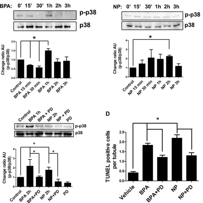
It has been reported that p38 MAPK binds and phosphorylates the cytoplasmic domain of ADAM17 under different conditions [10,11]. Since p38 MAPK is expressed by germ and Sertoli cells, we decided to evaluate the participation of this kinase in germ cell apoptosis in vivo and its relationship with ADAM17 translocation to the cell surface after NP and BPA treatment. For this purpose, 21-day-old rats were injected with 50 mg/kg of BPA or NP and sacrificed at different times (up to 3 hours) to detect the phosphorylated form of p38 MAPK (p-p38). The results showed that 1 h after BPA treatment (Fig. 7A) and 2 h after NP treatment (Fig. 7B), p-p38 was significantly increased. In order to evaluate whether the activation of p38 MAPK was involved in germ cell apoptosis, 5 mM of a pharmacological inhibitor, PD169316, was intra-testicularly injected 1 hour prior to BPA and NP treatment in 21-day-old rats. Levels of p-p38 were similar to those in controls and significantly reduced after treatment with BPA or NP in the presence of PD169316 (Fig. 7C). In addition, PD169316 significantly reduced the percentage of cells harboring ADAM17 at the surface, by 71% and 69% in BPA and NP treatments, respectively (Fig. 4). In the same way, animals treated with PD169316 showed a significant reduction of TUNEL-positive cells from
1.82¡0.11 to 1.21¡0.11 in the case of BPA, and from 2.19¡0.18 to 1.30¡0.16 for NP (Fig. 7D); this represents a reduction of BPA- and NP-induced apoptosis by 31% and 39%, respectively, suggesting the participation of p38 MAPK in the apoptosis that is induced by these compounds in the rat testis.
Discussion
Previous studies have shown that endocrine disruptors induce germ cell apoptosis and reduce male fertility in animal models; however, their effect at the cellular levels seems to be different and the signaling pathways elicited by these
compounds are far from elucidated. In this work, we report a novel mechanism by which BPA and NP induce germ cell apoptosis mediated by the activation of p38 MAPK and ADAM17.
Figure 6. BPA and NP induce the sheddase activity in TM4 cells.A) Non-transfected TM4 cells do not show any detectable shed of AP activity in the culture medium when incubated with BPA. However, NP induces the increase of AP in non-transfected TM4 cells only at concentrations of 10 and 20mM (D). B, E) The activity of AP significantly increases in the culture medium of transiently transfected TM4 cell with (AP)-NGR-b1 vector, when they are incubated for 24 h in the presence of 0.01–20mM BPA (B) or NP (E). (C, F) Time course of AP activity release of transiently transfected TM4 cells with (AP)-NGR-b1 using 0.05mM BPA (C) or NP (F). G) Transfection of TM4 cells with 10mg of shRNA (clone 1, sh1 10) induces a robust decrease in the levels of mRNA and (H) protein levels of ADAM17. I, J) Silencing of ADAM17 with 10mg of shRNA1 (shRNA1 10) in transiently transfected TM4 cells with (AP)-NGR-b1 slightly reduces the basal levels of AP shedding and completely prevents the effect of BPA and NP. * p,0.05, n53.
Previous work has shown that the chronic administration of BPA and NP to neonatal rats results in histological alterations in the testes, decreased sperm count and morphology and an increase in germ cell apoptosis [31,32,44,55,56]. In this paper, we extended those results and showed that acute exposure (a single dose of
Figure 7. Participation of p38 MAPK in the apoptosis induced by EDC in rat testis.A) Time course of p38 MAPK phosphorylation in 21-day-old rats treated with 50 mg/kg of BPA. B) Time course of p38 MAPK phosphorylation in 21-day-old rats treated with 50 mg/kg of NP. C) Intra-testicular application of 5mM PD169316 reduces the phosphorylation of p38 MAPK 1 or 2 h afterin vivotreatment with BPA or NP. D) The pharmacological inhibitor of p38 MAPK (PD169316) prevents the increase of TUNEL-positive cells in testes of 21-day-old rats treated with 50 mg/kg of BPA or NP. * p,0.05, n53.
50 mg/kg) of BPA or NP was sufficient to produce a robust increase in germ cell apoptosis within 24 h, as evaluated by the sub-G1 cell population, active caspase-3 and TUNEL assay (Figs. 1and2). Interestingly, we observed that most of the dying germ cells during BPA exposure were cells undergoing meiosis (spermatocytes), whereas those exposed to NP are spermatogonia and few spermatocytes (Fig. 2). These results have been previously observed and suggest that these endocrine disruptors attack different cell types, and may have slightly different mechanisms of action [31,32]. In addition, these results suggest that chronic exposure to NP might have longer-lasting effects than BPA since it affects spermatogonia rather than spermatocytes. We have shown here that BPA or NP induce apoptosis in germ, but not in Sertoli cells [49,50,57–60]. However, previous studies have shown that BPA or NP induce apoptosis in Sertoli cells, this discrepancy could be because underin vitroconditions Sertoli cells are prone to dead stimuli or that in vivo they have strong survival signals (e.g. extracellular matrix) that protect them from apoptosis. Further studies may be interesting, as they could elucidate the different targets and/or pathways modified by NP and BPA in spermatogonia and spermatocytes.
ADAM17 is a widely expressed enzyme involved in para/juxtacrine signaling in different cell types, which can be activated by different physiological and
Thein vitroresults show that NP and BPA induce the shedding of TNF-afrom Sertoli cells, and that the knockdown of ADAM17 in TM4 cells completely prevents the shedding of an exogenous substrate (Neuregulin). Therefore, these results suggest that BPA and NP are able to induce ADAM17 activation, and that
in vivo this enzyme probably sheds one or more specific proteins in Sertoli and germ cells. In this context, it is worth noting that BPA and NP are able to induce ADAM17 activationin vitroat concentrations similar to those reported in human blood samples [27,28,54], suggesting that these and other similar xenoestrogens could affect physiological processes in humans, different from germ cell apoptosis, such as the release of pro-inflammatory cytokines (e.g. TNF-a) [1,33,62,63]. In addition, these results indicate that, in vitro, BPA and NP directly induce the activation of ADAM17 in Sertoli cells from two species (Primary cultures from rat Sertoli cells and the mouse cell line TM4), suggesting that these observation are not restricted to rats and that this effect could probably be extrapolated to humans. In addition, our results suggest that, at least in vitro, ADAM17 can be directly activated by BPA and NP and that this effect is not necessarily related to the metabolites of these compounds in vivo. Further studies will be need to address the question of whether BPA or NP directly induce the activation of ADAM17in vivo, or if this is accomplished indirectly by promoting the action of other well-known activators of this enzyme, such as Lysophosphatidic Acid (LPA) or Transforming growth factor-alpha (TGF-a) [9,10,64].
p38 MAPK could act upon ancillary proteins in order to promote its translocation to the cell surface. Alternatively, the translocation of ADAM17 to the cell surface could be related to other process such as cell addition or germ cell apoptosis. However, p38 MAPK could influence apoptosis by other pathways, which could be independent of ADAM17 activation. Thus, the precise link between ADAM17 and p38 MAPK activation remains to be elucidated.
We have previously shown that germ cells undergoing apoptosis lack the extracellular domain of the tyrosine kinase receptor c-kit, suggesting that
activation of ADAM17 would shed the extracellular domain of this receptor and this, in turn, will promote apoptosis. We have shown here, at least in vitro, that BPA and NP induce the activation and shedding of ADAM17 substrates using Sertoli cells as a model system in two different species. In vivo, BPA and NP affect all cell types in the testes, but they elicit different responses. We believe that inside the seminiferous tubules, BPA and NP induce the shedding of different ADAM17 substrates in Sertoli and germ cells. In Sertoli cells, they could act in an autocrine way (e.g. TNF-a) in order to promote the survival of these cells. On the other hand, in germ cells, the activation of ADAM17 would induce shedding of the extracellular domain of c-kit and induce apoptosis. Interestingly, TNF-a is released by germ cells after MEHP-induced Sertoli cell injury and then induces FasL expression, which, in turn, would promote germ cell apoptosis [70]. These previous results fit very well with our model and suggest that the release of TNF-a could act on both autocrine and paracrine pathways in order to promote germ cell apoptosis.
Author Contributions
Conceived and designed the experiments: RDM. Performed the experiments: PUM RLC. Analyzed the data: PUM RLC RDM. Contributed reagents/materials/ analysis tools: RDM. Wrote the paper: RLC RDM.
References
1. Jenkins LK, Ross WL, Young KA (2007) Increases in apoptosis and declines in Bcl-XL protein characterise testicular regression in American crows (Corvus brachyrhynchos). Reprod Fertil Dev 19: 461–469.
2. Ji G, Gu A, Hu F, Wang S, Liang J, et al.(2009) Polymorphisms in cell death pathway genes are associated with altered sperm apoptosis and poor semen quality. Hum Reprod 24: 2439–2446.
3. Weikert S, Schrader M, Muller M, Krause H, Miller K(2004) Expression of the apoptosis inhibitor survivin in testicular tissue of infertile patients. Int J Androl 27: 161–165.
4. Urriola-Munoz P, Lizama C, Lagos-Cabre R, Reyes JG, Moreno RD(2014) Differential expression and localization of ADAM10 and ADAM17 during rat spermatogenesis suggest a role in germ cell differentiation. Biol Res 47: 31.
5. Lizama C, Rojas-Benitez D, Antonelli M, Ludwig A, Moreno RD (2012) Involvement of TACE/ ADAM17 and ADAM10 in etoposide-induced apoptosis of germ cells in rat spermatogenesis. J Cell Physiol 227: 829–838.
7. Moreno RD, Urriola-Munoz P, Lagos-Cabre R(2011) The emerging role of matrix metalloproteases of the ADAM family in male germ cell apoptosis. Spermatogenesis 1: 195–208.
8. White JM(2003) ADAMs: modulators of cell-cell and cell-matrix interactions. Curr Opin Cell Biol 15: 598–606.
9. Schlondorff J, Blobel CP(1999) Metalloprotease-disintegrins: modular proteins capable of promoting cell-cell interactions and triggering signals by protein-ectodomain shedding. J Cell Sci 112 (Pt 21): 3603– 3617.
10. Hall KC, Blobel CP(2012) Interleukin-1 stimulates ADAM17 through a mechanism independent of its cytoplasmic domain or phosphorylation at threonine 735. PLoS One 7: e31600.
11. Killock DJ, Ivetic A(2010) The cytoplasmic domains of TNFalpha-converting enzyme (TACE/ADAM17) and L-selectin are regulated differently by p38 MAPK and PKC to promote ectodomain shedding. Biochem J 428: 293–304.
12. Borroto A, Ruiz-Paz S, de la Torre TV, Borrell-Pages M, Merlos-Suarez A, et al.(2003) Impaired trafficking and activation of tumor necrosis factor-alpha-converting enzyme in cell mutants defective in protein ectodomain shedding. J Biol Chem 278: 25933–25939.
13. Xu P, Liu J, Sakaki-Yumoto M, Derynck R(2012) TACE activation by MAPK-mediated regulation of cell surface dimerization and TIMP3 association. Sci Signal 5: ra34.
14. Christova Y, Adrain C, Bambrough P, Ibrahim A, Freeman M(2013) Mammalian iRhoms have distinct physiological functions including an essential role in TACE regulation. EMBO Rep 14: 884–890.
15. Gutierrez-Lopez MD, Gilsanz A, Yanez-Mo M, Ovalle S, Lafuente EM, et al.(2011) The sheddase activity of ADAM17/TACE is regulated by the tetraspanin CD9. Cell Mol Life Sci 68: 3275–3292.
16. Nakayama H, Fukuda S, Inoue H, Nishida-Fukuda H, Shirakata Y, et al.(2012) Cell surface annexins regulate ADAM-mediated ectodomain shedding of proamphiregulin. Mol Biol Cell 23: 1964–1975.
17. Lizama C, Ludwig A, Moreno RD(2011) Etoposide induces apoptosis and upregulation of TACE/ ADAM17 and ADAM10 in an in vitro male germ cell line model. Biochim Biophys Acta 1813: 120–128.
18. Ortiz RJ, Lizama C, Codelia VA, Moreno RD(2009) A molecular evaluation of germ cell death induced by etoposide in pubertal rat testes. Mol Hum Reprod 15: 363–371.
19. Nassar N, Bower C, Barker A (2007) Increasing prevalence of hypospadias in Western Australia, 1980–2000. Arch Dis Child 92: 580–584.
20. Nelson CP, Park JM, Wan J, Bloom DA, Dunn RL, et al.(2005) The increasing incidence of congenital penile anomalies in the United States. J Urol 174: 1573–1576.
21. Grady R, Sathyanarayana S(2012) An update on phthalates and male reproductive development and function. Curr Urol Rep 13: 307–310.
22. Braun JM, Hauser R(2011) Bisphenol A and children’s health. Curr Opin Pediatr 23: 233–239.
23. Main KM, Mortensen GK, Kaleva MM, Boisen KA, Damgaard IN, et al.(2006) Human breast milk contamination with phthalates and alterations of endogenous reproductive hormones in infants three months of age. Environ Health Perspect 114: 270–276.
24. Inoue K, Wada M, Higuchi T, Oshio S, Umeda T, et al.(2002) Application of liquid chromatography-mass spectrometry to the quantification of bisphenol A in human semen. J Chromatogr B Analyt Technol Biomed Life Sci 773: 97–102.
25. Calafat AM, Kuklenyik Z, Reidy JA, Caudill SP, Ekong J, et al. (2005) Urinary concentrations of bisphenol A and 4-nonylphenol in a human reference population. Environ Health Perspect 113: 391–395.
26. Swan SH, Main KM, Liu F, Stewart SL, Kruse RL, et al.(2005) Decrease in anogenital distance among male infants with prenatal phthalate exposure. Environ Health Perspect 113: 1056–1061.
27. Zhang Y, Lin L, Cao Y, Chen B, Zheng L, et al.(2009) Phthalate levels and low birth weight: a nested case-control study of Chinese newborns. J Pediatr 155: 500–504.
28. Huang PC, Kuo PL, Chou YY, Lin SJ, Lee CC (2009) Association between prenatal exposure to phthalates and the health of newborns. Environ Int 35: 14–20.
30. Phillips KP, Tanphaichitr N (2008) Human exposure to endocrine disrupters and semen quality. J Toxicol Environ Health B Crit Rev 11: 188–220.
31. Liu C, Duan W, Li R, Xu S, Zhang L, et al.(2013) Exposure to bisphenol A disrupts meiotic progression during spermatogenesis in adult rats through estrogen-like activity. Cell Death Dis 4: e676.
32. McClusky LM, de Jager C, Bornman MS(2007) Stage-related increase in the proportion of apoptotic germ cells and altered frequencies of stages in the spermatogenic cycle following gestational, lactational, and direct exposure of male rats to p-nonylphenol. Toxicol Sci 95: 249–256.
33. Yao PL, Lin YC, Richburg JH(2009) TNF alpha-mediated disruption of spermatogenesis in response to Sertoli cell injury in rodents is partially regulated by MMP2. Biol Reprod 80: 581–589.
34. Obama T, Takayanagi T, Kobayashi T, Bourne AM, Elliott KJ, et al.(2014) Vascular Induction of a Disintegrin and Metalloprotease 17 by Angiotensin II Through Hypoxia Inducible Factor 1alpha. Am J Hypertens.
35. Endres K, Anders A, Kojro E, Gilbert S, Fahrenholz F, et al.(2003) Tumor necrosis factor-alpha converting enzyme is processed by proprotein-convertases to its mature form which is degraded upon phorbol ester stimulation. Eur J Biochem 270: 2386–2393.
36. Abcam website. Available: http://www.abcam.com/adam17-antibody-activation-site-ab39163.html. Accessed 2014 Jun 13.
37. Codelia VA, Cisternas P, Moreno RD(2008) Relevance of caspase activity during apoptosis in pubertal rat spermatogenesis. Mol Reprod Dev 75: 881–889.
38. Yan Y, Zhang J, Guo JL, Huang W, Yang YZ(2009) Multiple shRNA-mediated knockdown of TACE reduces the malignancy of HeLa cells. Cell Biol Int 33: 158–164.
39. Daidoji T, Inoue H, Kato S, Yokota H(2003) Glucuronidation and excretion of nonylphenol in perfused rat liver. Drug Metab Dispos 31: 993–998.
40. Pottenger LH, Domoradzki JY, Markham DA, Hansen SC, Cagen SZ, et al. (2000) The relative bioavailability and metabolism of bisphenol A in rats is dependent upon the route of administration. Toxicol Sci 54: 3–18.
41. Nakamura D, Yanagiba Y, Duan Z, Ito Y, Okamura A, et al. (2010) Bisphenol A may cause testosterone reduction by adversely affecting both testis and pituitary systems similar to estradiol. Toxicol Lett 194: 16–25.
42. Richter CA, Birnbaum LS, Farabollini F, Newbold RR, Rubin BS, et al.(2007) In vivo effects of bisphenol A in laboratory rodent studies. Reprod Toxicol 24: 199–224.
43. Manikkam M, Tracey R, Guerrero-Bosagna C, Skinner MK (2013) Plastics derived endocrine disruptors (BPA, DEHP and DBP) induce epigenetic transgenerational inheritance of obesity,
reproductive disease and sperm epimutations. PLoS One 8: e55387.
44. Salian S, Doshi T, Vanage G(2009) Neonatal exposure of male rats to Bisphenol A impairs fertility and expression of sertoli cell junctional proteins in the testis. Toxicology 265: 56–67.
45. vom Saal FS, Akingbemi BT, Belcher SM, Birnbaum LS, Crain DA, et al.(2007) Chapel Hill bisphenol A expert panel consensus statement: integration of mechanisms, effects in animals and potential to impact human health at current levels of exposure. Reprod Toxicol 24: 131–138.
46. Moreno RD, Lizama C, Urzua N, Vergara SP, Reyes JG(2006) Caspase activation throughout the first wave of spermatogenesis in the rat. Cell Tissue Res 325: 533–540.
47. Swindall AF, Stanley JA, Yang ES (2013) PARP-1: Friend or Foe of DNA Damage and Repair in Tumorigenesis? Cancers (Basel) 5: 943–958.
48. Peiretti F, Canault M, Deprez-Beauclair P, Berthet V, Bonardo B, et al.(2003) Intracellular maturation and transport of tumor necrosis factor alpha converting enzyme. Exp Cell Res 285: 278–285.
49. Choi MS, Park HJ, Oh JH, Lee EH, Park SM, et al.(2014) Nonylphenol-induced apoptotic cell death in mouse TM4 Sertoli cells via the generation of reactive oxygen species and activation of the ERK signaling pathway. J Appl Toxicol 34: 628–636.
51. Horiuchi K, Zhou HM, Kelly K, Manova K, Blobel CP(2005) Evaluation of the contributions of ADAMs 9, 12, 15, 17, and 19 to heart development and ectodomain shedding of neuregulins beta1 and beta2. Dev Biol 283: 459–471.
52. Mather JP(1980) Establishment and characterization of two distinct mouse testicular epithelial cell lines. Biol Reprod 23: 243–252.
53. Jin P, Wang X, Chang F, Bai Y, Li Y, et al.(2013) Low dose bisphenol A impairs spermatogenesis by suppressing reproductive hormone production and promoting germ cell apoptosis in adult rats. J Biomed Res 27: 135–144.
54. Susiarjo M, Hassold TJ, Freeman E, Hunt PA(2007) Bisphenol A exposure in utero disrupts early oogenesis in the mouse. PLoS Genet 3: e5.
55. de Jager C, Bornman MS, Oosthuizen JM(1999) The effect of p-nonylphenol on the fertility potential of male rats after gestational, lactational and direct exposure. Andrologia 31: 107–113.
56. Li YJ, Song TB, Cai YY, Zhou JS, Song X, et al.(2009) Bisphenol A exposure induces apoptosis and upregulation of Fas/FasL and caspase-3 expression in the testes of mice. Toxicol Sci 108: 427–436.
57. Qian W, Zhu J, Mao C, Liu J, Wang Y, et al.(2014) Involvement of CaM-CaMKII-ERK in bisphenol A-induced Sertoli cell apoptosis. Toxicology 324: 27–34.
58. Gong Y, Wu J, Huang Y, Shen S, Han X(2009) Nonylphenol induces apoptosis in rat testicular Sertoli cells via endoplasmic reticulum stress. Toxicol Lett 186: 84–95.
59. Gong Y, Pan X, Huang Y, Gao Z, Yu H, et al. (2008) NP-induced biophysical and biochemical alterations of rat testicular Sertoli cell membranes related to disturbed intracellular Ca(2+) homeostasis. Toxicol Lett 183: 10–20.
60. Gong Y, Han XD(2006) Nonylphenol-induced oxidative stress and cytotoxicity in testicular Sertoli cells. Reprod Toxicol 22: 623–630.
61. Kakiashvili E, Dan Q, Vandermeer M, Zhang Y, Waheed F, et al.(2011) The epidermal growth factor receptor mediates tumor necrosis factor-alpha-induced activation of the ERK/GEF-H1/RhoA pathway in tubular epithelium. J Biol Chem 286: 9268–9279.
62. Feng HL, Sandlow JI, Sparks AE, Sandra A, Zheng LJ (1999) Decreased expression of the c-kit receptor is associated with increased apoptosis in subfertile human testes. Fertil Steril 71: 85–89.
63. Lee J, Lim KT(2010) Expression of and IL-6 in HMC-1 cells treated with bisphenol A is attenuated by plant-originating glycoprotein (75 kDa) by blocking p38 MAPK. Naunyn Schmiedebergs Arch Pharmacol 382: 51–61.
64. Sahin U, Weskamp G, Kelly K, Zhou HM, Higashiyama S, et al.(2004) Distinct roles for ADAM10 and ADAM17 in ectodomain shedding of six EGFR ligands. J Cell Biol 164: 769–779.
65. Diaz-Rodriguez E, Montero JC, Esparis-Ogando A, Yuste L, Pandiella A(2002) Extracellular signal-regulated kinase phosphorylates tumor necrosis factor alpha-converting enzyme at threonine 735: a potential role in regulated shedding. Mol Biol Cell 13: 2031–2044.
66. Soond SM, Everson B, Riches DW, Murphy G(2005) ERK-mediated phosphorylation of Thr735 in TNFalpha-converting enzyme and its potential role in TACE protein trafficking. J Cell Sci 118: 2371– 2380.
67. Sato K, Nagai F, Aoki N (2001) Several environmental pollutant have binding affinities for both androgen receptor and estrogen receptor alpha. Journal of Health Scienice 47: 495–501.
68. Blair RM, Fang H, Branham WS, Hass BS, Dial SL, et al.(2000) The estrogen receptor relative binding affinities of 188 natural and xenochemicals: structural diversity of ligands. Toxicol Sci 54: 138–153.
69. Lizama C, Lagos CF, Lagos-Cabre R, Cantuarias L, Rivera F, et al.(2009) Calpain inhibitors prevent p38 MAPK activation and germ cell apoptosis after heat stress in pubertal rat testes. J Cell Physiol 221: 296–305.