SMAD-Independent Down-Regulation of
Caveolin-1 by TGF-
β
: Effects on Proliferation
and Survival of Myofibroblasts
Yan Y. Sanders1‡, Zongbin Cui2‡¤, Claude Jourdan Le Saux3, Jeffrey C. Horowitz2, Sunad Rangarajan1, Ashish Kurundkar1, Veena B. Antony1, Victor J. Thannickal1*
1Division of Pulmonary, Allergy and Critical Care, Department of Medicine, University of Alabama at Birmingham, Birmingham, Alabama, 35294, United States of America,2Division of Pulmonary and Critical Care Medicine, Department of Internal Medicine, University of Michigan Medical School, Ann Arbor, Michigan, 48109, United States of America,3Division of Cardiology, Department of Medicine, University of Texas Health Science Center at San Antonio, San Antonio, Texas, 78229, United States of America
‡These authors contributed equally to this work.
¤ Current address: Institute of Hydrobiology, Chinese Academy of Sciences, Wuhan, HuBei, China *[email protected]
Abstract
Transforming growth factor-β(TGF-β) mediates growth-inhibitory effects on most target cells via activation of the canonical SMAD signaling pathway. This growth-inhibitory activity may be coupled with cellular differentiation. Our studies demonstrate that TGF-β1 inhibits proliferation of primary, non-transformed human lung fibroblasts in association with the in-duction of myofibroblast differentiation. Differentiated myofibroblasts maintain the capacity to proliferate in response to exogenous mitogenic stimuli and are resistant to serum depriva-tion-induced apoptosis. These proliferative and anti-apoptotic properties of myofibroblasts are related, in part, to the down-regulation of caveolin-1 (Cav-1) by TGF-β1. Cav-1 down-regulation is mediated by early activation of p38 MAPK and does not require SMAD signal-ing. In contrast, myofibroblast differentiation is dependent on activation of the SMAD path-way, but not on p38 MAPK. Thus, combinatorial signaling by TGF-β1 of myofibroblast differentiation and down-regulation of Cav-1 by SMAD and p38 MAPK pathways, respec-tively, confer proliferative and apoptosis-resistant properties to myofibroblasts. Selective targeting of this SMAD-independent, p38-MAPK/Cav-1-dependent pathway is likely to be effective in the treatment of pathological conditions characterized by TGF-βsignaling and myofibroblast activation.
INTRODUCTION
Transforming growth factor-β1 (TGF-β1) regulates cell growth, differentiation and apoptosis in a cell- and context-specific manner; thus, both tumor-promoter and tumor-suppressive ac-tions have been described [1,2]. TGF-β1 mediates cytostatic effects on most target cells, includ-ing B and T lymphocytes [3,4], epithelial cells [5] and endothelial cells [6,7]. In contrast, OPEN ACCESS
Citation:Sanders YY, Cui Z, Le Saux CJ, Horowitz JC, Rangarajan S, Kurundkar A, et al. (2015) SMAD-Independent Down-Regulation of Caveolin-1 by TGF-β: Effects on Proliferation and Survival of Myofibroblasts. PLoS ONE 10(2): e0116995. doi:10.1371/journal.pone.0116995
Academic Editor:Rajasingh Johnson, University of Kansas Medical Center, UNITED STATES
Received:August 7, 2014
Accepted:December 17, 2014
Published:February 6, 2015
Copyright:© 2015 Sanders et al. This is an open access article distributed under the terms of the Creative Commons Attribution License, which permits unrestricted use, distribution, and reproduction in any medium, provided the original author and source are credited.
Data Availability Statement:All relevant data are within the paper.
Funding:This work was supported in part by a National Institutes of Health grant P01 HL114470 and R01 AG046210 (VJT). The funders had no role in study design, data collection and analysis, decision to publish, or preparation of the manuscript.
several studies have demonstrated the ability of TGF-β1 to promote mesenchymal cell prolifer-ation, an effect that appears to be mediated primarily by indirect mechanisms involving the au-tocrine production of mitogenic growth factors [8–10] and/or their receptor(s) up-regulation [11,12]. Furthermore, over-expression of TGF-β1 in rat lung results in the emergence and pro-liferation of myofibroblasts in association with prolonged severe fibrosis [13]. Similarly, direct transfer of TGF-β1 gene into arteries stimulates fibrocellular hyperplasia [14]. Thus, under-standing cellular/molecular mechanisms by which TGF-β1 promotes growth of mesenchymal cells, in particular myofibroblasts, is likely to be important in various pathological conditions characterized by myofibroblasts accumulation and activation [15,16].
Caveolin proteins are the principal components of caveolae, morphologically distinct plas-ma membrane invaginations present on plas-many cell types, that regulates a number of cellular physiological functions [17]. Caveolin-1 (Cav-1) was identified as the original member of the caveolin gene family and is expressed primarily in non-muscle cells. Overexpression of Cav-1 in cells lacking endogenous caveolae results in thede novoformation of caveolae [18,19]; while targeted down-regulation of Cav-1 in cells containing caveolae results in loss of caveolae [20,21]. Cav-1 gene is primarily recognized as a tumor-suppressor [22,23], although tumor-promoter activities have been described in some contexts [24,25]. The phenotype of Cav-1 knock-out mice has recently been described and is most remarkable for distinct pulmonary de-fects characterized by endothelial cell hyperproliferation and fibrosis [26]. The potential roles of fibroblasts/myofibroblasts, the major extracellular matrix (ECM)-producing cells in mam-mals, in the context of Cav-1 deficiency, is less clear.
We have previously shown that TGF-β1 is a potent inducer of myofibroblast differentiation by mechanisms involving cell adhesion and activation of focal adhesion kinase (FAK) [27]. TGF-β1 also promotes an apoptosis-resistant phenotype by the p38 MAPK-dependent auto-crine production of soluble growth factor(s) [28]. Furthermore, exogenous receptor tyrosine ki-nases (RTKs)-activating fibroblast growth factors mediate enhanced mitogenic responses in TGF-β1-differentiated myofibroblasts [12]. Interestingly, the apoptotic resistant phenotype of fibroblasts in idiopathic pulmonary fibrosis (IPF) also results from the down-regulation of Cav-1 via a PTEN/Akt-dependent pathway [29]. Cav-1 is typically expressed at high levels in terminally differentiated or quiescent cells; however, the regulation of Cav-1 during the induc-tion of myofibroblast differentiainduc-tion is not well defined. Recently it has been shown that TGF-β1 can induce miRNA-199a, which controls the down-regulation of Cav1 in TGF-β1 treated fibroblasts [30]. In this study, we examined the regulation of Cav-1 expression in non-trans-formed human lung fibroblasts that undergo myofibroblast differentiation in response to TGF-β1 stimulation. We describe, for the first time, a novel action of TGF-TGF-β1 to down-regulate Cav-1 expression via SMAD-independent and p38 MAPK-dependent mechanisms; this occurs con-comitantly with the induction of myofibroblast differentiation via canonical SMAD signaling. Furthermore, we explore potential role(s) of Cav-1 in the regulation of myofibroblast prolifera-tion and apoptosis resistance.
EXPERIMENTAL PROCEDURES
Reagents
DNA (ssDNA) was from Chemicon International, Temecula, CA. Secondary horseradish per-oxidase (HRP)-conjugated anti-rabbit or Alexa flour 594 goat conjugated anti-mouse antibod-ies were obtained from Pierce, Rockford, IL or Life Technologantibod-ies, Grand Island, NY. SB203580 was purchased from Calbiochem, La Jolla, CA. PD98059 was from Cell Signaling Technology. SB431542 was purchased from TOCRIS Bioscience, Avonmouth, UK. The concentrations used in this study were determined by our previous published studies [31]. All other reagents were obtained from Sigma, unless otherwise stated.
Cell culture
Studies were performed in non-transformed early passage of human fetal lung fibroblasts (IMR-90, Institute for Medical Research, Camden, NJ); these cells typically undergo cellular senescence after 40–50 replications, supporting their“normal”, non-transformed phenotype. IMR-90 fibroblasts were grown in DMEM (GIBCO, Grand Island, NY) supplemented with 10% fetal calf serum (FBS; Hyclone Laboratories, Logan, UT), 100 units/ml penicillin, 100μg/ml streptomycin, and 1.25μg/ml
amphotericin B (GIBCO). Cells were plated on 35 mm cell culture dishes or 96-well plates and in-cubated at 37°C in 5% CO2, 95% air. Medium containing 10% serum was changed every other day.
Generation of stably over-expressing Cav-1
α
, Cav-1 shRNA, SMAD2
shRNA and p38 MAPK mutant cell lines
A PCR-based strategy for site-specific mutagenesis was used to generate a construct pRC/CMV-Cav1 for overexpression of Cav-1αin IMR-90 cells. The codon of methionine at position 32 in human Cav-1 cDNA was changed to leucine (ATG to TTG) without affecting the function of Cav-1αand to eliminate transcription of Cav-1β[32]. PCR primers used for this mutation in-clude Cav1α-M1, 50-AACAAGGCCTTGGCAGACGAGCT-GAGCGAGAAG-30and Cav1α
-M2, 50-CGTCTGCCAAGGCCTTGTTGTTGGGCTTG-TAG-30. The sequence near the start
codon of Cav-1αwas replaced with a sequence containing a standard Kozak (GCCGCCATGG, start codon is underlined) to increase expression of Cav-1. The Cav-1αcDNA was then sub-cloned into theHind III/XbaI site of an expression vector, pRC/CMV2 from Invitrogen. Primers used for PCR were: Cav1α-1, 50
-TTTTAAGCTTGCC-GCCATGGCTGGGGGCAAATACGTAG-30and Cav1α-2, 50-GGGGTCTAGATTATAT-TTCTTTCTGCAAGTTGATG-30.
A DNA-basedsiRNA expression vector with hygromycin resistance, pSilencer2.1-U6 hygro from Ambion, was used to generate short hairpin RNA (shRNA) for knockdown of Cav-1 and SMAD2 in human lung fibroblasts. The pSilencer2.1-U6 hygro negative control plasmid sup-plied with the kit is a circular plasmid encoding ashRNA whose sequence is not found in the mouse, human, or rat genome databases. To choose an effective target on Cav-1 and SMAD2 mRNA, threeshRNA expression constructs for distinctive targets were tested for each gene. Western blotting data showed thatshRNAs from three constructs including pSU6H-shCav1 and pSU6H-shSMAD2 are able to knock down protein expression of Caveolin-1 and SMAD2 by 80–90% in IMR-90 cells. Oligonucleotides used for generating the construct pSU6H-shCav1 were: CA-2f: 50-GATCCCACACCTCAACGATGACGTGTTCAAGAGACACGTCATCGTT
GAGGGTTTTTTTGGAAA-30, and CA-2r, 50-AGCTTTTCCAAAAAAACACCTCAACGAT
GACGTGTCTCTTGAACACGTCATCGTTGAGGTGTGG-30. Oligonucleotides used for
generating the construct pSU6H-shSMAD2 were: SMAD2-3f, 50-GATCCCGTACACCAAAT
ACGATAGATTCAAGAGATCTATCGTATTTGGTGTACTTTTTTGGAAA-30and SMAD2-3r,
50-AGCTTTTCCAAAAAAGTACACCAAATACGATAGATCTCTTGAATCTATCGTATTT
GGTGTACGG-30. Sense and anti-sense strand sequences of Cav-1 and SMAD2 are underlined.
The mammalian expression plasmid, pcDNA-38KM encoding mutant p38 MAPK, was pro-vided by Dr. Kun Liang Guan, Department of Biological Chemistry, University of Michigan, Ann Arbor. This p38 MAPK mutant protein is able to be phosphorylated, but is not catalytical-ly active and unable to phosphorylate its downstream substrates such as ATF-2 [28]. Plasmid transfections of IMR-90 cells using the cationic lipid reagent, Lipofectamine (Invitrogen, Carls-bad, CA) were performed according to manufacturer’s instructions. Optimal ratio of DNA (μg) to Lipofectamine (μl) was determined to be*1:5 for IMR-90 cells. Cells were incubated with DNA-lipid complexes in serum-free Opti-MEM I medium (Invitrogen) for 4–5 h prior to introducing 10% FBS for 16–20 h. The next day, transfection medium was replaced by DMEM supplemented with 10% FBS and geneticin or hygromycin B (Invitrogen). Geneticin con-centrations were 200–400μg/ml for selection of stable transfectants and 150μg/ml was
the maintenance dose. Hygromycin B concentrations were 25–30μg/ml for selection and
2μg/ml was used as the maintenance dose. All cells were studied as pooled clones selected in
geneticin or hygromycin and cells were then treated with/without TGF-β1 in the absence of selection reagents.
Superarray cDNA microarray analyses and real-time quantitative RT-PCR:RNA was isolated from cells using TRIzol reagent from Invitrogen according to the manufacturer’s pro-tocol. Human extracellular matrix & adhesion molecules cDNA arrays from Superarray were used for the analyses of gene expression profiling in cells treated with/without TGF-β1 (2 ng/ml) for 24 h, according to manufacturer’s protocol (Superarray, Fredrick, MD). Cyclophilin A mRNA served as controls on these Superarray analyses.
Real-time quantitative RT-PCR was performed using iQ SYBR Green Supermix (BioRad, Palo Alto, CA) and using an iCylcer (BioRad) real-time detection system and normalized to 18S. Primers sequences are: Cav-1 F, 50-GAGCTGAGCGAGAAGCAAGT-30, Cav-1 R, 50
-TCCCTTCTGGTTCTGCAATC-30. 18S F, 50-GTCTGCCCTATCAACTTTCG-30, 18S R, 50
-ATGTGGTAGCCGTTTCTCA-3’as before [33]. Assays were performed in triplicate with 10ng of cDNA and 200 nM primers in a total reaction volume of 25μl. Thermal cycling
condi-tions were 95°C for 3 min, and 40 cycles of 95°C for 30 s, 60°C for 30 s.
Apoptosis assays
Apoptosis was quantitated with the use of an ELISA-based assay forssDNA (Apoptosis ELISA Kit, Chemicon International) according to the manufacturer’s instructions with minor modifi-cations as previously described [28].
Proliferation assay
Normal or stably transfected IMR-90 cells were seeded at 2 × 10⁴cells per 35 mm dishes and grown in 10% FBS until 50–60% confluence. Cells were then serum-deprived for 24 h and then treated with/without TGF-β1 (2 ng/ml) for 48 h followed by stimulation with 10% FBS for 24 h. Cell counts were assessed both prior to and after serum stimulation with an automated series Z Coulter counter (Coulter Electronics, Hialeah, FL).
BrdU incorporation assays were performed using an assay kit purchased from Oncogene, San Diego, CA, according to manufacturer’s instructions. The“background”absorbance of cells receiving no BrdU label was subtracted and BrdU incorporation index was calculated by dividing the corrected absorbance by cell counts obtained prior to fixing the cells.
Immunofluorescence staining and Western blotting
Statistical Analysis
Statistical analysis was performed using Student’sttest when comparing two groups and one-way analysis of variance with Bonferonni post-test when comparing three or more experi-mental conditions. This analysis was done using GraphPad Prism version 3.0 for Windows, GraphPad Software (San Diego, CA). Statistical significance was defined atp<0.05. Densito-metric analyses of Western blots were performed using the public domain NIH Image program available athttp://rsb.info.nih.gov/nih-image.
RESULTS
TGF-
β
1 mediates growth inhibition and induction of myofibroblast
differentiation in non-transformed human lung fibroblasts
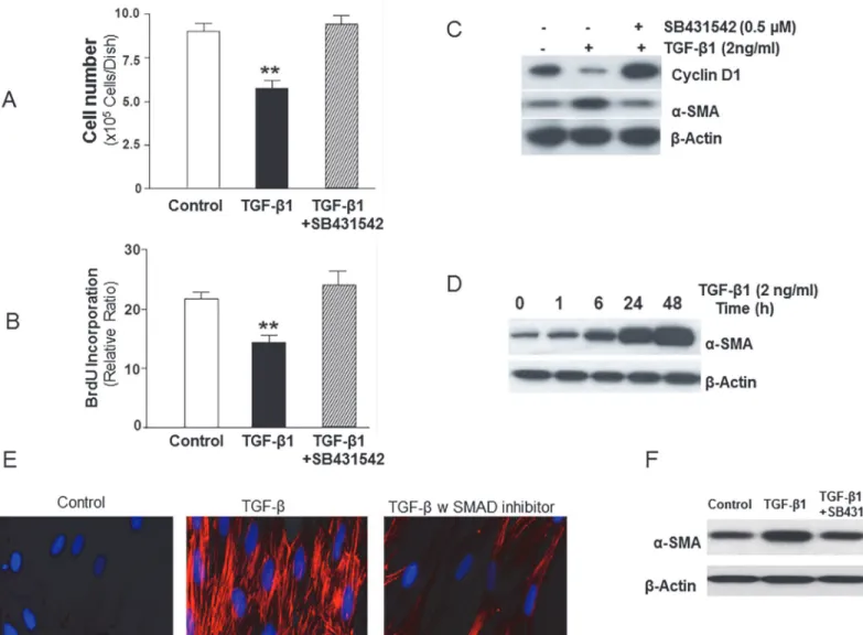
TGF-βis known to inhibit the proliferation of most target cells by SMAD-mediated signaling [34,35]. Cellular proliferative responses to TGF-βin mesenchymal cells have been described, although mechanisms are not well defined. We examined the effect of TGF-β1 on the prolifer-ative responses of early passage non-transformed human lung fibroblasts (IMR-90). Our stud-ies show that TGF-β1 (2 ng/ml) consistently inhibits the proliferation of IMR-90 fibroblasts grown in the presence of serum (10% FBS) as assessed by cell number/Coulter counter
(Fig. 1A) and by BrdU incorporation at 48 h following TGF-β1 stimulation (Fig. 1B). Pre-treat-ment with the ALK5/SMAD inhibitor (SB431542; 0.5μM) blocked the growth-inhibitory effect
of TGF-β1 (Fig. 1A, B). Under the same conditions, TGF-β1 mediates the down-regulation of cyclin D1 in these cells, an effect that is also abrogated by ALK5/SMAD inhibition (Fig. 1C).
Inhibition of cell proliferation may be associated with, and may promote/facilitate, the in-duction of cellular differentiation responses [36]. We have previously shown that TGF-β1 is a potent inducer of myofibroblast differentiation in quiescent, serum-starved IMR-90 fibroblasts [27]. To determine if inhibition of serum-stimulated fibroblast proliferation by TGF-β1 is asso-ciated with the stimulation of myofibroblast differentiation, we assessed cellular expression of α-smooth muscle actin (α-SMA), a marker of myofibroblast differentiation. TGF-β1 induced time-dependent protein expression ofα-SMA (Fig. 1D) and the formation of α-SMA-contain-ing stress fibers (Fig. 1E). As with the observed effects of ALK5/SMAD blockade (with SB431542) on cyclin D1 and associated growth-inhibition induced by TGF-β1, the up-regula-tion ofα-SMA was also inhibited by SB431542 (Fig. 1C; middle panel). Immunofluorescent staining demonstrated robust formation ofα-SMA-containing stress fibers in TGF-β differen-tiated cells, an effect that inhibited in the presence of SB431542 (Fig. 1E); this inhibition was also observed inα-SMA protein expression by western blotting (Fig. 1F). Together, these re-sults demonstrate that the“primary”effect of TGF-β1 on cultured non-transformed human lung fibroblasts is to induce a program of growth-arrest and cellular differentiation that require ALK5/SMAD signaling.
Cav-1 gene and protein expression are down-regulated during the
induction of myofibroblast differentiation by TGF-
β
1
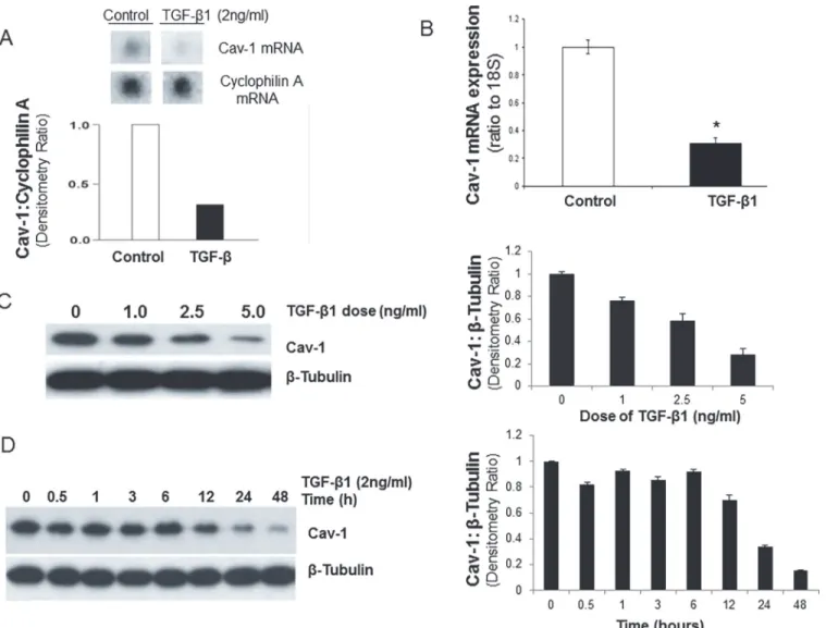
reduction in Cav-1 mRNA by 24 h in IMR-90 cells when assessed by cDNA Superarray analysis (Fig. 2A; Cav-1 and Cyclophilin A cDNA wells from the array are shown). This was confirmed with real-time RT-PCR (Fig. 2B). Dose-dependent effects of TGF-β1 (0, 1, 2.5 and 5 ng/ml) on the down-regulation of Cav-1 expression were confirmed by Western blot analysis (Fig. 2C). This effect of TGF-β1 (2 ng/ml) was time-dependent with a steady decline in Cav-1 protein Figure 1. TGF-β1 induces growth suppression in association with induction of myofibroblast differentiation of human lung fibroblasts.A, Early passage human lung fibroblasts (IMR-90), seeded at equal density on 35 mm dishes and grown to 50% confluence, were treated with or without TGF-β1 (2 ng/ml) in the presence/absence of the ALK5 inhibitor (SB431542, 0.5μM) in 10% FBS for 48 h prior to assessments of cell numbers by Coulter counting (n = 6 per group). Results are averages of at least three independent experiments. Data are presented as mean±S.E.M.**indicatesp<0.01 vs. control
cells or cells treated with TGF-βand SB431542.B, IMR-90 cells as described in (A) were grown in 96-well plates and labeled with BrdU for 24 h prior to assays for BrdU incorporation (n = 6 per group). Results are averages of at least three independent experiments. Data are presented as mean±S.E.M. **indicatesp<0.01 vs. control cells or cells treated with TGF-βand SB431542.C, IMR-90 cells grown in 10% FBS were stimulated with or without TGF-β1 (2 ng/ml) in the presence/absence of SB431542 (0.5μM) for 48 h. Cell lysates were subjected to SDS-PAGE and immunoblotted with an antibody against Cyclin D1; the blot was then stripped and probed forα-smooth muscle actin (α-SMA) andβ-actin.D, IMR-90 cells as described in (A) were stimulated with or without TGF-β1 (2 ng/ml) for the indicated times. Western immunoblotting was performed with a monoclonal antibody againstα-SMA; the blot was stripped and probed forβ-actin.E, Immunofluorescent images of IMR-90 cells treated 24h with vehicle only control, TGF-βonly (2ng/ml) and TGF-β(2ng/ml) with SMAD inhibitor (SB431542, 0.5μM). Cells were stained with antibody specific forα-SMA (1:100) with Alexa flour 594 conjugated secondary antibody (red); and 40,6-diamidino-2-phenylindole (DAPI, 300nM in PBS) staining was used to identify the nucleus (blue). Representative microscopy images are shown.
Magnification of ×40, figures were obtained with a Zeiss Axiovert fluorescence microscope.F,α-SMA expression by western blot in IMR-90 cells treated 24h with vehicle only control, TGF-βonly (2ng/ml) and TGF-β(2ng/ml) with SMAD inhibitor (SB431542, 0.5μM).β-actin is used as loading control.
expression up to 48 h when Cav-1 protein expression is reduced to*80% of baseline levels (Fig. 2D). Thus, TGF-β1 mediates a dose- and time-dependent down-regulation of Cav-1 ex-pression in cells that are undergoing myofibroblast differentiation.
TGF-
β
1-induced myofibroblast differentiation is SMAD-dependent, while
Cav-1 down-regulation is p38 MAPK-dependent and
SMAD-independent
TGF-βreceptor(s) signaling activates both SMAD-dependent and–independent pathways [2,35]. We have previously demonstrated rapid activation of p38 MAPK by TGF-β1 in human Figure 2. TGF-β1 induces dose- and time-dependent down-regulation of CAV-1 in human lung fibroblasts.A and B, Cultured IMR-90 cells were
treated with or without TGF-β1 (2 ng/ml) for 24 h and total RNA was isolated. A, cDNA Superarray analysis was performed and mRNA expression levels of Cav-1 and cyclophilin A are shown. Densitometric ratios of Cav-1:cyclophilin A are also shown. B, Histogram of real-time RT-PCR for Cav-1 expression after treated with TGF-β1 (2 ng/ml for 24 h) using triplicate samples from at least three individual experiments, normalized to 18S as mean±S.E.M.*p<0.05
compared to control.C, IMR-90 cells were treated with TGF-β1 (0–5 ng/ml) for 24 h and cell lysates extracted. Cell lysates were subjected to SDS-PAGE and immunoblotted with an antibody against Cav-1; the blot was stripped and probed forβ-tubulin. Densitometric ratios of Cav-1:β-tubulin are also shown on the right, as mean±S.E.M.D, IMR-90 cells were stimulated with TGF-β1 (2 ng/ml) for the indicated times. Cell lysates were subjected to SDS-PAGE and immunoblotted with an antibody against Cav-1; the blot was then stripped and probed forβ-tubulin. Densitometric ratios of Cav-1:β-tubulin are shown on the right, as mean±S.E.M. Results are averages of at least three independent experiments.
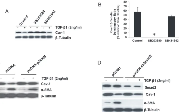
lung fibroblasts/mesenchymal cells, including IMR-90 fibroblasts [28]. Here, we determined the potential role(s) of early TGF-βreceptor(s)-activated SMAD-dependent and–independent p38 MAPK activation in the down-regulation of Cav-1 expression by TGF-β1. First, we as-sessed the effects of pharmacologic inhibitors of p38 MAPK (SB203580; 6μM) and ALK5/
SMAD (SB431542; 0.5μM) on TGF-β1-mediated down-regulation of Cav-1 expression.
Phar-macologic inhibition of p38 MAPK blocked TGF-β1-induced down-regulation of Cav-1 pro-tein expression, while inhibition of ALK5/SMAD signaling did not (Fig. 3A, B). Inhibitors of ERK-1/2 MAPK (with the MEK-1 inhibitor, PD98059, 20μM), c-Jun N-terminal kinase
(JNK-II, 0.1μM) and Src family protein tyrosine kinases (PP2, 10μM and SU6656, 2.7μM) had no
effect (data not shown). To confirm the role of p38 MAPK in TGF-β1-induced down-regula-tion of Cav-1 expression, we employed IMR-90 cells over-expressing a kinase-mutant p38 MAPK that is phosphorylated on Thr180/182, but is not catalytically active as previously de-scribed [28]. In cells expressing mutant p38 MAPK (p38-KM), TGF-β1 failed to down-regulate Cav-1 protein expression, while this effect was well-preserved in control vector-transfected
Figure 3. Down-regulation of Cav-1 by TGF-β1 is mediated by p38 MAPK-dependent and SMAD-independent mechanisms.A, IMR-90 cells were
treated with inhibitors of p38 MAPK (SB203580; 6μM) or ALK5 (SB431542; 0.5μM) for 30 min prior to treatment with or without TGF-β1 (2 ng/ml) for a period of 48 h. Cell lysates were extracted and Western immunoblotting performed with an antibody against Cav-1; the blot was then stripped and probed forβ -tubulin.B, Densitometric analyses of blots in (A) showed as % inhibition of baseline Cav-1 protein expression levels treated with TGF-β1.*indicates effect of SB203580 to completely block the inhibitory effect of TGF-β1 on Cav-1 expression. Results are averages of at least three independent experiments. Data are presented as mean±S.E.M.C, IMR-90 cells stably transfected with a kinase-deficient p38 MAPK (pcDNA-p38KM) or control vector (pcDNA) were treated
with or without TGF-β1 (2 ng/ml) for 24 h. Cell lysates were obtained and subjected to SDS-PAGE and immunoblotted for Cav-1 andα-smooth muscle actin (α-SMA); blots were stripped and probed forβ-tubulin.D, IMR-90 cells stably expressing SMAD2shRNA (pSU6H-shSMAD2) or control vector (pSU6H) were treated with/without TGF-β1 (2 ng/ml) for 24 h. Cell lysates were immunoblotted for SMAD2, Cav-1 andα-SMA. The blots were stripped and probed forβ -tubulin.
cells (pcDNA) (Fig. 3C). In contrast to effects on Cav-1 expression, blockade of p38 MAPK sig-naling doesnotalter the ability of TGF-β1 to induceα-SMA expression (Fig. 3C).
To then define the role of SMAD signaling in these TGF-β1 effects, cells were stably trans-fected with a plasmid encodingshRNA against SMAD2 (pSU6H-shSMAD2). Knock-down of SMAD2 in IMR-90 fibroblasts completely blocks the up-regulation ofα-SMA, but has no effect on Cav-1 down-regulation by TGF-β1 (Fig. 3D). Similar results were noted in SMAD3 knock-down cells (data not shown). These results demonstrate that myofibroblast differentiation and Cav-1 down-regulation are independently regulated via the SMAD and p38 MAPK signaling pathways, respectively.
Overexpression of Cav-1 blocks the
“
priming
”
effect of TGF-
β
on
proliferation and resistance to apoptosis of myofibroblasts
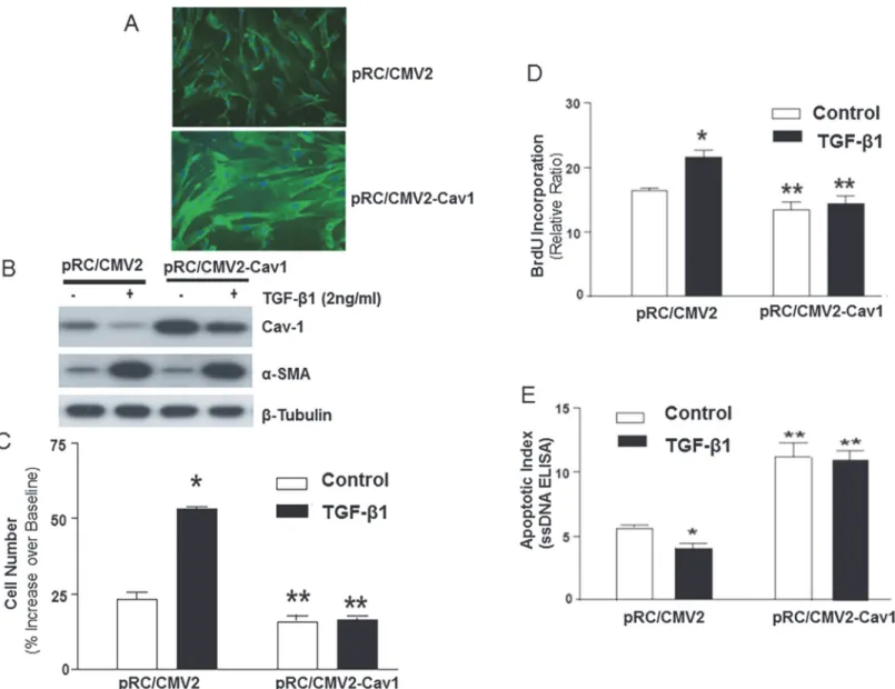
Cav-1 down-regulation may“prime”cells for enhanced proliferation in response to exogenous mitogens and the cells acquire an apoptosis-resistant phenotype. We have previously shown that TGF-β1-treated IMR-90 cells proliferate more robustly in response to exogenous mitogens [12] and that they are more resistant to serum deprivation-induced apoptosis [28]. Whether these effects may, in part, be mediated by the down-regulation of Cav-1 is not known. To examine the role(s) of Cav-1 down-regulation in these physiological responses to TGF-β1, Cav-1 protein was stably over-expressed in IMR-90 fibroblasts. In these cells, Cav-1 protein expression is primarily localized to the plasma membrane by immunofluorescence staining (Fig. 4A). Cav-1 over-expressing cells (pRC/CMV2-Cav1) maintained their ability to up-regulateα-SMA in response to TGF-β1 (Fig. 4B), suggesting that Cav-1 over-expression does not interfere with TGF-β1-induced myofibroblast differentiation. We noted down-regulation of Cav-1 protein expression by TGF-β1 even in pRC/CMV2-Cav1 cells; however, Cav-1 pro-tein levels do not decline below baseline/control levels in cells expressing the empty vector (pRC/CMV2) (Fig. 4B).
We then determined if over-expression of Cav-1 can mitigate the hyper-proliferative re-sponse and the apoptosis-resistant phenotype of myofibroblasts. Cav-1 over-expressing cells (pRC/CMV2-Cav1) and control vector-expressing cells (pRC/CMV2) were treated with (or without) TGF-β1 (2 ng/ml) in the absence of serum for 48 h to induce (or not induce) myofi-broblast differentiation; all cells were then stimulated with serum (10% FBS) for 24 h. In pRC/ CMV2 (control) cells, serum stimulation induced proliferative responses in both fibroblasts and myofibroblasts as assessed by cell number/Coulter counting and BrdU incorporation; this proliferative response was significantly greater in TGF-β1-differentiated myofibroblasts (Fig. 4C, D). Over-expression of Cav-1 abrogated the accentuated proliferative response in myofibroblasts (Fig. 4C, D), suggesting that down-regulation of Cav-1 represents an important mechanism for sensing and regulating extracellular serum-stimulated mitogenic signals in myofibroblasts. Cav-1 over-expression alone also suppressed, although to a lesser degree, the proliferative response to serum in undifferentiated fibroblasts (Fig. 4C, D).
Stable shRNA knockdown of Cav-1 promotes myofibroblast proliferation
and survival
We next assessed the effects of Cav-1 knockdown (by stable expression ofshRNA to Cav-1) on fibroblast/myofibroblast proliferation and resistance to apoptosis. Knockdown of Cav-1 at the protein level was confirmed by Western immunoblotting (Fig. 5A, top panel). TGF-β1 induced Figure 4. Over-expression of Cav-1 inhibits proliferation of human lung fibroblasts/myofibroblasts and abrogates the anti-apoptotic effects of TGF-β1.A, Human lung fibroblasts (IMR-90) were stably transfected with a plasmid encoding Cav-1 (pRC/CMV2-Cav1) or with empty vector (pRC/CMV2). Localization of Cav-1 protein was then analyzed by immunofluoresence staining with a rabbit polyclonal antibody to Cav-1.B, Stably-transfected cells described in (A) were treated with/without TGF-β1 (2 ng/ml) for 24 h. Cell lysates were obtained and subjected to immunoblotting for Cav-1,α-smooth muscle actin (α-SMA), andβ-tubulin.C, Stably-transfected cells described in (A) were serum-deprived for 24 h and then treated with/without TGF-β1 (2 ng/ml) for 48 h followed by stimulation with 10% fetal bovine serum for 24 h. Cell numbers were assessed both prior to and after serum stimulation with an automated Coulter counter (n = 6 per group) as mean±S.E.M.*indicatesp<0.05 vs. control pRC/CMV2“fibroblasts”.**indicatesp<0.05 vs. pRC/CMV2
“myofibroblasts”(TGF-β1 pre-treated). Similar results were obtained from 3 independent experiments.D, Cells in (C) were labeled with BrdU during the 24 h
of serum stimulation (n = 6 per group). BrdU assays were performed as described in“Methods”.*indicatesp<0.05 vs. control pRC/CMV2“fibroblasts”. **indicatesp<0.05 vs. pRC/CMV2“myofibroblasts”(TGF-β1 pre-treated). Similar results were obtained from 3 independent experiments.E, Quiescent stably transfected IMR-90 cells described in (A) were treated with/without TGF-β1 (2 ng/ml) for 5 days in serum-free medium. Apoptosis assays using an ELISA forssDNA (n = 6 for each group) were performed as described in“Methods”.*indicatesp<0.05 vs. control pRC/CMV2“fibroblasts”.**indicates p<0.01 vs. pRC/CMV2“myofibroblasts”(TGF-β1 pre-treated). Similar results were obtained from 3 independent experiments.
myofibroblast differentiation, as determined byα-SMA expression, inbothcontrol cells (pSU6H; transfected with empty vector) and Cav-1 knock-down cells (pSU6H-shCav1) (Fig. 5A). Proliferative responses to serum stimulation (10% FBS for 24 h) were significantly augmented in Cav-1 knock-down cells, both in control fibroblasts and myofibroblasts (cell pre-treated with TGF-β1 for 48 h) when assessed by cell numbers (Fig. 5B) and BrdU incorpo-ration (Fig. 5C). Serum deprivation-induced apoptosis of both fibroblasts and myofibroblasts was suppressed in Cav-1 knock-down cells (Fig. 5D). Interestingly, TGF-β1 confers additional protection from apoptosis even in cells deficient in Cav-1 (Fig. 5D), which may be related, in part, to further a decrease in Cav-1 expression observed in these cells (Fig. 5A). These results further support the notion that down-regulation of Cav-1 represents an important regulatory Figure 5.shRNA knock-down of Cav-1 enhances fibroblast/myofibroblast proliferation and protects against serum deprivation-induced apoptosis.A, IMR-90 cells stably transfected with a plasmid encodingshRNA targeted against Cav-1 (pSU6H-shCav1) or with control plasmid (pSU6H) were treated with/without TGF-β1 (2 ng/ml) for 24 h. Cell lysates were obtained and Western blots for Cav-1,α-smooth muscle actin (α-SMA) andβ-tubulin performed.B, Stably-transfected cells described in (A) were serum-deprived for 24 h and treated with/without TGF-β1 (2 ng/ml) for 48 h followed by stimulation with 10% FBS for 24 h. Cell counts were assessed both prior to and after serum stimulation with an automated Coulter counter (n = 6 per group) shown as mean±S.E.M.*indicatesp<0.05 vs. control pSU6H“fibroblasts”.**indicatesp<0.05 vs. pSU6H“myofibroblasts”(TGF-β1 pre-treated). Similar results were obtained from 3 independent experiments.C, Stably transfected cells described in (A) were serum-deprived for 24 h and treated with/without
TGF-β1 (2 ng/ml) for 48 h followed by BrdU labeling for 24 h in the presence of 10% FBS (n = 6 per group) shown as mean±S.E.M.*indicatesp<0.05 vs.
control pSU6H“fibroblasts”. Similar results were obtained from 3 independent experiments.D, Quiescent stably-transfected cells described in (A) were treated with/without TGF-β1 (2 ng/ml) for 5 days and apoptotic assay forssDNA performed as described in“Methods”(n = 6 per group) shown as mean±S.E. M.*indicatesp<0.05 vs. control pSU6H“fibroblasts”.**indicatesp<0.05 vs. pSU6H“myofibroblasts”(TGF-β1 pre-treated). Similar results were obtained
from 3 independent experiments.
mechanism for enhanced proliferative and apoptosis-resistant phenotypes of mesenchymal cells.
Activation of p38 MAPK, but not SMAD signaling, mediates augmented
proliferative responses to serum in myofibroblasts induced by TGF-
β
The TGF-β/SMAD pathway is well recognized to mediate growth-inhibitory and tumor-sup-pressive effects [2]. In some contexts, growth-promoting effects may be observed [8–12]. Our data indicate that early activation of the p38 MAPK pathway by TGF-β1 mediates the down-regulation of Cav-1 expression during the process of fibroblast to myofibroblast differentiation. Based on our model, blockade of the upstream activation of p38 MAPK should inhibit the pro-liferative responses of myofibroblasts to mitogenic stimulation. We first treated IMR-90 fibro-blasts stably expressing a control vector (pcDNA) or kinase-deficient p38 MAPK (pcDNA-p38KM) with or without TGF-β1 (2 ng/ml × 48 h) to differentiate them into myofibroblasts or maintain them as undifferentiated fibroblasts. We have already shown that blockade of the p38 MAPK pathway does not alter myofibroblast differentiation, but is critical for the down-regula-tion of Cav-1 in TGF-β1-stimulated IMR-90 cells (Fig. 3). Following differentiation into myofi-broblasts, cells were stimulated with/without serum (10% FBS) for 24 h to assess proliferative responses. Both fibroblasts and myofibroblasts responded robustly to serum stimulation; how-ever, the proliferative rates were consistently greater in myofibroblasts compared to fibroblasts (Fig. 6A, B: pcDNA control cell lines; andFig. 6C, D: pSU6H control cell lines). This“growth advantage”was completely lost in p38KM cells, supporting a role for p38 MAPK in this proliferative response of myofibroblasts (Fig. 6A, B). A statistically significant decrease in pro-liferative capacity was also noted in fibroblasts (non-TGF-β1-treated) expressing p38KM, sug-gesting that constitutive p38 MAPK (in the absence of TGF-β1 signaling) may also regulate this growth-promoting pathway. In support of this concept, baseline levels of Cav-1 expression were noted to be elevated in p38KM cells (Fig. 3C, compare 3rdand 4thlanes to 2ndlane).To examine the role of SMAD in proliferative signaling of mesenchymal cells, we generated IMR-90 cells stably expressing shRNA for SMAD2 (pSU6H-shSMAD2) and the control vector (pSU6H). In contrast to the observed effects of blockade of p38 MAPK signaling, knock-down of SMAD2 did not significantly alter the proliferative rate of myofibroblasts by both cell count-ing and BrdU incorporation (Fig. 6C, D), suggesting that p38 MAPK and not the SMAD2 sig-naling is required for the accentuated proliferation of myofibroblasts. In fact, SMAD2 knock-down cells demonstrated slightlyhigherbaseline rates of cell proliferation (Fig. 6C, D), consis-tent with the role of the TGF-β/SMAD pathway in cell cycle arrest and tumor suppression. We observed no further augmentation of cell proliferation in TGF-β1 pre-treated cells in the context of stable SMAD2-deficieny (Fig. 6C, D), suggesting that the growth advantage acquired as a result of the blockade of SMAD signaling is not further enhanced by down-regulation of Cav-1.
DISCUSSION
pathway in non-transformed human lung fibroblasts, secondary effects on myofibroblast pro-liferation and survival are mediated by an alternate SMAD-independent, and p38 MAPK-de-pendent, pathway that mediates the concomitant down-regulation of Cav-1 in differentiated myofibroblasts (Fig. 7). This provides a novel mechanism by which TGF-β1 mediates“ growth-suppressive”and“growth-promoting”effects on thesametarget cell, underdifferentcontexts. Figure 6. Blocking p38 MAPK inhibits myofibroblast proliferation.A, IMR-90 cells stably expressing a p38 MAPK dominant negative (pcDNA-p38KM)
and cells stably transfected with an empty vector (pcDNA) were serum-starved for 24 h and treated with/without TGF-β1 (2 ng/ml) for 48 h followed by stimulation with 10% fetal bovine serum for 24 h (n = 6 per group), data shown as mean±S.E.M.*indicatesp<0.05 vs. control pcDNA“fibroblasts”. **indicatesp<0.01 vs. control pcDNA“myofibroblasts”(TGF-β1 pre-treated). Similar results were obtained from 3 independent experiments.B, Cells
described in (A) were grown in 96-well plates and serum-starved for 24 h and treated with/without TGF-β1 (2 ng/ml) for 48 h followed by BrdU labeling for 24 h in the presence of 10% fetal bovine serum (n = 6 per group), data shown as mean±S.E.M.*indicatesp<0.05 vs. control pcDNA“fibroblasts”.**indicates p<0.01 vs. control pcDNA“myofibroblasts”(TGF-β1 pre-treated). Similar results were obtained from 3 independent experiments.C, IMR-90 cells stably expressing SMAD2shRNA and cells stably transfected with an empty vector (pSU6H) were serum-starved for 24 h and treated with/without TGF-β1 (2 ng/ml) for 48 h followed by stimulation with 10% fetal bovine serum for 24 h (n = 6 per group) data shown as mean±S.E.M.*indicatesp<0.05 vs. control
pSU6H“fibroblasts”(no TGF-β1 pre-treatment).**indicatesp<0.05 vs. control pcDNA“myofibroblasts”(TGF-β1 pre-treated).D, Cells described in (C) were grown in 96-well plates and serum-starved for 24 h and treated with/without TGF-β1 (2 ng/ml) for 48 h followed by BrdU labeling for 24 h in the presence of 10% fetal bovine serum (n = 6 per group) data shown as mean±S.E.M.*indicatesp<0.05 vs. control pSU6H“fibroblasts”(no TGF-β1 pre-treatment). **indicatesp<0.05 vs. control pcDNA“myofibroblasts”(TGF-β1 pre-treated).
There is growing recognition that plasma membrane caveolae microdomains regulate di-verse cellular functions [17]. Insights into the in-vivo physiological functions of caveolae have been gained from targeted disruption in mice of Cav-1, the main protein component of caveolae [26]. A prominent phenotype of Cav-1-deficient mice are the pulmonary abnormali-ties characterized by thickened alveolar septae and fibrotic changes involving the interstitium of the lung [26]; yet current understanding of Cav-1 function in fibroblasts/myofibroblasts, key effector cells of the fibrotic response, is limited. Although Cav-1 plays an important role in lung diseases [41], regulatory mechanisms that control Cav-1 gene/protein expression by growth factors have not been well defined. TGF-β1 is a central regulator of tissue fibrosis in most mammalian organ systems, including the lung [16,42,43]. In this study, we show that TGF-β1 down-regulates Cav-1 expression in association with myofibroblast differentiation in non-transformed human lung fibroblasts. Few growth factor ligands have been shown to di-rectly regulate gene/protein expression of Cav-1 in mammalian cells. Our study is similar to another report that TGF-β1 mediates the down-regulation of Cav-1 in human lung fibroblasts [44,45] and studies showed the decreased Cav-1 in IPF fibroblasts [44,45]. Activation of the Ras-MAPK pathway and protein kinase A has been shown to suppress Cav-1 gene expression [46]; thus, mitogenic factors in serum may be capable of inducing this effect. Chronic exposure to epidermal growth factor (EGF) has been recently reported to decrease Cav-1 expression in epithelial cancer cells that leads to the loss of E-cadherin and an epithelial-mesenchymal transi-tion [47]. Interestingly, TGF-β1 is a major regulator and inducer, of EMT [48,49].
TGF-βfamily members signal via heteromeric transmembrane complexes of type I and type II serine-threonine receptor kinases. The well-known direct effectors of TGF-βreceptor(s) sig-naling are SMAD proteins that, when activated, function as transcriptional regulators [2]. More recently, early post-receptor signaling via SMAD-independent pathways have been in-creasingly recognized [35]. SMAD-independent activation of p38 MAPK by TGF-β1 has been previously reported in epithelial cells [50,51] and mesenchymal cells [28]. Our current studies clearly implicate p38 MAPK activation in the down-regulation of Cav-1 by TGF-β1 in human lung fibroblasts. Previous studies from our laboratory have also implicated early activation of the p38 MAPK pathway in the autocrine induction of growth factors that“protect” mesenchy-mal cells from serum deprivation-induced apoptosis [28]. Given the new findings in this study showing p38 MAPK-dependent down-regulation of Cav-1, we speculate that multiple“ anti-apoptotic/pro-survival”mechanisms that coordinately interact with each other are activated by this SMAD-independent pathway. Our studies also support the concept that myofibroblast dif-ferentiation is regulated by a SMAD-dependent, yet p38 MAPK-independent, mechanism. Sig-naling via SMAD proteins have been previously implicated in myofibroblast differentiation induced by TGF-β1 [52,53]. Taken together, these data provide evidence for divergent signal transduction pathways involving a SMAD-dependent pathway that mediates growth-suppres-sion and myofibroblast differentiation, on the one hand, and a p38 MAPK pathway that medi-ates growth-promoting and anti-apoptotic effects by autocrine production of growth factors in concert with the down-regulation of Cav-1, on the other (Fig. 7).
differentiationper se. Moreover, preservation of SMAD signaling is likely to be critical for im-portant homeostatic functions of TGF-βon other cell types:“tumor-suppression”in epithelial cells and“immune-suppression”in immune cells [42].
In the current study, we have not explored the mechanisms by which p38 MAPK down-reg-ulates Cav-1. There are some plausible mechanisms; for example, previous studies demonstrate that GATA-6 binds to the Cav-1 promoter to down-regulate its transcription/gene expression [54], and that p38 MAPK can directly regulate the levels of GATA-6 via miR17-92 [55]. Other possible mechanisms may involve p38 MAPK-activated epigenetic regulation of Cav-1 expres-sion [56], which requires further investigation.
In summary, our study proposes a novel mechanism for the proliferation and survival of myofibroblasts that involves coordinate, and independently regulated, suppression of Cav-1 by TGF-β1. Cav-1 is typically up-regulated in terminally differentiated cells [39]; the down-regu-lation of Cav-1 in“differentiated”myofibroblasts may explain the unusual capacity of these cells to continue to proliferate and, in some contexts, to escape apoptosis [57]. Such mecha-nisms are likely to be important in chronic injury and repair processes characterized by the persistence of myofibroblasts in injured/fibrotic tissues that eventually progresses to end-organ failure. The potential roles for TGF-β1 regulation of Cav-1 in tumor biology and fibrosis re-quire further study.
Figure 7. Schematic representation of TGF-β1-activated signaling pathways mediating mesenchymal cell growth -suppressive and -promoting effects.TGF-β1 activates the cell surface TGF-βreceptor(s) complex that leads to rapid activation of the canonical SMAD pathway as well as the SMAD-independent p38 MAPK pathway. Activation of the SMAD pathway is required for the induction of a cellular program of growth-arrest and myofibroblast differentiation. In contrast, activation of the p38 MAPK pathway, independently of SMAD2/3, is required the down-regulation of Cav-1 by TGF-β1. Down-regulation of Cav-1 by TGF-β1“primes”differentiated myofibroblasts for enhanced proliferative responses to mitogens and resistance to apoptosis. These divergent TGF-βsignaling pathways may explain, in part, the contextual effects of TGF-β1 as both a growth-inhibitor and–promoter on the same target (mesenchymal) cells.
Acknowledgments
We thank Dr. Kun Liang Guan, Department of Biological Chemistry, University of Michigan, for providing the p38 MAPK kinase-deficient and control plasmid constructs used in these studies.
Author Contributions
Conceived and designed the experiments: YYS ZC CJLS VJT. Performed the experiments: ZC AK. Analyzed the data: YYS ZC CJLS JCH SR VBA VJT. Contributed reagents/materials/analy-sis tools: YYS ZC CJLS VJT. Wrote the paper: YYS ZC CJLS VJT.
REFERENCES
1. Derynck R, Akhurst RJ, Balmain A (2001) TGF-beta signaling in tumor suppression and cancer pro-gression. Nat Genet 29: 117–129. PMID:11586292
2. Shi Y, Massague J (2003) Mechanisms of TGF-beta signaling from cell membrane to the nucleus. Cell 113: 685–700. PMID:12809600
3. Kehrl JH, Roberts AB, Wakefield LM, Jakowlew S, Sporn MB, et al. (1986) Transforming growth factor beta is an important immunomodulatory protein for human B lymphocytes. J Immunol 137: 3855–3860. PMID:2878044
4. Kehrl JH, Wakefield LM, Roberts AB, Jakowlew S, Alvarez-Mon M, et al. (1986) Production of trans-forming growth factor beta by human T lymphocytes and its potential role in the regulation of T cell growth. J Exp Med 163: 1037–1050. PMID:24659787
5. Tucker RF, Shipley GD, Moses HL, Holley RW (1984) Growth inhibitor from BSC-1 cells closely related to platelet type beta transforming growth factor. Science 226: 705–707. PMID:6093254
6. Muller G, Behrens J, Nussbaumer U, Bohlen P, Birchmeier W (1987) Inhibitory action of transforming growth factor beta on endothelial cells. Proc Natl Acad Sci U S A 84: 5600–5604.
7. Takehara K, LeRoy EC, Grotendorst GR (1987) TGF-beta inhibition of endothelial cell proliferation: al-teration of EGF binding and EGF-induced growth-regulatory (competence) gene expression. Cell 49: 415–422. PMID:3494524
8. Battegay EJ, Raines EW, Seifert RA, Bowen-Pope DF, Ross R (1990) TGF-beta induces bimodal prolif-eration of connective tissue cells via complex control of an autocrine PDGF loop. Cell 63: 515–524. PMID:2171777
9. Khalil N, Xu YD, O’Connor R, Duronio V (2005) Proliferation of pulmonary interstitial fibroblasts is medi-ated by transforming growth factor-beta1-induced release of extracellular fibroblast growth factor-2 and phosphorylation of p38 MAPK and JNK. J Biol Chem 280: 43000–43009. PMID:16246848
10. Leof EB, Proper JA, Goustin AS, Shipley GD, DiCorleto PE, et al. (1986) Induction of c-sis mRNA and activity similar to platelet-derived growth factor by transforming growth factor beta: a proposed model for indirect mitogenesis involving autocrine activity. Proc Natl Acad Sci U S A 83: 2453–2457. PMID:
3010310
11. Rosenbaum J, Blazejewski S, Preaux AM, Mallat A, Dhumeaux D, et al. (1995) Fibroblast growth factor 2 and transforming growth factor beta 1 interactions in human liver myofibroblasts. Gastroenterology 109: 1986–1996. PMID:7498665
12. Thannickal VJ, Aldweib KD, Rajan T, Fanburg BL (1998) Upregulated expression of fibroblast growth factor (FGF) receptors by transforming growth factor-beta1 (TGF-beta1) mediates enhanced mitogenic responses to FGFs in cultured human lung fibroblasts. Biochem Biophys Res Commun 251: 437–441. PMID:9792792
13. Sime PJ, Xing Z, Graham FL, Csaky KG, Gauldie J (1997) Adenovector-mediated gene transfer of ac-tive transforming growth factor-beta1 induces prolonged severe fibrosis in rat lung. J Clin Invest 100: 768–776. PMID:9259574
14. Nabel EG, Shum L, Pompili VJ, Yang ZY, San H, et al. (1993) Direct transfer of transforming growth fac-tor beta 1 gene into arteries stimulates fibrocellular hyperplasia. Proc Natl Acad Sci U S A 90: 10759–
10763. PMID:8248168
15. Desmouliere A, Guyot C, Gabbiani G (2004) The stroma reaction myofibroblast: a key player in the con-trol of tumor cell behavior. Int J Dev Biol 48: 509–517. PMID:15349825
17. Razani B, Woodman SE, Lisanti MP (2002) Caveolae: from cell biology to animal physiology. Pharma-col Rev 54: 431–467. PMID:12223531
18. Engelman JA, Wykoff CC, Yasuhara S, Song KS, Okamoto T, et al. (1997) Recombinant expression of caveolin-1 in oncogenically transformed cells abrogates anchorage-independent growth. J Biol Chem 272: 16374–16381. PMID:9195944
19. Fra AM, Williamson E, Simons K, Parton RG (1995) De novo formation of caveolae in lymphocytes by expression of VIP21-caveolin. Proc Natl Acad Sci U S A 92: 8655–8659. PMID:7567992
20. Galbiati F, Volonte D, Engelman JA, Watanabe G, Burk R, et al. (1998) Targeted downregulation of caveolin-1 is sufficient to drive cell transformation and hyperactivate the p42/44 MAP kinase cascade. Embo J 17: 6633–6648. PMID:9822607
21. Liu J, Lee P, Galbiati F, Kitsis RN, Lisanti MP (2001) Caveolin-1 expression sensitizes fibroblastic and epithelial cells to apoptotic stimulation. Am J Physiol Cell Physiol 280: C823–835. PMID:11245599
22. Capozza F, Williams TM, Schubert W, McClain S, Bouzahzah B, et al. (2003) Absence of caveolin-1 sensitizes mouse skin to carcinogen-induced epidermal hyperplasia and tumor formation. Am J Pathol 162: 2029–2039. PMID:12759258
23. Williams TM, Lee H, Cheung MW, Cohen AW, Razani B, et al. (2004) Combined loss of INK4a and caveolin-1 synergistically enhances cell proliferation and oncogene-induced tumorigenesis: role of INK4a/CAV-1 in mammary epithelial cell hyperplasia. J Biol Chem 279: 24745–24756. PMID:
15044451
24. Ho CC, Huang PH, Huang HY, Chen YH, Yang PC, et al. (2002) Up-regulated caveolin-1 accentuates the metastasis capability of lung adenocarcinoma by inducing filopodia formation. Am J Pathol 161: 1647–1656. PMID:12414512
25. Yang G, Truong LD, Timme TL, Ren C, Wheeler TM, et al. (1998) Elevated expression of caveolin is as-sociated with prostate and breast cancer. Clin Cancer Res 4: 1873–1880. PMID:9717814
26. Drab M, Verkade P, Elger M, Kasper M, Lohn M, et al. (2001) Loss of caveolae, vascular dysfunction, and pulmonary defects in caveolin-1 gene-disrupted mice. Science 293: 2449–2452. PMID:11498544
27. Thannickal VJ, Lee DY, White ES, Cui Z, Larios JM, et al. (2003) Myofibroblast differentiation by trans-forming growth factor-beta1 is dependent on cell adhesion and integrin signaling via focal adhesion ki-nase. J Biol Chem 278: 12384–12389. PMID:12531888
28. Horowitz JC, Lee DY, Waghray M, Keshamouni VG, Thomas PE, et al. (2004) Activation of the pro-sur-vival phosphatidylinositol 3-kinase/AKT pathway by transforming growth factor-beta1 in mesenchymal cells is mediated by p38 MAPK-dependent induction of an autocrine growth factor. J Biol Chem 279: 1359–1367. PMID:14576166
29. Nho RS, Peterson M, Hergert P, Henke CA (2013) FoxO3a (Forkhead Box O3a) deficiency protects Idi-opathic Pulmonary Fibrosis (IPF) fibroblasts from type I polymerized collagen matrix-induced apoptosis via caveolin-1 (cav-1) and Fas. PLoS One 8: e61017. doi:10.1371/journal.pone.0061017PMID:
23580232
30. Lino Cardenas CL, Henaoui IS, Courcot E, Roderburg C, Cauffiez C, et al. (2013) miR-199a-5p Is upre-gulated during fibrogenic response to tissue injury and mediates TGFbeta-induced lung fibroblast acti-vation by targeting caveolin-1. PLoS Genet 9: e1003291. doi:10.1371/journal.pgen.1003291PMID:
23459460
31. Horowitz JC, Rogers DS, Sharma V, Vittal R, White ES, et al. (2007) Combinatorial activation of FAK and AKT by transforming growth factor-beta1 confers an anoikis-resistant phenotype to myofibroblasts. Cell Signal 19: 761–771. PMID:17113264
32. Fujimoto T, Kogo H, Nomura R, Une T (2000) Isoforms of caveolin-1 and caveolar structure. J Cell Sci 113 Pt 19: 3509–3517. PMID:10984441
33. Sanders YY, Hagood JS, Liu H, Zhang W, Ambalavanan N, et al. (2014) Histone deacetylase inhibition promotes fibroblast apoptosis and ameliorates pulmonary fibrosis in mice. Eur Respir J 43: 1448–
1458. doi:10.1183/09031936.00095113PMID:24603818
34. Carcamo J, Weis FM, Ventura F, Wieser R, Wrana JL, et al. (1994) Type I receptors specify growth-in-hibitory and transcriptional responses to transforming growth factor beta and activin. Mol Cell Biol 14: 3810–3821. PMID:8196624
35. Derynck R, Zhang YE (2003) Smad-dependent and Smad-independent pathways in TGF-beta family signalling. Nature 425: 577–584. PMID:14534577
36. Halevy O, Novitch BG, Spicer DB, Skapek SX, Rhee J, et al. (1995) Correlation of terminal cell cycle ar-rest of skeletal muscle with induction of p21 by MyoD. Science 267: 1018–1021. PMID:7863327
38. Galbiati F, Volonte D, Liu J, Capozza F, Frank PG, et al. (2001) Caveolin-1 expression negatively regu-lates cell cycle progression by inducing G(0)/G(1) arrest via a p53/p21(WAF1/Cip1)-dependent mecha-nism. Mol Biol Cell 12: 2229–2244. PMID:11514613
39. Williams TM, Lisanti MP (2005) Caveolin-1 in oncogenic transformation, cancer, and metastasis. Am J Physiol Cell Physiol 288: C494–506. PMID:15692148
40. Muraoka-Cook RS, Dumont N, Arteaga CL (2005) Dual role of transforming growth factor beta in mam-mary tumorigenesis and metastatic progression. Clin Cancer Res 11: 937s–943s. PMID:15701890
41. Jin Y, Lee SJ, Minshall RD, Choi AM (2011) Caveolin-1: a critical regulator of lung injury. Am J Physiol Lung Cell Mol Physiol 300: L151–160. PMID:21097526
42. Blobe GC, Schiemann WP, Lodish HF (2000) Role of transforming growth factor beta in human dis-ease. N Engl J Med 342: 1350–1358.
43. Chapman HA (2004) Disorders of lung matrix remodeling. J Clin Invest 113: 148–157.
44. Wang XM, Zhang Y, Kim HP, Zhou Z, Feghali-Bostwick CA, et al. (2006) Caveolin-1: a critical regulator of lung fibrosis in idiopathic pulmonary fibrosis. J Exp Med 203: 2895–2906. PMID:17178917
45. Xia H, Khalil W, Kahm J, Jessurun J, Kleidon J, et al. (2010) Pathologic caveolin-1 regulation of PTEN in idiopathic pulmonary fibrosis. Am J Pathol 176: 2626–2637. doi:10.2353/ajpath.2010.091117
PMID:20395445
46. Engelman JA, Zhang XL, Razani B, Pestell RG, Lisanti MP (1999) p42/44 MAP kinase-dependent and -independent signaling pathways regulate caveolin-1 gene expression. Activation of Ras-MAP kinase and protein kinase a signaling cascades transcriptionally down-regulates caveolin-1 promoter activity. J Biol Chem 274: 32333–32341. PMID:10542274
47. Lu Z, Ghosh S, Wang Z, Hunter T (2003) Downregulation of caveolin-1 function by EGF leads to the loss of E-cadherin, increased transcriptional activity of beta-catenin, and enhanced tumor cell invasion. Cancer Cell 4: 499–515.
48. Prunier C, Howe PH (2005) Disabled-2 (Dab2) is required for transforming growth factor beta-induced epithelial to mesenchymal transition (EMT). J Biol Chem 280: 17540–17548. PMID:15734730
49. Thiery JP (2003) Epithelial-mesenchymal transitions in development and pathologies. Curr Opin Cell Biol 15: 740–746.
50. Dai C, Yang J, Liu Y (2003) Transforming growth factor-beta1 potentiates renal tubular epithelial cell death by a mechanism independent of Smad signaling. J Biol Chem 278: 12537–12545. PMID:
12560323
51. Yu L, Hebert MC, Zhang YE (2002) TGF-beta receptor-activated p38 MAP kinase mediates Smad-in-dependent TGF-beta responses. Embo J 21: 3749–3759. PMID:12110587
52. Hu B, Wu Z, Phan SH (2003) Smad3 mediates transforming growth factor-beta-induced alpha-smooth muscle actin expression. Am J Respir Cell Mol Biol 29: 397–404. PMID:12702545
53. Qiu P, Feng XH, Li L (2003) Interaction of Smad3 and SRF-associated complex mediates TGF-beta1 signals to regulate SM22 transcription during myofibroblast differentiation. J Mol Cell Cardiol 35: 1407–
1420. PMID:14654367
54. Boopathi E, Gomes CM, Goldfarb R, John M, Srinivasan VG, et al. (2011) Transcriptional repression of Caveolin-1 (CAV1) gene expression by GATA-6 in bladder smooth muscle hypertrophy in mice and human beings. Am J Pathol 178: 2236–2251. doi:10.1016/j.ajpath.2011.01.038PMID:21514437
55. Oeztuerk-Winder F, Guinot A, Ochalek A, Ventura JJ (2012) Regulation of human lung alveolar multipo-tent cells by a novel p38alpha MAPK/miR-17-92 axis. EMBO J 31: 3431–3441. doi:10.1038/emboj. 2012.192PMID:22828869
56. Chen ST, Lin SY, Yeh KT, Kuo SJ, Chan WL, et al. (2004) Mutational, epigenetic and expressional analyses of caveolin-1 gene in breast cancers. Int J Mol Med 14: 577–582. PMID:15375584