Production or Protect Neurons against Inflammatory
Insult in In Vitro and In Vivo Mouse Models
Bin Xing1, Adam D. Bachstetter1, Linda J. Van Eldik1,2*
1Sanders-Brown Center on Aging, University of Kentucky, Lexington, Kentucky, United States of America,2Department of Anatomy and Neurobiology, College of Medicine, University of Kentucky, Lexington, Kentucky, United States of America
Abstract
The p38 MAPK pathway plays a key role in regulating the production of proinflammatory cytokines, such as TNFaand IL-1b, in peripheral inflammatory disorders. There are four major isoforms of p38 MAPK (p38a,b,d,c), with p38aand p38bthe targets of most p38 MAPK inhibitor drugs. Our previous studies demonstrated that the p38aMAPK isoform is an important contributor to stressor-induced proinflammatory cytokine up-regulation and neurotoxicity in the brain. However, the potential role of the p38b MAPK isoform in CNS proinflammatory cytokine overproduction and neurotoxicity is poorly understood. In the current studies, we used primary microglia from wild type (WT) and p38bknockout (KO) mice in co-culture with WT neurons, and measured proinflammatory cytokines and neuron death after LPS insult. We also measured neuroinflammatory responses in vivo in WT and p38b KO mice after administration of LPS by intraperitoneal or intracerebroventricular injection. WT and p38bKO microglia/neuron co-cultures showed similar levels of TNFaand IL-1b
production in response to LPS treatment, and no differences in LPS-induced neurotoxicity. The in vitro results were confirmedin vivo, where levels of TNFaand IL-1bin the CNS were not significantly different between WT or p38bKO mice after LPS insult. Our results suggest that, similar to peripheral inflammation, p38ais critical but p38bMAPK is dispensable in the brain in regards to proinflammatory cytokine production and neurotoxicity induced by LPS inflammatory insult.
Citation:Xing B, Bachstetter AD, Van Eldik LJ (2013) Deficiency in p38bMAPK Fails to Inhibit Cytokine Production or Protect Neurons against Inflammatory Insult in In Vitro and In Vivo Mouse Models. PLoS ONE 8(2): e56852. doi:10.1371/journal.pone.0056852
Editor:Malu´ G. Tansey, Emory University, United States of America
ReceivedDecember 15, 2012;AcceptedJanuary 15, 2013;PublishedFebruary 15, 2013
Copyright:ß2013 Xing et al. This is an open-access article distributed under the terms of the Creative Commons Attribution License, which permits unrestricted use, distribution, and reproduction in any medium, provided the original author and source are credited.
Funding:NIH R01 NS064247; NIH F32 AG037280; Alzheimer’s Association ZEN-09-134506. The funders had no role in study design, data collection and analysis, decision to publish, or preparation of the manuscript.
Competing Interests:The authors have declared that no competing interests exist.
* E-mail: linda.vaneldik@uky.edu
Introduction
Neuroinflammation and overproduction of proinflammatory cytokines are implicated in the pathological process of chronic neurodegenerative disorders such as Alzheimer’s disease (AD) (for recent review see: [1]) and acute trauma such as spinal cord injury (for recent review see: [2]). One of the most well established intracellular signal transduction cascades that regulate the production of proinflammatory cytokines is the p38 mitogen activated protein kinase (MAPK) family [3,4]. Consisting of four major isoforms (p38a, b, d, c), these kinases are encoded by separate genes, expressed in different tissues and cell types, and are often functionally distinct (for review see: [5]). For example, p38a
is expressed in most tissues and cell types, whereas the other isoforms are expressed in a more restricted distribution, with p38b
high in brain, p38d in endocrine glands, and p38c in skeletal muscle [6,7]. The roles of specific p38 isoforms in various physiological and pathological processes are under active investi-gation, but available evidence supports the idea that there can be both functional redundancies among the p38 isoforms as well as isoform-specific functions.
In terms of inflammatory pathways, the p38aisoform plays a major role in cytokine up-regulation in both CNS inflammatory disorders and peripheral inflammatory diseases [8,9]. We previ-ously showed, using both a pharmacological approach with a
selective small molecule p38a MAPK inhibitor and a genetic approach with primary microglia that are deficient in p38a, that this isoform is critical for the production of cytokines from activated microglia [8]. Moreover, pharmacological inhibition of p38a MAPK in an AD mouse model decreases brain proin-flammatory cytokine production, and attenuates synaptic protein loss [10]. Our more recent study further demonstrated that the deficiency of microglial p38aMAPK rescues neurons and reduces synaptic protein loss via suppressing LPS-induced TNFa overpro-duction [11]. These data demonstrated that microglia p38a
MAPK-mediated cytokine overproduction is critical to inflamma-tion-induced neurotoxicity. However, the role of p38bMAPK in CNS proinflammatory cytokine production and neurotoxicity is poorly understood.
p38bMAPK may have specific functions in the CNS that are not seen in the periphery. The goal of the current study was to explore, in a CNS context, whether p38bMAPK signaling contributes to LPS-induced proinflammatory cytokine production, as well as LPS-induced neuronal damage. In agreement with the peripheral inflammatory models, we found that p38bdoes not play a role in LPS-induced neuroinflammation and neurotoxicity.
Materials and Methods
Ethics Statement
All mouse experiments were conducted in accordance with the principles of animal care and experimentation in the Guide For the Care and Use of Laboratory Animals. The Institutional Animal Care and Use Committee of the University of Kentucky approved the use of animals in this study (protocol#2010-0615).
Reagents
Lipopolysaccharides (LPS) from Salmonella enterica serotype typhi-murium was obtained from Sigma-Aldrich, St. Louis, MO (Cat. no. L6143-1MG; EU/MG of LPS is 600,000).
Animals
C57BL/6 mice were obtained from Harlan Laboratories. The p38bMAPK global KO mice were generated [13] and obtained from Dr. Stephen J. O’Keefe at Merck Research Laboratories. Genotyping was performed by Transnetyx, Inc (Cordova, TN).
Primary neuronal culture
Primary neuronal cultures were derived from embryonic day 18, C57BL/6 mice, as previously described [11]. Briefly, cerebral cortices were dissected and the meninges were removed. Cells were dissociated by trypsinization for 20–25 min at 37uC and triturated, followed by passing through a 70mm nylon mesh cell strainer. The neurons were plated onto poly-D-lysine-coated 12-mm glass coverslips at a density of 56104/well in 24 well plates. Neurons were grown in neurobasal medium (Invitrogen) contain-ing 2% B27 supplement (Invitrogen), 0.5 mM L-glutamine (Mediatech), and 100 IU/ml penicillin, 100mg/ml streptomycin (Mediatech); no serum or mitosis inhibitors were used. Every 3 days, 50% of the media was replenished with fresh medium. The purity of the primary neuronal cultures was verified as 93% by immunocytochemistry for the neuronal marker NeuN, astrocyte marker GFAP, and microglia marker Iba-1 (data not shown).
Microglia culture
Microglia cultures were prepared as previously described [11]. Briefly, mixed glial cultures (,95% astrocytes, ,5% microglia)
were prepared from the cerebral cortices of 1–3 day old mice. The tissue was trypsinized as above, and the cells were resuspended in glia complete medium [a-minimum essential medium (a-MEM; Mediatech) supplemented with 10% fetal bovine serum (FBS) (US Characterized FBS; Hyclone; Cat no. SH30071.03), 100 IU/ml penicillin, 100mg/ml streptomycin, and 2 mM L-Glutamine]. After 10–14 days in culture, microglia were isolated from the mixed glial cultures by the shake-off procedure [19]. Specifically, loosely attached microglia were shaken off in an incubator shaker at 250 rpm for 2 hr at 37uC, the cell-containing medium was centrifuged at 1806gfor 3 min, and the cells were seeded onto 12-mm glass coverslips at a density of 26104 in 24 well plates, unless otherwise specified. Prior to plating the microglia on the coverslip, three equally spaced 1 mm glass beads (Borosilicate; Sigma) were attached to the coverslip with paraffin wax. The microglia cultures were verified to be $99% microglia by
immunocytochemistry. Microglia were incubated for one day before placing into co-culture with neurons.
Primary microglia/neuron co-culture and cell treatments After 7 days in culture, neurons on coverslips were co-cultured with mouse microglia as previously described [20], by placing the microglia-containing coverslips cell side down into the neuron-containing wells. In this co-culture system, the microglia and neurons are in close apposition and share the same neurobasal/ B27 culture media, but are separated by the 1 mm glass beads and do not have direct cell-cell contact. LPS was resuspended in sterile 0.9% sodium chloride at 100 mg/ml, and was used at a final concentration of 3 ng/ml for allin vitroexperiments.
Neuronal Viability Assay
Neuron viability was assayed by trypan blue exclusion [20]. Briefly, neuron-containing coverslips were incubated with 0.2% trypan blue in Hanks’ Balanced Salt Solution (HBSS) for 2 min in 37uC incubator and then gently rinsed 3 times with HBSS. Neurons were viewed under bright field microscopy at 2006final magnification. Three to five fields were chosen randomly per coverslip, and a total of 364 to 592 cells were counted per coverslip. Trypan blue-positive and negative neurons were counted per field and the ratio of negative cells to the total cells was taken as the index of neuronal survival rate.
ELISA assays
After 72 hr in the co-cultures, 20ml conditioned medium was harvested for TNFaand IL-1bELISA assay, using kits from Meso Scale Discovery (MSD) according to the manufacturer’s instruc-tions.
In vivo systemic and CNS inflammatory models
Adult mice (3–4 month old,,50% male and female) were used
Tissue processing and cytokine measurements
Brain cortex or hippocampus was homogenized using high shear homogenizer (Omni TH115), in a 1:10 (w/v) of ice-cold freshly prepared lysis buffer consisting of PBS containing 1mg/ml Leupeptin, 1 mM PMSF, and 1 mM EDTA. The brain lysates were centrifuged at 14,0006 g for 20 min at 4uC in a microcentrifuge. Fifty microliters of the resulting supernatant (,100mg of protein) was loaded per well of the MSD plate, and IL-1b and TNFa levels determined by MSD ELISA assay. Cytokine levels were normalized to the total amount of protein in the sample loaded as determined by BCA Protein Assay (Pierce), and data expressed as pg/ml. The detection limits of the MSD assays are 3.4 pg/ml for TNFaand 1.5 pg/ml for IL-1b.
Gene expression
RNA was isolated from dissected neocortical tissues stored at 280uC, or from primary microglia cultures using RNeasy mini-columns (Qiagen, Cat no. 74104) with on-column DNase treat-ment (Qiagen, Cat no. 79254) according to the manufacturer’s protocol. RNA quantity and quality were determined using A260/
A280 readings by NanoDrop (Thermo Scientific). Reverse
tran-scription (RT) was done following the manufacturer’s protocol using High Capacity cDNA Reverse Transcription Kit (Applied Biosystems, Cat no. 4368814). A no template and a no RT control were conducted to control for contamination. Real-time PCR was performed using the TaqMan Gene Expression assay kit (Applied Biosystems, Cat no. 4444964) according to the manufacturer’s instructions on a ViiATM 7 Real-Time PCR System (Applied Biosystems). The following TaqMan probes (Applied Biosystems)
were used: MAPK 11 (Mm00440955_m1); MAPK12
(Mm00443518_m1); MAPK13 (Mm00442488_m1); MAPK 14 (Mm00442507_m1); 18S rRNA (Hs99999901_s1). Relative gene expression was calculated by the 22DDCTmethod.
Statistics
Statistical analysis was conducted using GraphPad prism software V.5 (GraphPad Software, La Jolla, CA). Unless otherwise indicated, values are expressed as mean6SEM. Groups of two were compared by unpaired t-Test. One-way ANOVA followed by Bonferroni’s multiple comparison test was used for comparisons among three or more groups. Statistical significance was defined as p,0.05.
Results
Verification of p38b KO in microglia and brain
As a control for our studies, it was important to confirm the deletion of p38b MAPK in both the microglia cultures and the mouse brain tissue, and determine that significant compensatory changes in the other p38 isoforms were not present. Therefore, RNA was prepared from primary microglia cultures derived from WT or p38bKO mice (Figure 1A), and from lysates of WT and p38b KO mouse cortical tissue (Figure 1B), and the expression levels of the four p38 MAPK isoforms (p38a, p38b, p38d, p38c) were determined by qPCR. In both the microglia cultures and the brain tissue, p38b mRNA was readily measureable in WT mice but was not detected in the p38bKO mice. The mRNA level of p38a MAPK is ,10–100 fold higher than that of the other
isoforms in both WT and p38b KO microglia and brain tissue, and there was no significant difference between the levels of p38a
MAPK in either WT or p38b KO mice. The levels of p38d
mRNA were very low to undetectable in both WT and p38bKO mice. The expression of p38c mRNA was slightly higher in microglia cultures from p38b KO mice compared to microglia
from WT mice, but this difference was not seen in the cortical tissue samples. Altogether, these data verify that, as expected, p38b is deficient in microglia cultures and brain tissue from the p38bKO mice.
p38bKO in microglia did not inhibit LPS-induced cytokine production in microglia/neuron co-culture
As an initial step to determine the effect of microglial p38b
deficiency on LPS-induced proinflammatory cytokine production, primary microglia from WT or p38bKO mice were co-cultured with WT primary neurons and treated with either vehicle or LPS Figure 1. Verification of p38bKO in microglia and brain.Primary microglia from mouse cortex were prepared as described in Methods and plated at 16104 cells/well in 48 well plates. Total RNA from
microglia cultures (A) or from mouse cortical tissue (B) derived from WT (white bars) or p38bKO (black bars) mice was isolated, and the mRNA levels of different p38 MAPK isoforms were determined by qPCR. In both the microglia cultures and the brain tissues, p38b mRNA was readily measureable in WT mice but was not detected in the p38bKO mice. The p38a MAPK isoform in both microglia and cortex was expressed at much higher levels than any of the other isoforms, and there was no significant difference between the levels of p38ain either WT or p38b KO mice. The levels of p38d mRNA were very low to undetectable in both WT and p38bKO mice. The expression of p38c mRNA was slightly higher in microglia cultures from p38bKO mice compared to microglia from WT mice, but this difference was not seen in the cortical tissue samples. Results are expressed as fold change compared to p38b levels, and represent the mean 6 SEM of two determinations.
(3 ng/ml) for 72 hr. LPS induced a highly significant increase in the production of TNFa, and similar levels of TNFa induction were seen in WT or p38bKO microglia (Figure 2A). In a similar fashion, LPS stimulated a ,100-fold increase in IL-1b levels in
both the WT and p38bKO groups, with no differences detected between these two groups upon LPS challenge (Figure 2B). These results document that p38bin microglia is not required for LPS-induced cytokine production.
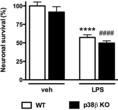
p38bKO in microglia failed to protect neurons against LPS-induced neurotoxicity in microglia/neuron co-culture
Previously we found p38a MAPK signaling is important for activated microglia-induced neuronal death in the microglia/ neuron co-culture, and that p38aMAPK deficiency in microglia protects against LPS-induced neurotoxicity [11]. However, the potential contribution of microglial p38bMAPK to LPS-induced neurotoxicity is not known. To determine if p38b deletion in microglia can provide similar neuroprotective effects against LPS-induced neuron damage, we isolated microglia from either WT or
p38b KO mice, placed them in co-culture with WT primary cortical neurons, and tested whether the absence or presence of microglia p38b would affect LPS-induced neurotoxicity. Treat-ment of WT microglia/neuron co-cultures with LPS for 72 hr led to significant neuronal death, as determined by trypan blue exclusion assay (Figure 3). In addition, a similar percentage (45%) of LPS-induced neuronal death was found when neurons were co-cultured with p38b KO microglia (Figure 3), showing that p38b
MAPK deficiency in microglia does not protect against LPS-induced neurotoxicity.
p38bKO failed to block CNS proinflammatory cytokine response to systemic and focal LPS insult in vivo
To test the effect of global p38bdeletion on cytokine production in the brain after inflammatory insult, we administered LPS to WT or p38b KO mice by two paradigms: systemic insult by IP injection (Figure 4A), and direct CNS focal insult by ICV injection (Figure 4B).
To induce systemic inflammation, we administered a single dose of LPS (1 mg/kg; IP) to WT and p38b KO mice, harvested cortical tissue at 6 hr after injection, and measured TNFaand IL-1b levels by ELISA. At the dose of LPS used, TNFalevels were near the limits of detection of the assay, but the TNFa levels reached were similar in the WT and p38bKO mice. IL-1blevels increased significantly after LPS treatment, and no significant differences in LPS-induced cytokine levels were seen between the WT and p38bKO mice (Figure 4A).
To determine whether p38b MAPK is important for proin-flammatory cytokine production induced by a direct CNS focal inflammation stimulus, LPS was injected into the right lateral ventricle of WT or p38b KO mice. The peak of the IL-1b
response in the ipsilateral hippocampus following ICV LPS occurs
,6 hr post-injection (data not shown); therefore this time point
was chosen for cytokine analysis. The result showed that the levels of TNFaand IL-1bincreased substantially in both WT and p38b
KO mice administered LPS ICV. In addition, consistent with the
Figure 2. Microglial p38b MAPK deficiency failed to reduce LPS-induced TNFa and IL-1bproduction in microglia/neuron co-culture.Mouse primary cortical neurons at DIV7 (56104/well) were
co-cultured with microglia (26104/well) from WT or p38bKO mice, and
the cytokine levels of TNFaand IL-1bwere measured by ELISA after 72 hr exposure to vehicle or LPS (3 ng/ml). LPS induced a significant increase in TNFa(A) and IL-1b(B) production, and p38bdeficiency in microglia did not reduce the production of TNFa and IL-1b. Data represent the mean 6 SEM from 2–4 independent experiments. ****p,0.0001 WT-veh vs. WT-LPS;###
p,0.001, ####
p,0.0001 KO-veh vs. KO-LPS.
doi:10.1371/journal.pone.0056852.g002
Figure 3. Microglial p38b MAPK deficiency failed to protect cortical neurons against LPS-induced neurotoxicity in microg-lia/neuron co-culture. WT mouse primary cortical neurons were plated on coverslips at 56104/well, and were co-cultured with microglia
(26104/well) from WT or p38bKO mice. Cells were treated with either
vehicle or LPS (3 ng/ml) for 72 hr, followed by trypan blue exclusion assay to evaluate neuronal survival. LPS treatment induced significant neuronal death in WT microglia/WT neuron co-culture. Similar levels of neuronal death were seen in LPS-treated co-cultures of p38b KO microglia/WT neurons. Data represent the mean 6 SEM from 2–4 independent experiments. ****p,0.0001 WT-veh vs. WT-LPS;
####
results from systemic IP administration of LPS, no significant differences in LPS-induced cytokine levels were seen between WT and p38bKO samples (Figure 4B).
Discussion
In the current study, we used LPS-insulted microglia/neuron co-culturein vitro and LPS-induced neuroinflammation inin vivo mouse models (IP and ICV) to test a potential contribution of p38bMAPK to the production of proinflammatory cytokines and the survival of cortical neurons. Our results document that p38b
MAPK in the brain is not a critical player in LPS-induced neuroinflammation and neurotoxicity, since 1) p38b KO in microglia co-cultured with primary neurons failed to reduce the cytokine production, 2) p38bdeletion in microglia cannot rescue neurons against LPS toxicity in co-cultures, and 3) no differences in brain cytokine levels were found between WT and p38bglobal KO mice in both LPS IP or ICV models. The findings that the LPS-induced CNS responses were not compromised in p38bKO mice support a functionally dispensable role for p38b in LPS-induced neuroinflammation and neurotoxicity. This is in contrast to the critical role of microglial p38a in LPS-induced cytokine production and neuron death in microglia/neuron co-culture [8,11].
Few studies have examined the role of p38b in CNS disease models, even though this isoform is expressed at substantial levels in the brain [21]. In animal models of ischemia, p38aand p38b
levels and activity increased in different cell types and with different temporal kinetics in the post-ischemic brain. For example, in transient global ischemia, p38a was induced early after ischemic insult and was localized mainly to microglia, whereas p38binduction was delayed and more protracted and was localized to astrocytes [22]. In a model of transient focal ischemia, p38b increased early and transiently in neurons, followed by a later increase in astrocytes [23]. Delayed induction of p38b in astrocytes has also been reported in mice subjected to kainate-induced seizures [24], and p38 inhibitor treatment attenuated kainate-induced hippocampal neuron loss and astrocyte activation [25]. Down-regulation of p38b, but not p38a, was also found to reduce sensitivity to pain following spinal cord injury [18]. In contrast to the reports above that suggest a potentially detrimental role of p38bin certain CNS disease models, other studies suggest that p38b may have specific beneficial roles in the CNS. For example, a recent study showed that p38boverexpression provides protection against H2O2-induced apoptosis in an astrocyte cell line
by inducing a small heat shock protein and inhibiting caspase-3 activation [26]. The p38b isoform was also reported to play an important role in the process by which postmitotic sympathetic noradrenergic neurons change to cholinergic neurons, and this neurotransmitter switch mechanism was impaired in neurons from p38b KO mice [17]. Taken together, these previous reports suggest that p38a and p38b may play distinct roles in different CNS disease models, and that their redundant or specific functions may depend on the spatial and temporal features of their activation in response to specific stressors.
The results reported here that p38bdoes not play a major role in CNS inflammation in response to LPS insult are consistent with previous studies examining the role of p38b in peripheral inflammation models. For example, LPS-induced proinflamma-tory cytokine production in macrophages and plasma was found to be similar in WT and p38bKO mice, and the p38bKO mice also responded normally in animal models of rheumatoid arthritis and inflammatory bowel disease [12,13]. These data demonstrated that p38b was not required for these peripheral inflammatory responses. Combined with our findings reported here, the available evidence shows that the p38b isoform does not play a key role in either peripheral or CNS proinflammatory cytokine responses or the resultant neurotoxicity induced by LPS insult. Figure 4. Proinflammatory cytokine levels in WT and p38bKO
mouse brain in response to LPS insult. CNS inflammatory responses were induced in WT (white bars) or p38bKO (black bars) mice upon either systemic insult by LPS IP injection (A) or direct CNS focal insult by LPS ICV injection (B). In the systemic insult paradigm, WT mice and p38bKO mice received IP injection of saline vehicle or LPS (1 mg/kg). Brain samples were harvested at 6 hr post-injection, and the levels of TNFaand IL-1bwere determined by ELISA (A). At this dose of LPS, the levels of TNFain the brain were near the level of detection of the ELISA assay, whereas the IL-1blevels showed a significant increase upon LPS insult. The profile of cytokine response to LPS stimulus in p38bKO mice was very similar to the response in WT mice. In the direct CNS focal insult paradigm, saline vehicle or LPS (25 ng) was injected into the right lateral ventricle of WT and p38bKO mice. Brain samples were harvested 6 hr post-injection, and the levels of TNFaand IL-1b were determined by ELISA (B). Both TNFaand IL-1bwere substantially increased in the brain samples from WT mice upon LPS treatment, and the p38bKO mice showed a similar induction of these proinflammatory cytokines. Results represent the mean6SEM. Cytokine levels in vehicle-treated mice (gray bars) represent data from both WT and p38bKO mice. n = 6–8 per group for panels A; n = 7–8 per group for panels B. **p,0.01, ***p,0.001 vehicle vs. WT-LPS; ##
p,0.01, ###
p,0.001 vehicle vs. KO-LPS. N.D. = not detected.
Taken together with our previous studies documenting the critical role of p38aMAPK in mediating LPS-induced CNS proinflam-matory cytokine production and neuron survival [8,11], these results suggest that p38a and not p38b is the key p38 isoform involved in peripheral and central inflammatory responses. These findings also raise the obvious corollary that development of p38 inhibitors to target CNS inflammatory diseases may not need to consider retention of p38binhibitory activity, but should instead focus on selective targeting of the p38a MAPK isoform as a potential therapeutic strategy.
Acknowledgments
We thank Danielle Goulding for assistance with breeding and maintenance of the mouse colonies.
Author Contributions
Conceived and designed the experiments: BX ADB LVE. Performed the experiments: BX ADB. Analyzed the data: BX ADB LVE. Wrote the paper: BX ADB LVE.
References
1. Wyss-Coray T, Rogers J (2012) Inflammation in Alzheimer disease-a brief review of the basic science and clinical literature. Cold Spring Harb Perspect Med 2: a006346.
2. David S, Kroner A (2011) Repertoire of microglial and macrophage responses after spinal cord injury. Nature Rev Neurosci 12: 388–399.
3. Kaminska B, Gozdz A, Zawadzka M, Ellert-Miklaszewska A, Lipko M (2009) MAPK signal transduction underlying brain inflammation and gliosis as therapeutic target. Anat Rec 292: 1902–1913.
4. Saklatvala J (2004) The p38 MAP kinase pathway as a therapeutic target in inflammatory disease. Curr Opin Pharmacol 4: 372–377.
5. Bachstetter AD, Van Eldik LJ (2010) The p38 MAP kinase family as regulators of proinflammatory cytokine production in degenerative diseases of the CNS. Aging Dis 1: 199–211.
6. Cuadrado A, Nebreda AR (2010) Mechanisms and functions of p38 MAPK signalling. Biochem J 429: 403–417.
7. Risco A, Cuenda A (2012) New Insights into the p38gamma and p38delta MAPK pathways. J Signal Transduct 2012: 520289.
8. Bachstetter AD, Xing B, de Almeida L, Dimayuga ER, Watterson DM, et al. (2011) Microglial p38alpha MAPK is a key regulator of proinflammatory cytokine up-regulation induced by toll-like receptor (TLR) ligands or beta-amyloid (Abeta). J Neuroinfl 8: 79.
9. Kim C, Sano Y, Todorova K, Carlson BA, Arpa L, et al. (2008) The kinase p38 alpha serves cell type-specific inflammatory functions in skin injury and coordinates pro- and anti-inflammatory gene expression. Nature Immunol 9: 1019–1027.
10. Munoz L, Ralay Ranaivo H, Roy SM, Hu W, Craft JM, et al. (2007) A novel p38 alpha MAPK inhibitor suppresses brain proinflammatory cytokine up-regulation and attenuates synaptic dysfunction and behavioral deficits in an Alzheimer’s disease mouse model. J Neuroinfl 4: 21.
11. Xing B, Bachstetter AD, Eldik LJ (2011) Microglial p38alpha MAPK is critical for LPS-induced neuron degeneration, through a mechanism involving TNFalpha. Mol Neurodegener 6: 84.
12. Beardmore VA, Hinton HJ, Eftychi C, Apostolaki M, Armaka M, et al. (2005) Generation and characterization of p38beta (MAPK11) gene-targeted mice. Mol Cell Biol 25: 10454–10464.
13. O’Keefe SJ, Mudgett JS, Cupo S, Parsons JN, Chartrain NA, et al. (2007) Chemical genetics define the roles of p38alpha and p38beta in acute and chronic inflammation. J Biol Chem 282: 34663–34671.
14. Kim HP, Wang X, Zhang J, Suh GY, Benjamin IJ, et al. (2005) Heat shock protein-70 mediates the cytoprotective effect of carbon monoxide: involvement of p38 beta MAPK and heat shock factor-1. J Immunol 175: 2622–2629. 15. Schallner N, Schwemmers S, Schwer CI, Froehlich C, Stoll P, et al. (2011)
p38beta-regulated induction of the heat shock response by carbon monoxide releasing molecule CORM-2 mediates cytoprotection in lung cells in vitro. Eur J Pharmacol 670: 58–66.
16. Guo YL, Kang B, Han J, Williamson JR (2001) p38beta MAP kinase protects rat mesangial cells from TNF-alpha-induced apoptosis. J Cell Biochem 82: 556– 565.
17. Loy B, Apostolova G, Dorn R, McGuire VA, Arthur JS, et al. (2011) p38alpha and p38beta mitogen-activated protein kinases determine cholinergic transdif-ferentiation of sympathetic neurons. J Neurosci 31: 12059–12067.
18. Fitzsimmons BL, Zattoni M, Svensson CI, Steinauer J, Hua XY, et al. (2010) Role of spinal p38alpha and beta MAPK in inflammatory hyperalgesia and spinal COX-2 expression. Neuroreport 21: 313–317.
19. Petrova TV, Akama KT, Van Eldik LJ (1999) Cyclopentenone prostaglandins suppress activation of microglia: down-regulation of inducible nitric-oxide synthase by 15-deoxy-Delta12,14-prostaglandin J2. Proc Natl Acad Sci USA 96: 4668–4673.
20. Xie Z, Smith CJ, Van Eldik LJ (2004) Activated glia induce neuron death via MAP kinase signaling pathways involving JNK and p38. Glia 45: 170–179. 21. Lee SH, Park J, Che Y, Han PL, Lee JK (2000) Constitutive activity and
differential localization of p38alpha and p38beta MAPKs in adult mouse brain. J Neurosc Res 60: 623–631.
22. Piao CS, Che Y, Han PL, Lee JK (2002) Delayed and differential induction of p38 MAPK isoforms in microglia and astrocytes in the brain after transient global ischemia. Brain Res Mol Brain Res 107: 137–144.
23. Piao CS, Yu YM, Han PL, Lee JK (2003) Dynamic expression of p38beta MAPK in neurons and astrocytes after transient focal ischemia. Brain Res 976: 120–124.
24. Che Y, Yu YM, Han PL, Lee JK (2001) Delayed induction of p38 MAPKs in reactive astrocytes in the brain of mice after KA-induced seizure. Brain Res Mol Brain Res 94: 157–165.
25. Kim SW, Yu YM, Piao CS, Kim JB, Lee JK (2004) Inhibition of delayed induction of p38 mitogen-activated protein kinase attenuates kainic acid-induced neuronal loss in the hippocampus. Brain Res 1007: 188–191.