The Zebrafish
moonshine
Gene Encodes
Transcriptional Intermediary Factor 1
c
,
an Essential Regulator of Hematopoiesis
David G. Ransom2¤, Nathan Bahary2, Knut Niss2, David Traver2, Caroline Burns2, Nikolaus S. Trede2,
Noelle Paffett-Lugassy2, Walter J. Saganic2, C. Anthoney Lim2, Candace Hersey2, Yi Zhou2, Bruce A. Barut1,2, Shuo Lin3, Paul D. Kingsley4, James Palis4, Stuart H. Orkin1,2, Leonard I. Zon1,2*
1Howard Hughes Medical Institute, Chevy Chase, Maryland, United States of America,2Division of Hematology/Oncology, Children’s Hospital and Harvard Medical School, Boston, Massachusetts, United States of America,3Department of Molecular, Cell and Developmental Biology, University of California, Los Angeles, California, United States of America, 4Department of Pediatrics and Center for Human Genetics and Molecular Pediatric Disease, University of Rochester Medical Center, Rochester, New York, United States of America
Hematopoiesis is precisely orchestrated by lineage-specific DNA-binding proteins that regulate transcription in concert with coactivators and corepressors. Mutations in the zebrafish moonshine (mon) gene specifically disrupt both embryonic and adult hematopoiesis, resulting in severe red blood cell aplasia. We report that mon encodes the zebrafish ortholog of mammalian transcriptional intermediary factor 1c (TIF1c) (or TRIM33), a member of the TIF1 family of coactivators and corepressors. During development, hematopoietic progenitor cells inmonmutants fail to express normal levels of hematopoietic transcription factors, includinggata1,and undergo apoptosis. Three different monmutant alleles each encode premature stop codons, and enforced expression of wild-typetif1c mRNA rescues embryonic hematopoiesis in homozygousmonmutants. Surprisingly, a high level of zygotic tif1c mRNA expression delineates ventral mesoderm during hematopoietic stem cell and progenitor formation prior to gata1 expression. Transplantation studies reveal thattif1cfunctions in a cell-autonomous manner during the differentiation of erythroid precursors. Studies in murine erythroid cell lines demonstrate that Tif1cprotein is localized within novel nuclear foci, and expression decreases during erythroid cell maturation. Our results establish a major role for this transcriptional intermediary factor in the differentiation of hematopoietic cells in vertebrates.
Citation: Ransom DG, Bahary N, Niss K, Traver D, Burns C, et al. (2004) The zebrafishmoonshinegene encodes transcriptional intermediary factor 1c, an essential regulator of hematopoiesis. PLoS Biol 2(8): e237.
Introduction
Hematopoiesis involves the coordinated processes of cell proliferation and differentiation of a relatively small number of progenitor cells into billions of circulating red and white blood cells (Thisse and Zon 2002). Hematopoiesis in vertebrates, from zebrafish to humans, is an evolutionarily conserved program that produces two waves of stem or progenitor cells that differ both in their embryonic origins and in the lineages of differentiated blood cells produced (Palis and Yoder 2001; Orkin and Zon 2002; Galloway and Zon 2003). The first, or primitive, wave of hematopoiesis originates from ventral mesoderm and gives rise to progen-itor cells that differentiate in embryonic blood islands. The primitive wave of hematopoiesis produces a burst of embryonic erythrocytes and macrophages. The second, or definitive, wave of hematopoiesis arises from self-renewing stem cells that develop primarily in the intraembryonic aorta–gonad–mesonephros region. These definitive hemato-poietic stem cells seed the later developing marrow spaces, to produce all lineages of adult blood cells, including definitive erythrocytes, myeloid cells, and lymphocytes.
We have undertaken a genetic approach to characterize genes that control hematopoiesis using the zebrafish as a model system (Thisse and Zon 2002). As part of a large-scale forward genetic screen, we previously identified bloodless zebrafish mutants that failed to express the erythroid
tran-scription factorgata1 normally in embryonic hematopoietic precursors (Ransom et al. 1996). We named one of these zebrafish genesmoonshine (mon),and another group named a noncomplementing allelevampire(Weinstein et al. 1996).
Here, we have determined that mutations in themongene cause a disruption in both primitive embryonic and definitive adult hematopoiesis, resulting in a severe loss of erythroid cells. Erythroid progenitor cells inmon mutants are initially present, but fail to express normal levels of hematopoietic transcription factors and undergo apoptosis.
Received January 20, 2004; Accepted May 26, 2004; Published August 17, 2004 DOI: 10.1371/journal.pbio.0020237
Copyright:Ó2004 Ransom et al. This is an open-access article distributed under the terms of the Creative Commons Attribution License, which permits unrestricted use, distribution, and reproduction in any medium, provided the original work is properly cited.
Abbreviations: AFLP, amplified fragment length polymorphism; ENU, ethyl-nitro-sourea; EST, expressed sequence tag; GFP, green fluorescent protein; hpf, hours postfertilization; KRAB, Kru¨ppel-associated box;mon, moonshine;PAC, P1 bacterial artificial chromosome clone; PML, promyelocytic leukemia gene product; RBCs, red blood cells; scl, stem cell leukemia; SSCP, simple sequence conformation polymorphism; TIF, transcriptional intermediary factor; TU, Tu¨bingen background
Academic Editor: William Talbot, Stanford University
*To whom correspondence should be addressed. E-mail: [email protected]. edu
Positional cloning identifies themongene as the zebrafish ortholog of mammalian transcriptional intermediary factor 1c (TIF1c), a member of the TIF1 family of transcriptional coactivators and corepressors (Le Douarin et al. 1995; Friedman et al. 1996; Kim et al. 1996; Venturini et al. 1999; Peng et al. 2002). The three members of the vertebrate TIF1 family (a, b, and c) are large nuclear proteins that each contain an N-terminal RBCC or TRIM domain (Reymond et al. 2001) composed of a RING finger, two B-boxes, and a coiled-coil domain. TIF1 family members also contain a C-terminal plant homeodomain finger and bromodomain that are characteristic of chromatin remodeling factors. TIF1ahas been shown to associate with a variety of ligand-bound nuclear hormone receptors (Le Douarin et al. 1995) and function as a coactivator for retinoic acid receptors (Zhong et al. 1999). TIF1bhas been shown to act as a corepressor for the large family of Kru¨ppel-associated box (KRAB) domain zinc-finger transcription factors (Friedman et al. 1996; Abrink et al. 2001). In contrast, TIF1cdoes not associate directly with either nuclear receptors or KRAB domains that bind to the other TIF1 family members (Venturini et al. 1999; Abrink et al. 2001). Biochemical studies also demonstrate that TIF1c
forms both homo-oligomers and hetero-oligomers with TIF1a
but not with TIF1b(Peng et al. 2002). The murineTif1aand Tif1c genes have not yet been subjected to gene targeting experiments, whereas analysis of mouse mutants demon-strates thatTif1b is required for postimplantation embryo-genesis and mesoderm induction in particular (Cammas et al. 2000). Taken together, these studies suggest that a major function of TIF1 family members is to link DNA-binding proteins with other coactivators or corepressors during development.
Our studies establish that tif1c functions as an essential regulator of embryonic and adult hematopoiesis in verte-brates. Cell transplantation studies demonstrate thattif1cacts in a cell-autonomous manner during embryonic hematopoi-esis. The tif1c gene is expressed specifically in ventral mesoderm and hematopoietic progenitors, then downregu-lated as erythroid maturation occurs. Tif1cprotein localizes to a novel class of nuclear bodies in both primary mouse embryo fibroblasts and erythroleukemia cell lines. Taken together, our studies demonstrate that Tif1cis required for normal erythroid cell development and survival.
Results
The ZebrafishmonGene Is Essential for Both Primitive and Definitive Erythropoiesis
In order to determine when the mon gene is required in development, we first examined hematopoietic gene expres-sion and apoptosis in zebrafish homozygous mon mutant embryos. During embryogenesis, homozygous zebrafish mon mutants have no red blood cells (RBCs) visible in circulation (Ransom et al. 1996; Weinstein et al. 1996). Themonmutants initiate expression ofgata1in hematopoietic cells around the five-somite stage, similar to wild-type embryos (data not shown); however, based on TUNEL staining, the differ-entiating erythroid cells undergo programmed cell death from the 12-somite stage to 22 h postfertilization (hpf) (Figure 1A and 1B, arrows). At 12 somites,gata1expression is only slightly reduced. By 18–22 hpf, hematopoietic-specific markers such asgata1, scl, gata2,andikarosare not detected in
the embryonic blood island (Figure 1A and 1B; unpublished data). The hematopoietic cells are thus correctly specified early during the development of mon mutant embryos, but these precursors undergo cell death. Based on expression of c-mybandrag1(Figure 1B, arrows),monmutants have normal myeloid and lymphoid development, respectively. In addition to the deficit of RBCs inmonmutants, there is a prominent loss of fin-fold and tail mesenchyme (Ransom et al. 1996). TUNEL staining of mon mutants demonstrates extensive apoptosis of mesenchymal cells in the trunk and tail bud regions (Figure 1A and 1B, arrows). The mon gene is thus required for normal development and survival of both committed erythroid progenitor cells and posterior mesen-chymal cells.
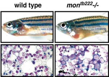
We next examined definitive hematopoiesis in rare surviv-ing homozygous adult zebrafish mon mutants. Mutations in mon are generally lethal by 10 to 14 d of development (Ransom et al. 1996), although raremonhomozygous mutants (approximately 1 in 500 bloodless embryos) of all tested alleles survive to adulthood. Adultmonmutants show cardiac hypertrophy, presumably due to the severe anemia leading to a high output state (Figure 2). In wild-type zebrafish, the adult site of hematopoiesis is the kidney (Al Adhami and Kunz 1977), which contains erythroid, lymphoid, and myeloid populations at various stages of differentiation (Bennett et al. 2001). Inmonhomozygous mutants, there is a severe block in maturation at the proerythroblast stage (Figure 2), whereas the differentiation of myeloid cells is normal (unpublished data). This demonstrates that the mon gene product acts during both primitive and definitive erythropoiesis.
Positional Cloning Identifiesmonas the Zebrafish Ortholog of Mammalian TIF1c
We identified the mongene by positional cloning using a panel of 2,200 diploid mutants collected from Tu¨bingen background (TU)/WIK strain hybrid parents carrying the montg234 allele. The mon mutant gene was positioned on Chromosome 8 between microsatellite markers z987 and z11001 (Figure 3A) (Knapik et al. 1998). For positional cloning purposes, over 12,000 polymorphic markers were screened using amplified fragment length polymorphism (AFLP) (Ransom and Zon 1999), and 36 markers within the interval were isolated. One of these, MA3, was found to be 0.3 cM from the gene (Figure 3A) and was utilized as the starting point of a chromosomal walk. A critical P1 bacterial artificial chromosome clone (PAC), 107N19, was obtained that spanned the genetic interval. Two simple sequence confor-mation polymorphism (SSCP) markers found on this PAC clone flank the critical genetic interval. The marker 80M12-T7 maps two recombinants out of 4,400 meioses telomeric of the mutation, and the marker 157J23-T7 maps one recombi-nant centromic of the mutation (Figure 3A). The end sequences and SSCP markers of PAC 107N19 are found in the zebrafish genomic sequence contig ctg23107 (http:// www.ensembl.org/Danio_rerio/) containing a predicted ze-brafish TIF1 family gene. This PAC was hybridized to a kidney cDNA library, resulting in the isolation of four clones that represented the same gene.
locations of exon boundaries are conserved between the zebrafish and human genes (unpublished data). Themonlocus on zebrafish Chromosome 8 is also predicted to be syntenic to the region of human Chromosome 1p that contains theTIF1c
gene based on the conserved locations of 12 other ortholo-gous gene pairs, includingNRAS,mapped to these regions in human and zebrafish (Barbazuk et al. 2000). Therefore, based on sequence similarity and chromosomal location, the zebrafishmongene is the likely ortholog of the humanTIF1c
gene.
We have identified ethyl-nitrosourea (ENU)-induced point mutations in three alleles ofmon(Figure 3C and 3D), each of which generates a premature stop codon. Themontb222band montg234 alleles have a severe phenotype with no circulating blood cells. In contrast, the monm262 allele has 10–100 circulating blood cells by 48 hpf, in comparison to the approximately 3,000 RBCs in the circulation of wild-type or heterozygous embryos at the same time point. Themonm262 allele was found to encode a premature stop codon at position E40, which would encode a putative protein of only
Figure 2.ZebrafishmonMutants Also Have Severe Defects in Definitive Hematopoiesis
Adult phenotype of wild-type and mon mutants. A rare surviving
montb222homozygous adult shows significant cardiomegaly in compar-ison to a wild-type age-matched control. Wright–Giemsa stained marrow of wild-type adult in comparison to a homozygous mutant. Note the dramatic reduction of terminally differentiated erythroid cells and the presence of abnormally large megaloblastic proerythro-blasts in themontb222mutant marrow.
DOI: 10.1371/journal.pbio.0020237.g002
Figure 1. Zebrafish mon Mutants Have Severe Defects in Primitive Hematopoiesis
(A) Whole-mount TUNEL assays reveal that ventral-posterior mesodermal cells undergo apoptosis in homozygousmontg234
mutant embryos. Whole-mount in situ hybridization ofgata1detected at the 12- and 18-somite stage in genotyped embryos. Posterior views, anterior to the left.
(B) Extensive apoptosis is visible in the trunk and tail (arrowhead) and also in hematopoietic cells of the embryonic blood island at 22 h of development (arrow). Whole-mount in situ hybridization at 22 hpf including scl, gata2, gata1, ikaros, and myb in montg234 mutants. Expression ofmybis greatly reduced in the blood islands because of a loss of erythroid cells, but embryonic macrophages are still present (arrows). The expression ofrag1in thymic T-cells appears normal in
monmutants at 5 d postfertilization (arrow heads). Lateral views of 22 hpf and 5-d-old embryos.
40 amino acids. Although this mutation would be expected to lead to a complete loss of mon gene product, another methionine is found downstream at amino acid position 104. In vitro translation experiments in reticulocyte lysates demonstrate reinitiation of translation from this methionine (unpublished data). Therefore, the hypomorphic larval phenotype of the monm262allele is likely due to partial loss ofmonfunction or expression. The presence of mutations in each of themonalleles indicates that defective Tif1cfunction is the cause of themonphenotype.
In order to determine whether tif1c is expressed in hematopoietic mesoderm, we next examined zebrafish embryos by whole-mount in situ hybridization (Figure 4A). tif1cmRNA is expressed maternally and is found throughout the embryo during blastula stages. During gastrulation and epiboly stages, zygotic expression of mon is highest in the mesendoderm of the germ ring. At tail bud and early somite stages a high level oftif1cexpression delineates a horseshoe-shaped population of ventral/lateral mesoderm that will give rise to blood and also expresses stem cell leukemia hemato-poietic transcription factor(scl)(Liao et al. 1997). This group of cells continues to expresstif1cand sclwhile it converges and forms the embryonic blood island (Detrich et al. 1995). Thetif1cgene is also highly expressed in the central nervous system as well as the mesenchyme of the trunk and tail. Homozygousmontg234mutants have a greatly reduced amount
of tif1c mRNA in all tissues consistent with nonsense-mediated message decay. Thus, zebrafish tif1cis specifically expressed in ventral mesoderm and putative hemangioblasts prior to and during the embryonic stages when hemato-poietic progenitors are undergoing apoptosis inmonmutants. We also compared the expression of zebrafishmonto mouse Tif1c(Figure 4A and 4B). MouseTif1cis highly expressed in erythroid blood islands of the yolk sac, and it is subsequently expressed in the fetal liver at a high level, and in other tissues, including the central nervous system. Taken together these results strongly suggest that zebrafish mon and mouse Tif1c
are orthologs that function during hematopoiesis.
Given that mammalian TIF1c has been shown to form hetero-oligomers with Tif1a (Peng et al. 2002), we searched for additional TIF1 family members in zebrafish to compare with tif1c. Using zebrafish expressed sequence tag (EST) sequences, we designed primers to RT-PCR amplify a TIF1-related cDNA from embryonic 10-hpf and 24-hpf RNA. This cDNA encodes a predicted zebrafish ortholog of human TIF1a based on predicted amino acid sequences (see Figure 3B). In addition, zebrafishtif1aESTs map to LG4 in a region predicted to be syntenic to the region of human Chromo-some 7 that contains theTIF1agene based on the conserved locations of eight other orthologous gene pairs, including SEMA3A, mapped to these regions in human and zebrafish (Barbazuk et al. 2000). We next compared the embryonic
Figure 3.Positional Cloning Identifies the
monGene as Zebrafishtif1c
(A) Physical map of the mon locus on zebrafish Chromosome 8. Microsatellite markers z987 and z11001 were used to initially identify recombinants in a panel of 2,200 diploid montg234
homozygous mutants. The AFLP marker MA3 was used to initiate a chromosomal walk in PAC libraries. The critical PACS that were isolated to encompass themonlocus are indicated by numbers above bar. The PAC 107N19 defines the critical interval for themongene. This PAC was used as a probe to screen cDNA libraries and to identify zebrafishtif1ccDNAs. Numbers below the bar indicate the number of recombinants identified by SSCP analy-sis.
(B) Clustal-W–generated phylogentic tree of zebrafish (Danio rerio[Dr]) Tif1c
and Tif1apeptide sequences in compar-ison to TIF1 family members: human (Hs) TIF1a, TIF1b, and TIF1c; mouse (Mm) Tif1a, Tif1b, and Tif1c;; and fly (Dm) bonus.
(C) Diagrams illustrating the structure of the Tif1c-predicted peptide and the three identified point mutants. RING finger (RING), B-boxes (B1 and B2), plant homeodomain finger (PHD) and bromo-domain (BROMO). Numbers below the first diagram indicate the percent iden-tity shared between each of these do-mains in zebrafish and human TIF1c. The predicted truncated proteins are indicated.
(D) DNA sequence chromatograms show-ing the three ENU-induced point mu-tants in comparison to wild-type control sequences
expression pattern of tif1a mRNA to tif1c by in situ hybridization. Like mammalian TIF1a (Le Douarin et al. 1995; Niederreither et al. 1999), the predicted zebrafishtif1c
gene is broadly expressed (see Figure 4A). At five somites, zebrafishtif1adoes not display the relatively high expression in the horseshoe-shaped region of hematopoietic mesoderm seen with tif1c. At later stages, tif1a is evenly expressed throughout most of the embryo, including the developing blood islands. Therefore,tif1ais coexpressed in the same cells with tif1c and may therefore be available to form hetero-oligomers in vivo.
Forced Expression oftif1cRescues Hematopoiesis inmon
Mutants
To further confirm that a mutation in the zebrafish tif1c
gene is responsible for the mon mutant phenotype we performed embryo rescue experiments (Figure 5A; Table 1). Microinjection of synthetic wild-typemonmRNA at the one-cell stage rescues the formation of embryonic erythrocytes in genotyped mutant embryos without causing obvious defects
in embryonic patterning or organogenesis. At 4 d of development, 70% (n = 10) of montg234 mutants show significant (greater than 200 cells in comparison to a wild-type estimate of 3,000 cells) rescue of circulating hemoglobinized RBCs in comparison to control sibling mutants (n = 75). Based on the correction of the jagged fin-fold phenotype (Ransom et al. 1996), the mesenchymal cells are rescued to a similar extent as the anemia (unpublished data). Overexpression ofmon did not result in expanded blood cell numbers in wild-type embryos and was not toxic at doses that rescue the phenotype ofmonmutants (unpublished data). Since there were no expanded or ectopic blood populations in the embryos, these rescue experiments suggest thatmonfunctions as a permissive factor required for hematopoiesis.
Marrow Transplantation Rescues Erythropoiesis inmon
Mutants
The high levels oftif1cexpression in erythroid cells suggest that it functions as a cell-autonomous regulator of gene
Figure 4. The mon/tif1c Gene Is Highly Expressed in Hematopoietic Mesoderm (A) In situ hybridization of zebrafish embryos demonstrating the embryonic expression of tif1c. tif1c is initially ex-pressed as a maternal mRNA. Increased expression of tif1c in ventral-lateral mesoderm begins between the one- to three-somite stages and increases through early development. By five somites, tif1c is strongly expressed in lateral stripes of mesoderm that also express scl.At 22 hpftif1cis expressed broadly in the brain, spinal cord, trunk, and tail mesenchyme, but is at much higher levels in hematopoietic cells of the blood island. Zebrafish tif1ais also broadly expressed but relatively more uniform in most tissues, in comparison totif1c. Tif1ais weakly expressed at early somite stages in hematopoietic meso-derm and uniformly expressed at 22 hpf, including expression in the blood is-lands. Expression of scl at five somites and 22 hpf highlights the embryonic blood island in comparison to tif1c
expression.
(B) In situ hybridization of mouse em-bryos detects broad expression ofTif1c
at embryonic day 8.5 including the yolk sac blood islands (arrow). AT embryonic day 12.5, there is high level expression in the fetal liver (arrow) and broad expression in the embryonic brain, spinal chord, gut, and muscle.
DOI: 10.1371/journal.pbio.0020237.g004
Figure 5.Overexpression of Wild-Typetif1c mRNA or Marrow Trans-plantation Rescues Embryonic Hematopoiesis inmonMutants (A) montg234mutants are rescued by injection of mRNA-encoding wild-type Tif1c protein. At 4 d of development, large numbers of RBCs are visible in the circulation of wild-type zebrafish, shown here by o-dianisidine staining of hemoglobin. Uninjected monttg234
homozygous mutants are completely bloodless. Injection of 100 pg of wild-typetif1cmRNA rescues erythropoiesis in mutant embryos. o-dianisidine-stained larvae are shown in ventral views to highlight blood in vessels.
(B) Transplantation of wild-type zebrafish marrow cells carrying a
gata1:GFPtransgene into 2-d-old embryos reconstitutes erythropoi-esis, but not viability, inmontg234homozygous mutants. Still frames from movies of live embryos at day 3 posttransplant highlight less than 100 GFPþ
RBCs in circulation (top). Transplanted cells were observed to proliferate resulting in thousands of donor-derived erythrocytes 7 d later (bottom). Arrows indicate the hearts of control and transplanted zebrafish. See Videos S1–S4.
expression in hematopoietic cells. In order to test this hypothesis, we transplanted wild-type adult zebrafish kidney marrow cells carrying a gata1:green fluorescent protein (GFP) transgene into 48-hpfmonmutant embryos (Figure 5B; Table 2). Thegata1:GFPtransgene makes use of thegata1promoter to drive GFP expression and can thus be used to mark donor-derived erythroid cells (Long et al. 1997). Untransplanted mutant embryos have no embryonic blood cells in circu-lation. Following transplantation, mutant host embryos were observed daily for 2 wk. Of 191 mutant embryos injected, 129 (68%) showed GFPþ
cells in circulation 2 d later. Many recipients showed robust increases in donor-derived cells over the observation period. Of 81 recipients initially scored as having less than ten GFPþ
cells at day 2 posttransplant, 13 (16%) of these demonstrated a marked increase in erythroid cells with 100–1,000 GFPþ
cells in circulation 6 d later. By day 10, these transplanted embryos showed approximately 3,000 cells in circulation, similar to the number of blood cells in normal embryos. Despite robust reconstitution of blood cells, mutant recipients did not inflate their swim bladders and thus failed to survive longer than nontransplanted sibling controls, all dying by 3 wk of age. In contrast, 13/35 (37%) heterozygousmontg234transplants survived to early adulthood. Similar transplants of wild-type cells can fully rescuevlad tepes (gata1)mutants (Traver et al. 2003). Therefore, the results of cell transplantations suggests that tif1c plays a cell-autono-mous role in erythroid cells, and its role in nonhematopoietic tissues, such as trunk mesenchyme or the nervous system, is also required for embryo survival.
Tif1cin Punctate Nuclear Foci Is Developmentally Regulated
In order to examine the subcellular distribution of Tif1c
protein, we generated an affinity-purified rabbit polyclonal antiserum directed against the C-terminal 15 amino acids conserved in human TIF1cand mouse Tif1c. Immunofluo-rescence of mouse embryo fibroblast nuclei with the anti-Tif1cantiserum demonstrates that Tif1cis localized in small nuclear foci (Figure 6A). The localization of Tif1c protein appears different from the more diffuse nuclear patterns typically seen in studies of Tif1a(Remboutsika et al. 2002) or TIF1b (Cammas et al. 2002). A recent report demonstrates that TIF1b associates with heterochromatin-containing foci after retinoic acid treatment or serum starvation (Cammas et al. 2002). Thus, localization or expression of the TIF1 proteins may be regulated during distinct developmental processes or by environmental cues. The nuclear foci that contain Tif1cdo not colocalize with two markers of heterochromatin, HP1a
protein and DAPI staining of DNA (Figure 6A). Furthermore, Tif1cdoes not colocalize with promyelocytic leukemia gene product (PML) nuclear bodies, DNA repair complexes that contain Mre11, or transcriptional complexes containing TFII-B (unpublished data). We next examined the expression of Tif1c protein during the differentiation of G1E cells, a murine erythroleukemia cell line that can terminally differ-entiate into erythrocytes when a Gata1:estrogen receptor fusion protein is stabilized in response to estrogen exposure (Weiss et al. 1997). Western blot analysis demonstrated that Tif1cprotein expression decreases with terminal erythroid Table 1.Overexpression oftif1cmRNA RescuesmonMutants: Hematopoietic Phenotypes
Embryo Genotypes
Rescue N Number Wild-Type
(Percent)
Number with 100–200 RBCs (Percent)
Number Bloodless (Percent)
Wild-type monmRNA 75 75 (100) 0 0
montg234/ None 75 0 0 75 (100)
montg234/ monmRNA 10 4 (40) 3 (30) 3 (30)
Synthetictif1cmRNA (100 pg) was injected at the one-cell stage into embryos of the indicated genotypes. For themonembryos, circulating cells where counted each day through 4 d, when the embryos were fixed and stained with o-dianisidine to detect hemoglobin in mature RBCs. Normal embryos contain approximately 3,000 circulating cells at these time points. Results are given as number of embryos with the indicated phenotype. Numbers in parentheses represent percentage of total embryos analyzed. DOI: 10.1371/journal.pbio.0020237.t001
Table 2.Marrow Transplantation Rescues Hematopoiesis But Not Survival inmonMutants: Embryos with Transplanted Erythroid Cells
Embryo Genotypes
Transplant N Number 2 d
Posttransplant (Percent)
Number 8 d
Posttransplant (Percent)
Number Surviving 3 Mo (Percent)
montg234þ/ gata1:GFP marrow 35 13 (37) 13 (37) 13 (37)
montg234/ gata1:GFP marrow 191 129 (68) 13 (16) 0
montg234/ None 50 0 0 0
Between 100 and 1,000 kidney marrow cells from adultgata1:EGFPtransgenic donors were injected per zebrafish embryo at 48 hpf. Individual transplanted embryos were anesthetized and visualized for GFPþ
erythroid cells. By 10 d posttransplantation the indicated number of embryos had an estimated 100 to 3,000 GFPþ
cells in circulation. At 3 mo the indicated number of fish were alive. The relative percentage of embryos is shown in parentheses.
differentiation (Figure 6B). Consistent with this finding, after 24 hpf, zebrafish mon mRNA expression falls during the terminal maturation of the primitive erythroid cells (unpub-lished data). In two different murine erythroleukemia cell lines (MEL and G1E), Tif1cis also expressed in nuclear foci, and even though the overall Tif1cprotein level is reduced, this nuclear foci localization does not change with differ-entiation (unpublished data). This provides further support for the hypothesis that Tif1cacts within novel nuclear foci, during erythroid differentiation.
Discussion
The zebrafish is an excellent model system to elucidate the molecular machinery controlling gene expression during hematopoiesis (Thisse and Zon 2002; Galloway and Zon 2003). As part of a large-scale forward genetic screen, we originally identified a complementation group of independent mutant alleles in the zebrafish gene that we namedmoonshine(Ransom et al. 1996). Positional cloning was used to identify themon gene, establishing a critical role for a transcriptional
intermediary factor, Tif1c, during hematopoietic develop-ment.
ThemonGene Encodes the Zebrafish Ortholog of Mammalian TIF1c
Our results strongly support the conclusion that we have positionally cloned the zebrafishmongene correctly, and it is the ortholog of mammalian Tif1c. Tif1c is present in the critical genetic interval encompassing a single approximately 50-kb PAC clone defined by linkage analysis (see Figure 3). Sequence analysis indicates that zebrafishtif1cis most similar in predicted amino acid sequence and intron/exon structure compared to the predicted orthologous human and mouse genes. Zebrafish tif1c is located in a region of zebrafish Chromosome 8 syntenic to the region of human Chromo-some 1 containing TIF1c.We identified point mutations in tif1c from three different alleles of mon that each result in premature stop codons and mRNA decay. In addition,tif1c/ Tif1cis highly expressed in hematopoietic cells throughout embryogenesis in both zebrafish and mouse (see Figure 4). And as predicted, forced expression of wild-typetif1cmRNA efficiently rescues hematopoiesis inmonmutants and does not perturb hematopoiesis in wild-type embryos (see Figure 5). We have also cloned the predicted zebrafish ortholog oftif1a, which is more uniformly expressed in zebrafish embryos like mammalianTIF1a(Le Douarin et al. 1995; Niederreither et al. 1999) (see Figures 3A and 4A) and may therefore be available to form hetero-oligomers with Tif1c protein in developing hematopoietic cells. Comparing available zebrafish and mammalian TIF1-predicted amino acid sequences, it appears that the Tif1corthologs are the most highly conserved family members while the Tif1a sequences are relatively more divergent. We have not found a Tif1b ortholog, thus far, in the zebrafish orfugugenome or EST sequences. It is possible that Tif1b, like the KRAB domain transcription factors it binds to, may be present only in tetrapods (Urrutia 2003). However, more complete genome sequences will be needed to confirm this hypothesis. Based on our analysis of zebrafish monmutants, it is reasonable to predict that Tif1c, the most evolutionarily conserved TIF1 family member, plays a similarly essential role in human and mouse hematopoiesis.
Mutations intif1cCause Apoptosis of Erythroid Progenitors
Our examination of hematopoietic gene expression, apoptosis, and marrow histology in mon mutants demon-strates that early erythroid progenitors are formed in homozygous mutants, but they fail to properly differentiate and instead undergo programmed cell death (see Figure 1). The expression of gata1 appears to initiate normally in the committed erythroid cells ofmonmutants. However, the cells are abnormal prior to the complete loss ofgata1expression. TUNEL-positive apoptotic cells are abundant by the 12-somite stage of development, and by 22 hpf all hematopoietic gene expression is extinguished. The expression of marker genes, includingsclandgata2,characteristic of hematopoietic stem cells and primitive hematopoietic progenitors, are also not detected in the embryonic blood islands of mutants at 22 hpf. This indicates that the mutant hematopoietic cells are not blocked prior to commitment to the erythroid lineage, but instead develop as abnormal erythroid cells and undergo apoptosis, similar togata1-deficient erythroid cells (Fujiwara
Figure 6.Mammalian Tif1cProtein Localizes to Nuclear Bodies Distinct from Heterochromatin
(A) Deconvolved immunofluorescence images of a mouse embryonic fibroblast cell nucleus stained with an anti-Tif1cantibody and stained with a monoclonal antibody directed against HP1a. This is also compared to DAPI staining. The merged images of the nucleus show that Tif1cdoes not colocalize with the HP1aor DAPI staining of heterochromatin while HP1aand DAPI staining overlap.
(B) G1ER mouse erythroleukemia cells express high levels of Tif1c
protein as detected by Western blot analysis. Expression of Tif1c
decreases during Gata1-dependent erythroid maturation induced by
et al. 1996; Lyons et al. 2002). Defective erythropoiesis and severe anemia were also observed in rare surviving homo-zygous mutant mon adults, demonstrating that tif1c is also required in definitive hematopoiesis (see Figure 2).
The zygotic phenotypes ofmonmutants may not reveal the function of maternally inherited Tif1c. Maternally expressed zebrafish Tif1c may play roles in hematopoiesis or other aspects of organogenesis that are not detectable due to the presence of wild-type mRNA in eggs laid by heterozygous mothers. Analysis of the offspring of homozygousmonmutant female zebrafish will aid in defining the function of this maternal mRNA. The present analysis of zygoticmonmutants provides data that are consistent with the conclusion that tif1c is essential for erythropoiesis but do not rule out essential functions in other hematopoietic lineages.
The hematopoietic phenotype of mon mutants resembles the loss of erythroid cells seen in both mouseGata1knockout embryos and zebrafish vlad tepes (gata1) mutant embryos (Fujiwara et al. 1996; Lyons et al. 2002). In an effort to determine if there is a genetic relationship betweenmonand gata1, we tested their ability to rescue erythropoiesis. Both injection of gata1 mRNA into mon homozygous mutant embryos and injection oftif1cmRNA intogata1knock-down embryos failed to rescue hematopoiesis (unpublished data). We also tested for a direct interaction between Tif1c and Gata1 proteins by coimmunoprecipitation and yeast two-hybrid assays and found no association (unpublished data). Although the mutations in each of these genes arrest cells at a similar stage of development, our results suggest that gata1 and tif1c act independently. This does not rule out the possibility that parallel genetic pathways involvinggata1and tif1c,operating together, regulate gene transcription within blood cells.
The Role of Tif1cin Primitive and Definitive Erythropoiesis
Taken together, our data suggest thattif1cis required as a permissive cofactor for the erythroid lineage-specific control of hematopoietic gene expression. We reasonably predict that Tif1cprotein functions as a transcriptional intermediary factor in hematopoietic progenitor cells given that both TIF1a (Zhong et al. 1999) and TIF1b (Friedman et al. 1996; Abrink et al. 2001) have been shown to act as intermediary factors that positively or negatively regulate gene tran-scription. These studies indicate that TIF1a and TIF1b act as scaffolds that link different classes of DNA-binding proteins and chromatin-associated proteins into larger regulatory complexes. Tif1cis detected within nuclear foci (see Figure 6), which, based on our analysis, do not appear to correspond to several types of previously described nuclear bodies, including PML bodies. Localization of Tif1cto these nuclear bodies may be regulated by posttranslational modification such as SUMO modification that is required for PML to form PML nuclear domains (Zhong et al. 2000a, 2000b; Best et al. 2002). These foci may serve as assembly points where Tif1cforms multisubunit complexes with DNA-binding transcription factors and their other essential coactivators or corepressors, during the early stages of erythroid differentiation. It will be important to determine the identity of Tif1c-interacting proteins in nuclear foci and establish how they function with Tif1cto regulate blood cell development.
Materials and Methods
Zebrafish and mouse strains and studies. Zebrafish were main-tained and staged as described (Westerfield 1998). The allelesmontb222b
andmontg234were generated in a large-scale screen for ENU-induced mutations (Ransom et al. 1996) on the TU, whereas themonm262allele was derived on the AB strain and was originally called vampire
(Weinstein et al. 1996). Mapping strains were constructed by mating to WIK or SJD polymorphic strains. Linkage analysis was performed on haploid or diploid embryos obtained from TU/SJD or TU/WIK hybrids. In situ hybridization and stainings of embryos were done as described (Thompson et al. 1998; Liao et al. 2002). In situ hybrid-ization of mouse embryos was performed as described (Kingsley et al. 2001). Genomic DNA isolation, genotyping, AFLP analysis, and chromosomal walking were each performed as previously described (Brownlie et al. 1998; Ransom and Zon 1999). A complete list of primers for genetic mapping, RT-PCR, and sequencing of monare available on request.
mRNA expression constructs, morpholinos, and microinjection. The full-lengthmoncDNA was subcloned into EcoRI and XhoI sites in the pCS2þ
vector. Synthetic mRNA was transcribed in vitro, and microinjection was performed essentially as described (Liao et al. 2002).
Cell transplantation. Whole kidney marrow cells were isolated from adult gata1:EGFP transgenic donors, resuspended in 0.9X phosphate-buffered salineþ5% fetal bovine serum, and injected into the sinus venosus of 2-d-old montg234/and control embryos.
Between 102and 103kidney marrow cells were injected per embryo.
Individual transplanted embryos were anesthetized and visualized daily under an inverted fluorescent microscope (DM-IRE2; Leica, Wetzlar, Germany) for GFPþ
cells over a span of 12 d. On day 13 posttransplant, all surviving larvae (12/129; 9%) were placed in tanks and monitored for survival.
Antibodies, immunostaining, and immunoblots. Antisera against the human C-terminal TIF1c sequence RRKRLKSDERPVHIK was generated in rabbits (Genemed Synthesis, South San Francisco, California, United States) and affinity purified. Mouse embryonic fibroblasts grown on coverslips were immunostained with HP1a
(Chemicon, Temecula, California, United States) and Tif1cantisera simultaneously. In brief, cells were fixed in 4% paraformaldehyde for 5 min, washed with phosphate-buffered saline, and blocked with 5% serum (PBST) for 30 min. After incubation with the primary antibodies (PBST, 60 min) cells were washed three times with PBST and incubated with secondary antibodies (Jackson Laboratory, Bar Harbor, Maine, United States) followed by three washes in PBST. Cells were embedded with Vectashield/DAPI and analyzed using an Axioplan 2 microscope (Zeiss, Jena, Germany). Digital images were processed using the Volocity 1.0 software (Improvision, Lexington, Massachusetts, United States). G1E cell differentiation experiments were performed essentially as described (Weiss et al. 1997).
Supporting Information
Transplantation of wild-type zebrafish marrow cells carrying a
gata1:GFPtransgene into 2-d-old embryos reconstitutes erythropoi-esis, but not viability, in montg234homozygous mutants. Movies of live
embryos at day 3 posttransplant highlight less than 100 GFPþ
RBCs in circulation. Transplanted cells were observed to proliferate, resulting in thousands of donor-derived erythrocytes 7 d later. Movies present GFP-fluorescent images of live zebrafish larvae.
Video S1. Untransplanted Controlmontg234
Homozygous Mutants Had No Fluorescent Cells in Circulation at 3 Days of Development Found at DOI: 10.1371/journal.pbio.0020237.sv001 (13.7 MB MOV).
Video S2. One Day after Transplantation, Less Than 100 GFPþ
Erythrocytes Were Visible in the Circulation of Three montg234
Homozygous Mutants
Found at DOI: 10.1371/journal.pbio.0020237.sv002 (11.3 MB MOV).
Video S3. Untransplanted Controlmontg234 Homozygous Mutants Had No Fluorescent Cells in Circulation at 9 Days of Development Found at DOI: 10.1371/journal.pbio.0020237.sv003 (7.9 MB MOV).
Video S4. Seven Days after Transplantation, Thousands of Donor-Derived Erythrocytes Were Visible in the Circulation of a Repre-sentativemontg234
Homozygous Mutant
Accession Numbers
The GenBank (http://www.ncbi.nlm.nih.gov/Genbank) accession num-bers for the genes and gene products discussed in this paper are fly bonus (AAF19646), human TIF1a(015164), human TIF1b(Q13263), human TIFc (Q9UPN9), human TIF1c (Q9UPN9), mon (AY59853), mouse Tif1a(Q64127), mouse Tif1b(AAH58391), and mouse Tif1c
(NP444400).
The cDNA sequences of zebrafish mon/tif1c and tif1a have been deposited in GenBank under the accession numbers AY598453 and AY598454, respectively.
Acknowledgments
We thank A. Davidson, J. Amatruda, and J. Christian for critical review of this manuscript; J. Postlethwait and W. Talbot for helpful discussions and experimental advice; B. Weinstein for the gift of the
m262allele ofmon;and D. Giarla for administrative assistance. DGR was funded by the American Cancer Society and an award to Oregon Health and Science University by the Howard Hughes Medical Institute (HHMI) Biomedical Research Support Program for Medical Schools. LIZ and SHO are investigators of the HHMI. This work was supported by grants from the National Institutes of Health.
Conflicts of interest.The authors have declared that no conflicts of interest exist.
Author contributions.DGR, NB, KN, DT, CB, NST, YZ, JP, SHO, and LIZ conceived and designed the experiments. DGR, NB, KN, DT, CB, NST, NPL, WJS, CAL, CH, BAB, and PDK performed the experiments. DGR, NB, KN, DT, CB, NST, NPL, YZ, JP, SHO, and LIZ analyzed the data. DGR, NB, KN, DT, NST, YZ, BAB, SL, and JP contributed reagents/materials/analysis tools. DGR, NB, KN, DT, and
LIZ wrote the paper. &
References
Abrink M, Ortiz JA, Mark C, Sanchez C, Looman C, et al. (2001) Conserved interaction between distinct Kruppel-associated box domains and the transcriptional intermediary factor 1 beta. Proc Natl Acad Sci U S A 98: 1422–1426.
Al Adhami MA, Kunz YW (1977) Ontogenesis of haematopoietic sites in
Brachydanio rerio. Dev Growth Differ 19: 171–179.
Barbazuk WB, Korf I, Kadavi C, Heyen J, Tate S (2000) The syntenic relationship of the zebrafish and human genomes. Genome Res 10: 1351– 1358.
Bennett CM, Kanki JP, Rhodes J, Liu TX, Paw BH, et al. (2001) Myelopoiesis in the zebrafish,Danio rerio. Blood 98: 643–651.
Best JL, Ganiatsas S, Agarwal S, Changou A, Salomoni P, et al. (2002) SUMO-1 protease-1 regulates gene transcription through PML. Mol Cell 10: 843–855. Brownlie A, Donovan A, Pratt SJ, Paw BH, Oates AC, et al. (1998) Positional cloning of the zebrafish sauternes gene: A model for congenital sideroblastic anaemia. Nat Genet 20: 244–250.
Cammas F, Mark M, Dolle P, Dierich A, Chambon P, et al. (2000) Mice lacking the transcriptional corepressor TIF1beta are defective in early postim-plantation development. Development 127: 2955–2963.
Cammas F, Oulad-Abdelghani M, Vonesch JL., Huss-Garcia Y, Chambon P, et al. (2002) Cell differentiation induces TIF1beta association with centromeric heterochromatin via an HP1 interaction. J Cell Sci 115: 3439–3448. Detrich HW III, Kieran MW, Chan FY, Barone LM, Yee K, et al. (1995)
Intraembryonic hematopoietic cell migration during vertebrate develop-ment. Proc Natl Acad Sci U S A 92: 10713–10717.
Friedman JR, Fredericks WJ, Jensen DE, Speicher DW, Huang XP, et al. (1996) KAP-1, a novel corepressor for the highly conserved KRAB repression domain. Genes Dev 10: 2067–2078.
Fujiwara Y, Browne CP, Cunniff K, Goff SC, Orkin SH (1996) Arrested development of embryonic red cell precursors in mouse embryos lacking transcription factor GATA-1. Proc Natl Acad Sci U S A 93: 12355–12358. Galloway JL, Zon LI (2003) Ontogeny of hematopoiesis: Examining the
emergence of hematopoietic cells in the vertebrate embryo. Curr Top Dev Biol 53: 139–158.
Kim SS, Chen YM, O’Leary E, Witzgall R, Vidal M, et al. (1996) A novel member of the RING finger family, KRIP-1, associates with the KRAB-A transcrip-tional repressor domain of zinc finger proteins. Proc Natl Acad Sci U S A 93: 15299–15304.
Kingsley PD, McGrath KE, Maltby KM, Koniski AD, Ramchandran R, et al. (2001) Subtractive hybridization reveals tissue-specific expression of ahnak during embryonic development. Dev Growth Differ 43: 133–143. Knapik EW, Goodman A, Ekker M, Chevrette M, Delgado J, et al. (1998) A
microsatellite genetic linkage map for zebrafish(Danio rerio). Nat Genet 18: 338–343.
Le Douarin B, Zechel C, Garnier JM, Lutz Y, Tora L, et al. (1995) The N-terminal part of TIF1, a putative mediator of the ligand-dependent activation function (AF-2) of nuclear receptors, is fused to B-raf in the oncogenic protein T18. EMBO J 14: 2020–2033.
Liao EC, Trede NS, Ransom D, Zapata A, Kieran M, (2002) Non-cell autonomous requirement for the bloodless gene in primitive hematopoiesis of zebrafish. Development 129: 649–659.
Liao W, Bisgrove BW, Sawyer H, Hug B, Bell B, et al. (1997) The zebrafish gene cloche acts upstream of a flk-1 homologue to regulate endothelial cell differentiation. Development 124: 381–389.
Long Q, Meng A, Wang H, Jessen JR, Farrell MJ, et al. (1997) GATA-1 expression pattern can be recapitulated in living transgenic zebrafish using GFP reporter gene. Development 124: 4105–4111.
Lyons SE, Lawson ND, Lei L, Bennett PE, Weinstein BM, et al. (2002) A nonsense mutation in zebrafish gata1 causes the bloodless phenotype in vlad tepes. Proc Natl Acad Sci U S A 99: 5454–5459.
Niederreither K, Remboutsika E, Gansmuller A, Losson R, Dolle P (1999) Expression of the transcriptional intermediary factor TIF1alpha during mouse development and in the reproductive organs. Mech Dev 88: 111–117. Orkin SH, Zon LI (2002) Hematopoiesis and stem cells: Plasticity versus
developmental heterogeneity. Nat Immunol 3: 323–328.
Palis J, Yoder MC (2001) Yolk-sac hematopoiesis: The first blood cells of mouse and man. Exp Hematol 29: 927–936.
Peng H, Feldman I, Rauscher FJ III (2002) Hetero-oligomerization among the TIF family of RBCC/TRIM domain-containing nuclear cofactors: A potential mechanism for regulating the switch between coactivation and corepres-sion. J Mol Biol 320: 629–644.
Ransom DG, Zon LI (1999) Mapping zebrafish mutations by AFLP. Methods Cell Biol 60: 195–211.
Ransom DG, Haffter P, Odenthal J, Brownlie A, Vogelsang E, et al. (1996) Characterization of zebrafish mutants with defects in embryonic hemato-poiesis. Development 123: 311–319.
Remboutsika E, Yamamoto K, Harbers M, Schmutz M (2002) The bromodomain mediates transcriptional intermediary factor 1alpha-nucleosome interac-tions. J Biol Chem 277: 50318–50325.
Reymond A, Meroni G, Fantozzi A, Merla G, Cairo S, et al. (2001) The tripartite motif family identifies cell compartments. EMBO J 20: 2140–2151. Thisse C, Zon LI (2002) Organogenesis—heart and blood formation from the
zebrafish point of view. Science 295: 457–462.
Thompson MA, Ransom DG, Pratt SJ, MacLennan H, Kieran MW, et al. (1998) The cloche and spadetail genes differentially affect hematopoiesis and vasculogenesis. Dev Biol 197: 248–269.
Traver D, Paw BH, Poss KD, Penberthy WT, Lin S, et al. (2003) Transplantation and in vivo imaging of multilineage engraftment in zebrafish bloodless mutants. Nat Immunol 4: 1238–1246.
Urrutia R (2003) KRAB-containing zinc-finger repressor proteins. Genome Biol 4: 231.
Venturini L, You J, Stadler M, Galien R, Lallemand V, et al. (1999) TIF1gamma, a novel member of the transcriptional intermediary factor 1 family. Oncogene 18: 1209–1217.
Weinstein BM Schier AF, Abdelilah S, Malicki J, Solnica-Krezel L, et al. (1996) Hematopoietic mutations in the zebrafish. Development 123: 303–309. Weiss MJ, Yu C, Orkin SH (1997) Erythroid-cell-specific properties of
transcription factor GATA-1 revealed by phenotypic rescue of a gene-targeted cell line. Mol Cell Biol 17: 1642–1651.
Westerfield M (1998) The zebrafish book, 4th ed. Eugene (Oregon): University of Oregon Press. 50 p.
Zhong S, Delva L, Rachez C, Cenciarelli C, Gandini D, et al. (1999) A RA-dependent, tumour-growth suppressive transcription complex is the target of the PML-RARalpha and T18 oncoproteins. Nat Genet 23: 287–295. Zhong S, Muller S, Ronchetti S, Freemont PS, Dejean A, et al. (2000a). Role of
SUMO-1-modified PML in nuclear body formation. Blood 95: 2748–2752. Zhong S, Salomoni P, Pandolfi PP (2000b). The transcriptional role of PML and