Liliana S. Batista Nascimento
Dissertation presented to obtain the Ph.D degree in Biology
Instituto de Tecnologia Química e Biológica | Universidade Nova de Lisboa
Oeiras,
Insert here an image
with rounded corners
regulation of metabolic pathways in mammals
1.
Arsenic exposure destabilizes the high-affinity iron uptake
system and induces iron deficiency .
2.
Human Heat Shock Transcription Factor 1 regulation via
Liliana S. Batista Nascimento
Dissertation presented to obtain the Ph.D degree in Biology
Instituto de Tecnologia Química e Biológica | Universidade Nova de Lisboa
Oeiras, June, 2012
Yeast as a model system to study genetic and
post-translational regulation of metabolic pathways in
mammals
1.
Arsenic exposure destabilizes the high-affinity iron
uptake system and induces iron deficiency.
2.
Human Heat Shock Transcription Factor 1 regulation via
Financial Support from Fundação para a Ciência e a
Tecnologia (No.SFRH/BD/39389/2007).
Co-financial support:
Cover Image
Source: Google Images
Work performed at:
Genomics and Stress Laboratory
Instituto de Tecnologia Química e Biológica
Av. da República, EAN
2781-901 Oeiras, Portugal,
Tel:. (+351) 214469624
Thiele Laboratory
Department of Pharmacology and Cancer Biology
Duke University Medical Center Box 3813
Research Drive, LSRC C134
Supervisor:
Professora Doutora Claudina Rodrigues-‐Pousada
Professora Catedrática Convidada
Head of the Genomics and Stress Laboratory
Instituto de Tecnologia Química e Biológica Universidade Nova de
Lisboa
Co-‐Supervisor:
Dennis J. Thiele, PhD
George Barth Geller Professor
Department of Pharmacology & Cancer Biology
Duke University Medical Center
Esta tese é dedicada
à
memória
do meu querido avô
João
This thesis is dedicated to
the memory of my loved grandfather João
Acknowledgments
This doctoral thesis makes part of a journey full of joy, frustration and
accomplishment. It would have been impossible for me to finish my PhD
study without the help of others.
My first debt of gratitude must go to my advisors, Prof. Dra. Claudina Rodrigues-‐Pousada and Professor Dennis J. Thiele, for their guidance and mentorship during the course of this study, and for providing me the
vision, encouragement and advises necessary for me to pursue my work. I
believe I have been extremely lucky to have you both as my advisors who
cared so much about my work, and who responded to my questions and
queries so promptly.
I whish to thank my colleagues in the Thiele Lab, the former and the
present ones: Sandra Vergara, Stefano Cottignoli, Scott McNaughton, Byung Kim, Kent Wood, Michelle Tursky, Kelli Cole, Dan Neef, Yasuhiro Nose, and Tracy Nevitt. To all of you thank you for your help when I started in the lab, for all your patience with me, for all your enthusiasm,
joy and friendship.
To Dan Neef, a deeply and truly thank you for accepting me as your lab partner, for sharing your knowledge and expertise with me and for all the
support.
To Sandra, a special thank you for all your support in Durham where we shared unforgettable moments.
Special thanks to all my other friends in the US, Portugal and in other parts of the World who were sources of laughter, joy, and support. In
many cases, my friendships with you extended well beyond our shared
time in Durham, NC.
My deep appreciation goes to my colleagues in the Genomics and Stress
laughter and for making work so much fun. I wish you all the best for your
future.
To Regina and Catarina Pimentel my deeply and truly thank you for your help and encouragement. Your presence in the lab was very important for
me.
I appreciate all the help and support from Glycobiology Lab at ITQB. I would like to thank Julia Costa for her technique support and good advices.
To all my colleagues from the ITQB PhD Program, thank you for all your support, for all the dinners we had and for all the conversations about
funny stories. I wish you all the best for your future.
I would like to thank my parents Secundino do Nascimento and Guiomar Batista and my brother Miguel. Their love provided my inspiration and was my driving force. I owe them everything I am and wish I could show
them just how much I love and appreciate them. I hope that this work
makes you proud.
Gostaria de agradecer aos meus pais Secundino do Nascimento e
Guiomar Batista, ao meu irmão Miguel e à avó Amélia. O vosso amor incondicional tem sido a minha inspiração e a minha força. A vós devo a
pessoa que sou e desejo que um dia vos possa mostrar o quanto vos amo
e aprecio. Espero que este doutoramento vos faça sentir orgulhosos.
My final important thank you goes to him for his love, support and patience. The scientist and the person that I am today I owe it also to him.
Table of Contents
ACKNOWLEDGMENTS ix
TABLE OF CONTENTS xi
LIST OF PUBLICATIONS xiii
FOREWORD xv
ABBREVIATIONS xvii
ABSTRACT 1
RESUMO 5
GENERAL INTRODUCTION 9
THE YEAST MODEL SYSTEM – SACCHAROMYCES CEREVISIAE Saccharomyces cerevisiae: An Experimental organism 11
The response to stress 13
CHAPTER 1 19
DEALING WITH ARSENIC
Arsenic exposure destabilizes the high-‐affinity iron uptake system and induces
iron deficiency 33
CHAPTER 2 63
HEAT SHOCK FACTOR 1 - THE MOLECULAR THERMOMETER
Deciphering Human Heat Shock Transcription Factor 1 Regulation Via
Post-‐translational Modification in Yeast 79
CHAPTER 3 111
DISCUSSION AND FUTURE PERSPECTIVES
Discussion 113 Future Perspectives 115
APPENDIX 119
Supplemental Material – Chapter 1 120
Published papers related with this thesis 133
Scientific articles in international peer reviewed journals
•
Ferreira RT, Silva ARC, Pimentel C,
Batista-‐Nascimento L
,
Rodrigues-‐Pousada C, Menezes, RA, 2012 Arsenic stress
elicits cytosolic Ca
2+-‐burst and Crz1 activation in
Saccharomyces cerevisiae
. MICROBIOLOGY (
in press
).
• Batista-‐Nascimento L, Pimentel C, Menezes R, Rodrigues-‐ Pousada C, 2012 Iron and Neurodegeneration: from cellular homeostasis to disease. Oxidative Medicine and Cellular Longevity Journal.
• Pimentel C, Batista-‐Nascimento L, Menezes R, Rodrigues-‐ Pousada C, 2012 Oxidative stress and neurodegenerative disorders: insights from the yeast Saccharomyces cerevisiae Oxidative Medicine and Cellular Longevity Journal.
• Batista-‐Nascimento L, Neef DW, Liu PCC, Rodrigues-‐Pousada C, Thiele DJ, 2011 Deciphering Human Heat Shock Transcription Factor 1 Regulation via Post-‐Translational Modification in Yeast. PLoS ONE 6(1).
• Menezes R, Amaral C, Batista-‐Nascimento L, Eleutherio E C A, Ferreira R B, Devaux F, Rodrigues-‐Pousada C. Contribution of Yap1 towards S. cerevisiae adaptation to arsenic mediated oxidative stress. Biochem J, 2008; 414(2):301-‐11.
• Azevedo D, Batista-‐Nascimento L, Labarre J, Toledano M, Rodrigues-‐Pousada C. "The S. cerevisiae Yap1 and Yap2 transcription factors share a common cadmium-‐sensing domain", FEBS Letters, 2007; 581(2):187-‐95.
T
his dissertation represents the research developed during mygraduation course.
Initially I spent some time in the laboratory of Professor Dennis J. Thiele,
Department of Pharmacology and Cancer Biology conducting studies
related to the importance of key repressive phosphorylation events in the regulation of human Heat Shock Transcription Factor 1 (HSF1). The results were published in PLos ONE in a paper that I co-‐authored and are described in Chapter 2.
At the laboratory of Professor Claudina Rodrigues-‐Pousada I have carried out an extensive transcriptomic analysis to address the response of Saccharomyces cerevisiae to arsenic stress. This work shows for the first time that arsenic, a toxic metalloid also used as a chemotherapy drug used to treat a specific type of acute promyelocytic leukemia (APL), disrupts the cellular iron homeostasis. This disruption also seems to take place in mammalian cells, which if confirmed could be a critical discovery for clinical application. This study is described on Chapter 1 and will soon be published.
mechanisms occurring in many human diseases, in particular, neurodegenerative disorders. This subject is addressed in more detail in two reviews that I co-‐author, presented in the appendix section of this dissertation.
ALS -‐ Amyotrophic Lateral Sclerosis
AP-‐1 – Activator Protein 1
APL – Acute Promyelocytic Leukemia
AQP-‐ aquaglyceroporins
As – Arsenic
ATO – Arsenic Trioxide
BCA -‐ Bicinchoninic Acid
BPS -‐
Bathophenanthrolinedisulfonate
CBP -‐ CREB-‐binding protein
CCS – Copper Chaperone SOD1
CREB -‐ cAMP response element-‐ binding
DABCO -‐ 1,4-‐
diazabicyclo[2.2.2]octane
DAPI -‐ 4',6-‐diamidino-‐2-‐ phenylindole
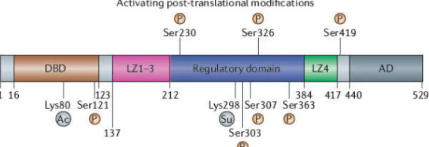
DBD – DNA Binding Domain
DFX -‐ Desferroxamine
DMEM -‐ Dulbecco's Modified Eagle Medium
DMSO -‐ Dimethyl Sulfoxide
DNA – deoxyribonucleic acid
EDTA -‐
Ethylenediaminetetraacetic Acid
EGS -‐ Ethylene Glycol Succinimidyl succinate
ER – Endoplasmic Reticulum
ERK -‐ Extracellular signal-‐ regulated kinases
FAC -‐ Ferric Ammonium Citrate
FBS -‐ Fetal Bovine Serum
Fe -‐ Iron
GAPDH -‐ Glyceraldehyde 3-‐ phosphate dehydrogenase
GEO -‐ Gene Expression Omnibus
GFP -‐ Green Fluorescent Protein
GSK3 -‐ Glycogen synthase kinase 3
HEPES -‐ 4-‐(2-‐hydroxyethyl)-‐1-‐ piperazineethanesulfonic acid
HIS -‐ Histidine
HOG1 – High Osmolarity
Glycerol
HS – Heat Shock
HSE – Heat Shock Element
HSF – Heat Shock Factor
HSP – Heat Shock Protein
HSR – Heat Shock Response
Abbreviations
IRP – Ireon Responsive Protein
JNK -‐ c-‐Jun N-‐terminal kinase
MAP -‐ Mitogen-‐activated
Protein
MAPK -‐ Mitogen-‐activated Protein Kinase
MEF-‐ Mouse Embrionic Fibroblast
MRP – Multi Drug-‐resistance Pump
NAD -‐ Nicotinamide adenine dinucleotide
NCBI -‐ National Center for Biotechnology Information
ONPG -‐ Ortho-‐Nitrophenyl-‐β-‐ Galactoside
PBS -‐ Phosphate Buffered Saline
PCR – Polymerase Chain Reaction
PDSM -‐ Phosphorylation-‐ Dependent Sumoylation Modifications
PKC -‐ Protein kinase C
PM – Plasma Membrane
RD – Regulatory Domain
RNA -‐ ribonucleic acid
ROS – Redox Oxygen Species
RT-‐ PCR – Real Time Polymerase Chain Reaction
SDS -‐ Sodium Dodecyl Sulfate
SDS – PAGE -‐ Sodium Dodecyl Sulfate Polyacrylamide Gel Electrophoresis
STRE-‐ Stress Response Element
SUMO -‐ Small Ubiquitin-‐like Modifier
TCA – Tricarboxylic Acid Cycle
UTR – Untranslated Region
WHO – World Health Organization
WT – Wild-‐Type
Yap – Yeast Activator Protein
YPD – Yeast Peptone Dextrose
T
he work presented in this thesis describes the use of yeastSaccharomyces cerevisiae as a model system to study two different stress
response processes and its extrapolation to higher eukaryotes.
In the Chapter 1 of this work we report the transcriptomic analysis of S.
cerevisiae response to arsenic, a highly toxic and carcinogenic metalloid.
This study shows that arsenic stress interferes with genes involved in iron
homeostasis including those encoding proteins that function in iron
uptake, incorporation into heme and Fe–S (iron–sulfur) clusters,
compartmentalization to and mobilization from intracellular stores and
other functions in iron balance. In S. cerevisiae iron metabolism is
balanced by two distinct iron transport systems, depending on the
bioavailability of the metal in the extracellular environment. Interestingly
mRNA levels encoding Fet3 and Ftr1, a complex required for high-‐affinity
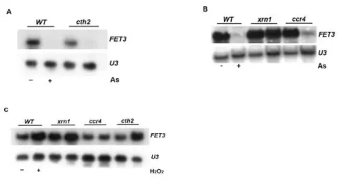
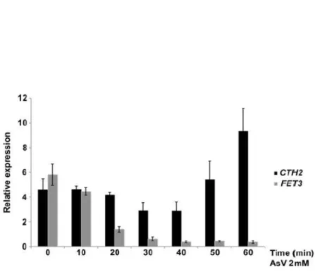
iron uptake, are drastically decreased upon arsenic exposure. Moreover
the FET3 mRNA decrease is mediated by the major pathway for mRNA
decay, the 5’-‐3’ exonuclease Xrn1 and is independent of the formation of
ROS. Strikingly, upon addition of arsenic Fet3 does not localize in the
plasma membrane but rather resides in the endoplasmic reticulum. Also,
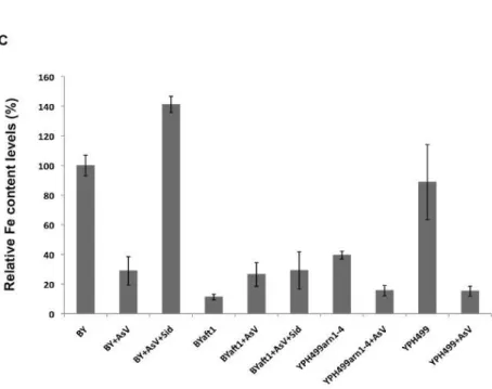
the fet3ftr1 mutant shows increased arsenic resistance over the wild-‐
type, suggesting that Fet3 plays a role in arsenic toxicity. Unexpectedly,
arsenic treatment seems to activate the non-‐reductive iron uptake
system. Finally, we provide data suggesting that arsenic-‐mediated
disruption of iron homeostasis also occurs in mammalian cells, an
observation that can be relevant for clinical applications.
In the Chapter 2 of this work we report on the repression mechanisms for
human Heat Shock Factor 1 (hHSF1) activity when expressed in S.
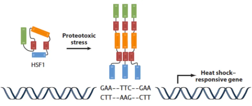
Cerevisiae. The heat shock transcription factor plays an important role in
the cellular response to proteotoxic stressors. Under normal conditions,
HSF1 is maintained as an inactive monomer and lacks the ability to
activate gene expression. In response to stress, HSF1 homotrimerizes,
accumulates in the nucleus, becomes hyper-‐phosphorylated, binds DNA
and activates gene transcription. Upon passage of the stress, HSF1
becomes hypo-‐phosphorylated and relocalizes to the cytoplasm
becoming re-‐established as an inactive monomer. While significant
advances have been made to understand the mechanisms involved in the
HSF1 activation, relatively little is known about the mechanisms
underlying repression. HSF1 is a highly conserved protein amongst
eukaryotes and previous data have shown that hHSF1 is unable to
complement for the loss of the essential yeast HSF, as hHSF1 is
maintained in an inactive monomer in yeast. Interestingly, many of the
proteins linked to the repression of HSF1 function, including the protein
chaperones HSP90 and HSP70, as well as the kinases JNK, ERK and GSK3,
are conserved in yeast. We hypothesized that gaining an understanding
of the mechanisms by which hHSF1 is maintained in an inactive state in
yeast would further our comprehension of HSF1 regulation in metazoans.
To explore these mechanisms we used a variety of biochemical and
genetic approaches to identify potential inhibitory phosphorylation sites
as well as a screen of mutant yeast strains to identify potential hHSF1
repressor proteins. Here we show that when hHSF1 is expressed in yeast
it is phosphorylated at S303 by the MAP-‐kinase Slt2 and that this
HSF1-‐dependent transactivation, we show in both yeast and mammalian
cells that S303 phosphorylation also represses HSF1 multimerization. Together, our data support the hypothesis that the yeast can be used to
simplify and elucidate complex regulatory mechanisms modulating
mammalian HSF1 activity as well as identify novel regulatory aspects
controlling HSF1 activity in yeast and mammalian cells.
Taken together, this work suggests that yeast cells can be a powerful
experimental tool for investigating aspects as arsenic toxicity and human
HSF1 regulation by providing a platform for the understanding of these
mechanisms in higher eukaryotes.
O
trabalho apresentado nesta tese descreve a utilização da leveduraSaccharomyces cerevisiae como um sistema modelo para estudar dois processos diferentes da resposta ao stress e a sua extrapolação para os
eucariotas superiores.
No Capítulo 1 deste trabalho, apresentamos a análise transcriptómica da
resposta S. cerevisiae ao arsénio, um metalóide altamente tóxico e cancerígeno. Este estudo demonstra que o stress pelo arsénio interfere
com os genes envolvidos na homeostase do ferro, em particular, os genes
que codificam as proteínas que funcionam na absorção de ferro,
incorporação nos clusters de heme Fe-‐S (ferro-‐enxofre), na
compartimentalização e mobilização das reservas intracelulares entre
outras funções. Em S. cerevisiae o equilibrio do metabolismo do ferro ocorre atraves de dois sistemas distintos de transporte do ferro,
dependendo da biodisponibilidade deste metal no ambiente extracelular.
Curiosamente os níveis de mRNA que codificam Fet3 e Ftr1, um complexo
de alta afinidade para a absorção de ferro, estão drasticamente
diminuidos após exposição ao arsénio. Além disso a diminuição do FET3 mRNA é mediada pela via principal de degradação do mRNA através da
5'-‐3 'exonuclease Xrn1 e é independente da formação de ROS.
Surpreendentemente, após a adição de arsénio Fet3 não está localizado
na membrana plasmática mas sim no retículo endoplasmático. Além
disso, o mutante fet3ftr1 apresenta mais resistência ao arsénio que a estirpe do tipo selvagem, sugerindo que o Fet3 desempenha um papel na
toxicidade do arsénio. Curiosamente, o tratamento com arsénio parece
apresenta dados que sugerem que a perturbação da homeostase do ferro
pelo arsénio também ocorre em células de mamífero, uma observação
que pode ser relevante para aplicações clínicas.
No Capítulo 2 deste trabalho descrevemos alguns dos mecanismos de
repressão da atividade do factor de transcrição humano “Heat Shock
Factor” (HSF1) expresso na S. cerevisiae. O HSF1 desempenha um papel muito importante na resposta celular a factores de stress proteotóxicos.
Em condições fisiológicas, o HSF1 é mantido como um monómero
inactivo e não tem a capacidade para activar a expressão de genes. Após
o stress, HSF1 homotrimeriza, desloca-‐se para o núcleo, torna-‐se
hiperfosforilado, liga-‐se DNA e activa a transcrição dos genes alvo. Após o
stress, HSF1 torna-‐se hipo-‐fosforilado e regressa para o citoplasma,
voltando à forma de monómero inativo. Embora já tenham sido feitos
avanços significativos para compreender os mecanismos envolvidos na
ativação do HSF1, relativamente pouco se sabe sobre os mecanismos
subjacentes à sua repressão. HSF1 é uma proteína altamente conservada
entre os eucariotas e dados anteriores demonstraram que o HSF1
humano (hHSF1) é incapaz de complementar a ausência do HSF, essencial na levedura, visto que o hHSF1 é mantido como um monómero inactivo.
Curiosamente, muitas das proteínas envolvidas na repressão da função
do HSF1, incluindo as proteínas chaperone Hsp90 e Hsp70, bem como a
JNK quinases, ERK e GSK3 estão conservados na levedura. Desta forma a
compreensão dos mecanismos pelos quais hHSF1 é mantido num estado
inativo na levedura poderá aprofundar a nossa compreensão da
regulação do HSF1 em metazoários. Para explorar estes mecanismos
proteínas repressoras do hHSF1. Neste trabalho mostra-‐se que quando
HSF1 humano é expresso em levedura está fosforilado na S303 pela Slt2
MAP-‐quinase e que esta fosforilação ocorre independentemente da
fosforilação prévia da S307 que até então se acreditava ser essencial.
Além disso, enquanto que estudos anteriores sugeriam que a fosforilação
na S303 reprime a transactivação dependente do HSF1, nós mostramos
em levedura e células de mamíferos que a fosforilação na S303 também
reprime a multimerização do HSF1. Em suma, os nossos resultados
apoiam a hipótese de que a levedura pode ser usada para simplificar e
elucidar mecanismos complexos de regulação da actividade do HSF1
humano, bem como identificar novos aspectos reguladores que
controlam a actividade do HSF1 na levedura e nas células de mamíferos.
Finalmente este trabalho sugere que as células de levedura podem
constituir uma poderosa ferramenta experimental para investigar
aspectos como a toxicidade de arsénio e regulação da actividade do HSF1
humano, fornecendo uma plataforma para a compreensão destes
mecanismos em eucariotas superiores.
General Introduction
The Yeast Model System – Saccharomyces cerevisiae
YEAST CELLS -‐ “OUR RELATIVES”
Sixteen years ago, in 1996, it was released the electronic version of the first complete DNA sequence of a eukaryotic genome, the yeast
Saccharomyces cerevisiae [1, 2], one of the oldest “domesticated”
organisms. Already at the time there was no doubt that this yeast would
be a fine “model organism”, useful for interpreting and understanding human DNA sequences [3].
The yeast genome contains 6000 genes and at least 60% have statistically
robust human homologues or one conserved domain with human genes.
Interestingly more than 30% of human disease genes have counterparts in yeast, and about 71 human genes complement yeast mutations (Table 1 in Appendix) [4]. Despite the tremendous achievement in sequencing the entire human genome (3 billion base pairs in human DNA), researchers would not be able to use this “treasure” if they did not have
access to the information provided by model organisms like yeast.
Indeed, their genomic homology shows the conservation of the fundamental cell biological processes between yeast and mammalian
cells. Furthermore yeast cells, just like our cells, have a nucleus containing
chromosomes and divide in a similar manner recapitulating fundamental aspects of the eukaryotic biology.
Therefore S. cerevisiae as a simple eukaryote with a tractable genome, a
short generation time, and a large network of researchers who have generated exquisite research tools, became a popular organism suited to help reveal the function of genes implicated in human biology. Many times the first clue about the function of higher eukaryotic genes has
biology, including the process of cell division and genetic transmission, transcriptional regulation, biogenesis and function of cellular organelles, protein targeting and secretion, cytoskeletal dynamics and regulation, and cellular metabolism [5].
Certainly, yeast does not have Parkinson or heart disease, but yet many
of the human genes that are malfunctioning in these and other disorders
have parallel genes in this model organism, where they can be more easily studied. Therefore many researchers are now developing yeast
models of many human diseases. Also, the modern tools available in S. cerevisiae make possible to perform systematic analysis of a cellular
process or phenotype, such us, genetic screening libraries,
transcriptomic, proteomic and metabolomic analysis, chemical genomics and chemical genetics, and phenotypic microarray analysis [6].
Despite yeast cells don't have brains enormous attention has been directed recently to their potential as an experimental model for neurodegenerative diseases, such as Alzheimer, Parkinson and
Huntington (see reviews in appendix). Many of the problems in these
diseases derive from problems in protein folding and trafficking, which is largely the same in yeast as it is in neurons [7]. Additionally yeast has
been extensively used to study cancer and age-‐related disorders.
repair are currently studied in yeast cells and then scaled up in more complex animals. Furthermore, screening libraries of yeast varieties and mutants is widely used to find potential gene targets for drug development.
Strikingly according to McGary K. L. et al work [8], that describes a new
system biology approach for identifying model organisms for human diseases, yeast could also be a powerful model organism for studying angiogenesis, although they don’t have blood vessels.
Throughout this dissertation we report the use of the yeast assay system to further understand two types of stress response. The first, regarding how arsenic stress induces a global metabolic reprogramming of the
cellular iron homeostasis both in S. cerevisiae and mammals and the
second concerning the mechanisms that regulate human Heat Shock Factor 1, a stress-‐activated transcription factor, via post-‐translational
modifications. Given the tools available in S. cerevisiae, this is an ideal
system to begin to decipher these biological phenomena.
T
he Response to Stress
All living organisms are subject to changes in the environment, to which they must adapt in order to survive. Yeast cell homeostasis is achieved through the activation of several transcription factors, each one acting
singly or in combination to perform specific functions. In the yeast S.
types of activators, the partially redundant zinc-‐finger transcription factors Msn2 and Msn4, the Heat Shock Factor (HSF), and the basic-‐ leucine zipper (b-‐Zip) transcription factors Yap family (Yeast-‐specific AP-‐1 like factors) [9].
The response mediated by the transcription factors Msn2 and Msn4 happens via stress response element (STREs) present on the target gene
promoters. The STRE was the first common cis-‐element identified in a set
of genes that sense a broad array of stress conditions determined by general stress response. It is estimated that over 186 genes (approximately 3% of the yeast genome) are potentially subject to STRE-‐ mediated regulation [10]. Msn2/4 are controlled by high osmolarity via the HOG signal pathway, which comprises a MAP kinase module. Some of the important aspects of Msn2/Msn4 regulation include nuclear-‐ cytoplasmic shuttling and targeted degradation of these factors at gene promoters during transcriptional activation [11]. The intracellular distribution of Msn2/4 is highly correlated with their phosphorylation state. It has been suggested that cAMP-‐dependent PKA inhibits nuclear import of Msn2/4, through direct phosphorylation of their nuclear localization signal (NLS). A second mechanism for PKA-‐mediated regulation of STRE-‐controlled gene expression involves the Ccr4-‐Not complex, a global transcriptional regulator that affects genes positively and negatively [12].
have demonstrated that both mammalian HSF1 and HSF2 expressed
together can rescue the viability defect of S. cerevisiae cells lacking
endogenous yeast HSF [14]. HSF is essential for cell viability in S.
cerevisiae, oogenesis and early development in Drosophila melanogaster,
extended life span in Caenorhabditis elegans, and extraembryonic
development and stress resistance in mammals.
In yeast Hsf1 is constitutively bound as a trimer to high affinity HSEs present on several promoters leading to a moderate expression of the
heat shock genes. Upon stress the occupancy of specific HSEs in the yeast
HSP82 promoter increases, suggesting that some yeast HSEs are inducibly bound, as observed in higher eukaryotes [15]. Moreover, the
identification of genome-‐wide Saccharomyces cerevisiae HSF targets by
chromatin immunoprecipitation, combined with DNA microarray analysis, demonstrated global heat-‐stimulated binding of HSF to many target genes [16]. Furthermore, similar to mammalian HSF1, yeast HSF is activated by multiple stresses, including heat shock, oxidative stress, and glucose starvation [17].
Recent studies have also provided novel insights into the role of HSF in growth, development, disease, and aging and in the complex metabolic reprogramming that occurs in all cells in response to stress [18].
in oxidative stress, Yap2 in cadmium stress, Yap4 and Yap6 in osmotic stress, and Yap8 in arsenic stress nevertheless there is also evidence of
cross-‐talk between the Yap members. For example, the yap1yap2 double
mutant is more sensitive to oxidative stress than either single mutant
alone, as is the yap1yap8 double mutant to arsenic stress [9]. Moreover
at least four Yap proteins (Yap1, Yap2, Yap4 and Yap5) bind most efficiently to the Yap Responsive Element (YRE) TTAC/GTAA. Both Yap1 and Yap8 bind to the sequence GATTTAATAATCA, in which the bases flanking the core sequence (underlined) are essential for Yap8 recognition [20]. So far, only the corresponding binding site for Yap3, Yap6 and Yap7 has not been characterized yet.
The three types of stress control seem to have overlapping, but distinct functions. Some stress proteins encoded by HSE-‐regulated genes are necessary for growth of yeast under moderate stress, products of STRE-‐ activated genes appear to be important for survival under severe stress and YRE-‐controlled genes may mainly function during oxidative stress and in the response to toxic conditions, such as caused by heavy metal ions. While our understanding of these processes in higher eukaryotes remains incomplete, earlier studies have suggested that many of these phenomena may be conserved in yeast.
1. Goffeau, A., et al., Life with 6000 genes. Science, 1996.
274(5287): p. 546, 563-‐7.
2. Guerreiro, P., et al., Sequencing of a 9.9 kb segment on the right
arm of yeast chromosome VII reveals four open reading frames, including PFK1, the gene coding for succinyl-‐CoA synthetase (beta-‐chain) and two ORFs sharing homology with ORFs of the yeast chromosome VIII. Yeast, 1997. 13(3): p. 275-‐80.
3. Chu, S., et al., The transcriptional program of sporulation in
budding yeast. Science, 1998. 282(5389): p. 699-‐705.
4. Chervitz, S.A., et al., Comparison of the complete protein sets of
worm and yeast: orthology and divergence. Science, 1998.
282(5396): p. 2022-‐8.
5. Botstein, D. and G.R. Fink, Yeast: an experimental organism for
21st Century biology. Genetics. 189(3): p. 695-‐704.
6. Khurana, V. and S. Lindquist, Modelling neurodegeneration in
Saccharomyces cerevisiae: why cook with baker's yeast? Nat Rev
Neurosci. 11(6): p. 436-‐49.
7. Winderickx, J., et al., Protein folding diseases and
neurodegeneration: lessons learned from yeast. Biochim Biophys
Acta, 2008. 1783(7): p. 1381-‐95.
8. McGary, K.L., et al., Systematic discovery of nonobvious human
disease models through orthologous phenotypes. Proc Natl Acad
Sci U S A. 107(14): p. 6544-‐9.
9. Rodrigues-‐Pousada, C., R.A. Menezes, and C. Pimentel, The Yap
family and its role in stress response. Yeast. 27(5): p. 245-‐58.
10. Moskvina, E., et al., A search in the genome of Saccharomyces
cerevisiae for genes regulated via stress response elements.
Yeast, 1998. 14(11): p. 1041-‐50.
11. Jacquet, M., et al., Oscillatory nucleocytoplasmic shuttling of the
general stress response transcriptional activators Msn2 and Msn4 in Saccharomyces cerevisiae. J Cell Biol, 2003. 161(3): p. 497-‐505.
12. Garreau, H., et al., Hyperphosphorylation of Msn2p and Msn4p in
response to heat shock and the diauxic shift is inhibited by cAMP in Saccharomyces cerevisiae. Microbiology, 2000. 146 ( Pt 9): p. 2113-‐20.
13. Hahn, J.S. and D.J. Thiele, Activation of the Saccharomyces
starvation conditions by Snf1 protein kinase. J Biol Chem, 2004.
279(7): p. 5169-‐76.
14. Liu, X.D., et al., Conservation of a stress response: human heat
shock transcription factors functionally substitute for yeast HSF.
EMBO J, 1997. 16(21): p. 6466-‐77.
15. Giardina, C. and J.T. Lis, Dynamic protein-‐DNA architecture of a
yeast heat shock promoter. Mol Cell Biol, 1995. 15(5): p. 2737-‐44.
16. Hahn, J.S., et al., Genome-‐wide analysis of the biology of stress
responses through heat shock transcription factor. Mol Cell Biol,
2004. 24(12): p. 5249-‐56.
17. Amoros, M. and F. Estruch, Hsf1p and Msn2/4p cooperate in the
expression of Saccharomyces cerevisiae genes HSP26 and HSP104 in a gene-‐ and stress type-‐dependent manner. Mol Microbiol,
2001. 39(6): p. 1523-‐32.
18. Hahn, J.S., D.W. Neef, and D.J. Thiele, A stress regulatory network
for co-‐ordinated activation of proteasome expression mediated by yeast heat shock transcription factor. Mol Microbiol, 2006. 60(1): p. 240-‐51.
19. Fernandes, L., C. Rodrigues-‐Pousada, and K. Struhl, Yap, a novel
family of eight bZIP proteins in Saccharomyces cerevisiae with distinct biological functions. Mol Cell Biol, 1997. 17(12): p. 6982-‐ 93.
20. Ilina, Y., et al., Characterization of the DNA-‐binding motif of the
arsenic-‐responsive transcription factor Yap8p. Biochem J, 2008.
415(3): p. 467-‐75.
This chapter includes data of a manuscript in preparation:
Batista-‐Nascimento, L. Thiele, D.J., Rodrigues-‐Pousada, C. (2012) Arsenic exposure induces iron deficiency and destabilizes the high-‐affinity iron uptake system.
Chapter 1
Introduction
Dealing with Arsenic
ARSENIC
The author of this dissertation had a major contribution in this work,
namely in the planning of the experimental work, in the execution and
analysis of the experiments and is the first author of the manuscript.
Abstract 33
Introduction 34
Material and Methods 36
Results 40
The Saccharomyces cerevisiae Genome-‐wide Response
to Arsenic Exposure 40
Aft1 is activated by arsenic stress 45
Arsenic triggers degradation of the FET3 mRNA by the
5’-‐3’ exonuclease Xrn1 47
Arsenic impairs high affinity Fe uptake by reducing
FET3 protein levels and plasma membrane expression 49
The fet3ftr1 double mutant is resistant to arsenic 52
Arsenic trioxide causes Fe uptake destabilization in
mammalian cells 55
Discussion 57
References 58
Acknowledgments 61
Footnotes 61
is prevalent. Since these metals are highly toxic, they pose a considerable
threat to nature and to human health. The main routes of poisoning are
through occupational exposure or through ingestion of contaminated
food and water. The contamination of drinking water by arsenic is a
major health concern because of the large number of contaminated sites
and people at risk of exposure. These metals are implicated in a broad
spectrum of degenerative conditions in humans, including neurotoxicity,
nephrotoxicity, genotoxicity and carcinogenesis. Chronic As exposure
induces cardiovascular diseases, neurological disorders and liver injury,
and is associated with cancers of the skin, bladder, liver and lung [1]. The
mechanism of As toxicity is not fully understood at the molecular level. In
general, it may act by targeting signaling or regulatory proteins that
control cell proliferation, differentiation and cell cycle regulation. Due to
its complex chemistry and ability to form many different compounds As is
an intricate element to understand. Arsenic is most commonly found in
two oxidation states, AsIII and AsV. The most common inorganic AsIII
compounds found are arsenic trioxide, sodium arsenite, and arsenic
trichloride. AsV compounds such as arsenic pentoxide, arsenic acid, and
arsenates are also quite common. Arsenic is methylated by
microorganisms, but inorganic AsIII and AsV compounds are considered
the most toxic [2]. A common property of As is its high reactivity with
sulphydryl groups. Hence, it can bind to and affect the activity of many
proteins. AsIII can inhibit more than 200 enzymes, in events such as DNA
repair, methylation of DNA, and increase radical formation and activation
attributed to its capability to induce formation of reactive oxygen species
(ROS). The damage caused by ROS to lipids, proteins and DNA are likely to
contribute to As toxicity [3]. Nevertheless, neither the exact details of
metal-‐induced ROS generation nor the full set of toxicity targets is known.
Drugs containing arsenicals such as arsenic trioxide (Trisenox®) have been
used to treat acute promyelocytic leukaemia (APL) and other
haematological and solid cancers [5]. However, the development of
resistance threatens the efficacy of medical treatment and hence, there is
an increasing demand to identify tolerance mechanisms. Tolerance and
detoxification mechanisms often involve extrusion of the toxic ions from
the cell, sequestration within internal organelles, chelation by metal-‐
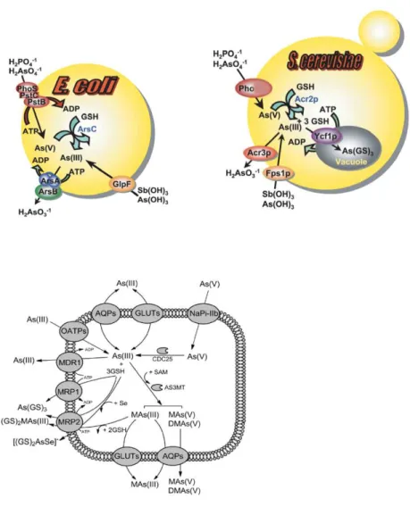
binding proteins, and reduction of uptake. As shown in Figure 1,
organisms take up AsV via phosphate transporters (Pho84 and Pho87)
and AsIII by aquaglyceroporins (GlpF in E. coli, Fps1 in yeast, and AQP7/9
in mammals) and hexose permeases (HXT family in yeast, and GLUT1/4 in
mammals). Once inside the cell AsV is reduced to AsIII by the Acr2
(arsenate-‐reductase) enzyme, with glutathione and glutaredoxin serving
as a source of reducing potential [7].
In vertebrates, five sodium/phosphate transporters NaPiIIa, NaPiIIb,
NaPiIIc, Pit-‐1 and Pit-‐2, which constitute the mammalian phosphate
uptake system, were recently identified as arsenic transporters. NaPiIIa,
NaPiIIc, Pit-‐1 and Pit-‐2 correspond to the low-‐affinity and NaPiIIb
represent the high-‐affinity AsV transport. Next, AsV is reduced to AsIII by
CDC25 phosphatases/arsenate reductases [8].
AsIII is then extruded from S. cerevisiae cells by the Acr3 (plasma
membrane arsenite-‐efflux protein), and compartmentalized into the
Studies with the budding yeast S. cerevisiae have demonstrated that in
response to arsenic stress cells utilize two b-‐Zip transcription factors,
Yap1 and Yap8, members of the Yap (Yeast AP-‐1 like) family. Yap8 is the
master regulator of this response, mediating the transcriptional
activation of ACR2 and ACR3 [10, 11] and Yap1 regulates YCF1 expression
and helps to maintain the cellular redox homeostasis [12]. In addition,
arsenic can be methylated [13], although this process may increase
arsenic toxicity rather than contributing toward detoxification [7].
Moreover, AsIII, the most toxic form of As, triggers increased ROS
production in mammals [14], but not to any large extent in wild-‐type
yeast [12].
Nevertheless, AsIII-‐induced oxidative stress and lipid peroxidation are
detected in mutants with impaired AsIII detoxification (yap8 mutant cells)
or oxidative stress defense (yap1 mutant cells) systems, indicating that
AsIII enhances ROS levels also in yeast [12],
Arsenic toxicity has therefore been suggested to be caused by oxidative
stress, impaired DNA repair, inhibition of enzyme function and by
disturbing the function of proteins that regulate proliferation, cell cycle
progression, apoptosis or differentiation [15-‐19] (Figure 2).
Figure 1: Arsenic detoxification in prokaryotes (E.coli) and eukaryotes (S.
cerevisiae and mammals). AsV is taken up by phosphate and
sodium/phosphate transporters, and AsIII is taken up by
aquaglyceroporins (GlpF in E. coli, Fps1p in yeast and Aqp7 and Aqp9 in
mammals). AsV is reduced to AsIII by the bacterial ArsC, the yeast Acr2p
or the mammal CDC25 enzymes. Glutathione and glutaredoxin serve as
the ABC superfamily of drug-‐resistance pumps that transports As(GS)3
into the vacuole. In mammals Mrp isoforms pump As(GS)3 out of cells.
Adapted form [6] and [7].
Figure 2: Arsenic response and toxicity mechanisms. Arsenic triggers


![Table 1: Heat shock factor 1 (HSF1) post-‐translational modifications (PTMs) events [15]](https://thumb-eu.123doks.com/thumbv2/123dok_br/15769987.641183/91.765.135.646.788.932/table-heat-shock-factor-translational-modifications-ptms-events.webp)
