American Journal of Animal and Veterinary Sciences 3 (1): 28-31, 2008 ISSN 1557-4555
© 2008 Science Publications
Corresponding Author: Dr. Mehrdad Jahanshahi, Department of Anatomy, Gorgan University of Medical Sciences, P.O. Box 49175-553, Gorgan, Iran Tel: 0098-171-4421651
28
Working Memory Learning Method and Astrocytes Number in Different
Subfields of Rat's Hippocampus
1
Jahanshahi Mehrdad,
2Sadeghi Yousef,
2Hosseini Ahmad,
3Naghdi Naser and
2Piriaie Abbas
1
Department of Anatomy, Gorgan University of Medical Sciences, Gorgan, Iran
2
Cellular and Molecular Research Center,
Shahid Beheshti University of Medical Sciences, Tehran, Iran
3
Department of Physiology, Institute of Pasteur, Tehran, Iran
Abstract: The aim of this study was evaluation of the astrocytes number in different subfields of rat's Hippocampus after spatial learning with usage of Morris Water Maze technique and working memory method. In this study, between 2005-2006 years in Pasteur institute of Iran-Tehran and histological department of Gorgan University with usage of Morris Water Maze and working memory technique, we used 14 male albino wistar rats. Seventh rats were in control group and 7 rats in working memory group. After histological preparation, the slides were stained with PTAH staining for showing the Astrocytes. Present results showed significant difference in astrocytes number in CA1, CA2 and CA3 areas of hippocampus between control and reference memory group. The number of astrocytes is increased in working memory group. Then we divided the hippocampus to three parts: Anterior, middle and posterior and with compare of different area (CA1, CA2 and CA3) of hippocampus, we found that the differences between Anterior-middle and Middle-Posterior of CA1 and CA2 area of hippocampus were significant, whereas the difference between Anterior-Posterior parts was not significant in CA1 and CA2 areas. In CA3 area, the difference between Middle and Anterior-Posterior parts was significant, whereas the difference between middle and posterior parts was not significant. We concluded that the number of astrocytes increased due to spatial learning and working memory technique.
Key words: Hippocampus, astrocytes, working memory, spatial learning
INTRODUCTION
The hippocampal formation plays an important role in memory and learning. The Morris Water Maze (MWM) is a test of spatial learning for rodents that relies on distal cues to navigate from start locations around the perimeter of an open swimming arena to locate a submerged escape platform. Spatial learning is assessed across repeated trials and reference memory is determined by preference for the platform area when the platform is absent[1].
Learning needs some instrument for information storage and information maintenances mechanisms resemble to memory. In the other hand, the memory always accompany with learning[2].
The hippocampal formation consists of the subiculum, the hippocampus and the dentate gyrus[3]. The hippocampus can be subdivided into three subfields: The CA1, CA2 and CA3 areas[4].
The principal cells in hippocampus are pyramidal neurons and in the dentate gyrus are the granule cells. Apart from principal neurons, the hippocampal formation contains different types of glial cells[5].
Astrocytes, strategically positioned between the capillaries and neurons, are thought to play a role in neuronal energy metabolism [6,7]. Glycogen is localized in the brain almost exclusively in astrocytes[8,9].
American J. Animal & Vet. Sci., 3 (1): 28-31, 2008
29 Recently, the researches showed that the astrocytes, not only receive the information from environment, but also send the signals to neurons [14]. According to our hypothesis, the number of astrocytes after spatial learning must be increased, because astrocytes have a closely relationship to synapses. The knowledge of changes in astrocytes number can help us that know what amount these cells involve in memory, therefore the aim of this study was evaluation of the astrocytes number in different subfields of rat's Hippocampus after spatial learning with usage of Morris Water Maze technique and working memory method.
MATERIALS AND METHOD
During 2005-2006, 14 male albinos Wistar rats (200-250 g.) obtained from Pasteur institute of Iran were used. Rats were housed in large plastic cage, food and water were available. Animals were maintained under standard conditions and 12/12 h light/dark cycle with lights on at 7.00 am. After accommodation with environment, we divided rats to Control and Working memory groups. We used of Morris Water Maze technique for spatial learning in working memory group.
MWM Testing: The rats were placed in a circular plastic pool (diameter, 120 cm) with white inside walls, located in a normally equipped laboratory room, uniformly lighted by four neon lamps (40 Weach) suspended from the ceiling (3 m). No care was taken to enhance (or, vice versa, to impoverish) extra-maze cues, which were held in constant spatial relations throughout the experiments.
The pool was filled with water (24°C), which was 50 cm deep and made opaque by the addition of 2 L of milk. A white, steel escape platform (10 cm in diameter) was placed in the middle of one cardinal quadrant (NW, NE, SW, SE) 30 cm from the side walls, it was either submerged 2 cm below or elevated 2 cm above the water level. Each rat was released gently into the water always from the same cardinal wall point (S) facing the center of the pool. The animal was allowed to swim around to find the platform. Blocks of four trials were presented to each rat, two blocks of trials per day[15].
Working memory testing in the water maze: On each trial, the rats were placed into the water at one of the four cardinal points of the compass (N, E, S, W), which varied from trial to trial in a quasi-random order. The rats had to swim until they climbed onto the escape
platform. If they failed to locate the platform within 60 sec, they were guided there. The rats were allowed to stay on the platform for 20 sec.
Two day after the reference memory pre-training phase, training on the working memory version of the navigation task started. Only two trials per day were given until performance stabilized in the first trial (acquisition), the animal had to find the platform in a new position. The rats were allowed to stay there for 20 sec before they were returned to the home cage. on the second trial (retrieval ), which was administrated 75 min later, the platform was in its previous position but the animals was started from a different place to the preceding trial[16,17].
After learning examinations, animals were decapitated after ether anesthesia and the brains were removed for histological verification, at first the brains fixed in formaldehyde 10% and two week later impregnated with paraffin wax. After histological processing, slices of 7 µm coronally (anterior to posterior of hippocampus) were produced with Leitz rotary microtome (One of 10 sections was selected for staining and morphometeric measurements). For astrocytes staining, we used PTAH (Phosphotanguestic Acid Haematoxylin) staining[18] because it is the special staining method for astrocyte cells and their processes. In this method the astrocytes appeared blue and the neurons appeared pink.
Morphometric measurement was carried out using on Olympus DP 12 digital camera and BX51 microscope. We selected a field (75000 µm2) within the pyramidal layer of hippocampal subfield CA2. Randomly selected, non-overlapping photographs using a ×40 objective lens were taken from the designated areas. Images were saved by the Bioreporter program and further processed using the Adobe Photoshop 6.0 program (Adobe System Inc., San Jose, CA, USA).
For cell counts, photographs at a magnification of ×40 (objective lens) were taken throughout the longitudinal axis of the hippocampal subfields and further processed as described above. All of the astrocytes shown on this field counted and then the mean and SD of astrocytes number were measured.
Statistical analysis: Data was expressed as mean ±SD differences among areas were statistically evaluated using the one-way analysis of variance (ANOVA). Probabilities of p<0.05 were considered significant.
RESULTS AND DISCUSSION
American J. Animal & Vet. Sci., 3 (1): 28-31, 2008
30
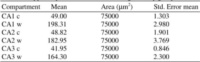
Table 1: The mean of astrocytes number in different areas of hippocampus in control and working memory groups Compartment Mean Area (µm2) Std. Error mean
CA1 c 49.00 75000 1.303
CA1 w 198.31 75000 2.980
CA2 c 48.82 75000 1.901
CA2 w 182.95 75000 3.769
CA3 c 41.95 75000 0.846
CA3 w 164.30 75000 2.300
c = Control w = Working
Table 2: The mean of astrocytes number in different parts (Anterior, Middle and Posterior) of control and working memory groups
Compartment Mean Area (µm2) Std. Error mean
CA1 wa 195.67 75000 5.21
CA1 wm 208.92 75000 6.05
CA1 wp 190.53 75000 3.25
CA2 wa 192.15 75000 7.19
CA2 wm 165.18 75000 5.74
CA2 wp 190.00 75000 5.68
CA3 wa 155.28 75000 3.89
CA3 wm 169.60 75000 4.49
CA3 wp 167.00 75000 2.99
C = Control and w = Working
The mean and SD of the number of Astrocytes in shown area of hippocampus (per 75000 µm2) is depicted in Table 1. In control group, the mean of astrocytes number in CA1 and CA2 was similar and more than CA3 subfield[19]. In working memory group, the number of astrocytes in CA1 and CA2 was similar and it was more than CA3 subfield.
Then we divided the hippocampus to three parts: Anterior, Middle and Posterior one-thirds, according of their functional differences[20], the mean and SD of the number of Astrocytes in different parts (per 75000 µm2) is shown in Table 2.
The differences of astrocytes number between all areas (CA1, CA2, CA3) of hippocampus in control and working memory groups were significant. In all area, the number of astrocytes increased. Also, after the diviation of hippocampus to three parts: Anterior, Middle and Posterior one-thirds, because their functional differences[20], we showed that in CA1 area of working memory group, the most number of astrocytes was in middle one-third, in CA2 area it was in anterior one-third and in CA3 area the most number of astrocytes was in middle one-third of hippocampus.
These results indicated that the working memory method of spatial learning can cause increasing of astrocytes number in posterior two-third of hippocampus, especially in CA3 area.
Physiologically, present results similar and resemble too many researches that worked on the spatial learning[16,17,21,22,23].
Many studies provided the relationship between exercise and neurogenesis in hippocampus and especially in dentate gyrus[24]. Physical exercise increases the neurogenesis in hippocampus as well as genetic factors[24,25]. One of the exercise and learning method is the Morris Water Maze, that it can increase neurogenesis in dentate gyrus[26].
Keuker in 2003 with usage of water maze technique and Reference and working method (similar to present research), said that: The working memory in aged animals significantly differs from the young animals, whereas the reference memory doesn't changes with ages[27].
Rusakov in 1997 said that: Memory formation is believed to alter neural circuitry at the synaptic level. Although the hippocampus is known to play an important role in spatial learning, no experimental data exist on the synaptic correlates of this process at the ultrastructural level. Analysis of synaptic spatial distribution showed a training-associated increase in the frequency of shorter distances (i.e., clustering) between synaptic active zones in CA1, but not dentate, thus indicating alterations in local neural circuitry. This finding indicates subtle changes in synaptic organization in area CA1 of the hippocampus following a learning experience, suggesting that spatial memory formation in mammalian hippocampus may involve topographical changes in local circuitry without synapse formation de novo[28].
In conclusion these researches almost are resemble to our study and showed that spatial learning can increase the synaptic location and indirectly we showed that the increase of synaptic number, can increase the number of astrocytes.
REFERENCES
1. Vorhees, C.V. and M.T. Williams, 2006. Morris water maze: Procedures for assessing spatial and related forms of learning and memory. Nat. Protocols, 1: 848-858.
2. Markowitsch, H.J., 1995. Anatomical Basis of Memory Disorders. In: Cognitive Neurosci, Gazzaniga, M.S. (Ed.). Cambridge, MA: Mit press, pp: 665-679.
3. Knowles, W.D., 1992. Normal anatomy and neurophysiology of hippocampal formation. J. Clin. Neurophysiol., 9(2): 252-263.
American J. Animal & Vet. Sci., 3 (1): 28-31, 2008
31 5. Williams, P.L., L.H. Bannister and
M.M. Berry et al., 1995. Gray's Anatomy. In: Nervous System. 38th Edn. London. Churchill. Livingston, pp: 1123-1129.
6. Pellerin, L. and J. Magistrettip, 2003. Food for thought: Challenging the dogmas. J. Cereb. B1F1 Metab., 23: 1282-1286.
7. Forsyth, R., A. Fray and M. Boutelle et al., 1996. A role for astrocytes in glucose delivery to neurons? Dev. Neurosci., 18: 360-370.
8. Gruetter, R., 2003. Glycogen: The forgotten cerebral energy store. J. Neurosci. Res., 74: 179-183.
9. Tsacopolos, M. and P.J. Magistretti, 1996. Metabolic coupling between glia and neurons. J. Neurosci., 16: 877-885.
10. Rabcheusky, A.G., 2002. Influences of activated microglia/brain macrophages on spinal cord injury and regeneration. In: Microglia in the Regeneration and Degenerating Cerebral Nervous System. Streit, W.J. (Ed.). Springer-Verlag, New York. 209-226.
11. Bechmann, I. and R. Nitsch, 1997. Astrocytes and microglia cells in corporate degenerating fibers following enthorhinal lesion. Glia, 20: 145-154. 12. Teter, B. and J.W. Ashford, 2002. Neuroplasticity
in Alzheimer's disease. J. Neurosci. Res., 70: 402-437.
13. Laming, P.R. et al., 2000. Neuronal-glial interactions and behaviour. Neurosci. Biobehav. Rev., 24: 295-340.
14. Caudle, 2006. Memory in Astrocytes: A hypothesis. Theor. Biol. Med. Modelling, 3: 2. 15. Leggio, M.G., M. Molinari and P. Neri et al., 2000.
Representation of actions in rats: The role of cerebellum in learning spatial performances by observation. PNAS., 97 (5): 2320-2325.
16. Naghdi, N. and A. Asadollahi, 2004. Genomic and nongenomic effects of intrahippocampal microinjection of testosterone on long-term memory in male adult rats. Behav. Brain Res., 153: 1-6.
17. Sarihi, A., F. Motamedi, N. Naghdi and A. Rashidy, 2000. Lidocaine reversible inactivation of the median raphe nucleus has no effect on reference memory but enhances working memory versions of the Morris water maze task. Behav. Brain Res., 114: 1-9.
18. Bancroft, J.B. and A. Stevens, 1990. Theory and Practice of Histological Techniques. Churchil Livinsgstone, Edinburgh.
19. Jahanshahi, M., Y. Sadeghi and A. Hosseini, 2006. Estimation of Astrocyte number in different subfield of rat hippocampus. Pak. J. Biol. Sci., 9 (8): 1595-1597.
20. Moser, M.B. and E.I. Moser, 1998. Functional differentiation in the hippocampus. J. Hippocampus, 8: 608-619.
21. Bronders, R., S.Y. Brandy and S. Yehuda, 1989. The use of the MWM in the study of memory and learning. J. Neurosci., 48: 29-62.
22. Isgor, C. and D.R. Sengelaub, 1998. Prenatal gonadal steroids affect adult spatial behavior, CA1 and CA3 pyramidal cell morphology in rats. Horm. Behav., 34: 183-198.
23. Redish, A.D. and D. Touretzky, 1998. The role of the hippocampus in solving the morris water maze. Neural. Comp., 10 (1): 39-73.
24. Van Praag, H., B.R. Christic, T.J. Sejnowski and F.H. Gage, 1999. Runing enhances neurogenesis, learning and long term potentiation in mice. Proceedings of the National Academy of Sciences, USA, 96: 13427-13431.
25. Madsen, T.M. and D.D. Yeh et al., 2005. Electroconvulsive seizure treatment increases cell proliferation in rat frontal cortex. Neuropsychopharmacology, 30: 27-34.
26. Rosenzweig, E.S., A.D. Redish, B.L. McNaughton and C.A. Barnes, 2003. Hippocampal map realignment and spatial learning. Nature Neurosci., 6 (6): 609-615.
27. Keuker, J.I.H. and G. Biurrun et al., 2004. Preservation of hippocampal neuron numbers and hippocampal subfield volumes in behaviorally characterized aged tree shrews. J. Comp. Neurol., 468: 509-517.