ORIGINAL ARTICLE
SPECTRUM OF ORCHIDECTOMY LESIONS: 5 YRS STUDY
T. Sundari Devi1, B. Vijaya Nirmala2, N. Srivani3, O. Shravan Kumar4
HOW TO CITE THIS ARTICLE:
T. Sundari Devi, B. Vijaya Nirmala, N. Srivani, O. Shravan Kumar. ”Spectrum of Orchidectomy Lesions: 5 Yrs Study”. Journal of Evidence based Medicine and Healthcare; Volume 2, Issue 26, June 29, 2015; Page: 3880-3892.
ABSTRACT: AIM: To study the incidence of different testicular lesions both neoplastic and non-neoplastic lesions in orchidectomy specimens at Gandhi Hospital, Hyderabad A.P. OBJECTIVE:
To study the various morphological patterns of neoplastic and non-neoplastic lesions of testis.
MATERIALS AND METHODS: One hundred and nineteen orchidectomy specimens were received in the Pathology Department of Gandhi Medical College from July 2008 to June 2013. The patients clinical details were noted. The gross examination findings of the excised specimens along with the histopathological findings were analysed. RESULTS: Out of 119 orchidectomy specimens 7 are neoplastic lesions& 112 non-neoplastic lesions. Among non-neoplastic lesions 24 are chronic non-specific epididymo orchitis, 5- caseating granulomatous orchitis, 1-filarial orchitis, 1-syphilitic orchitis, 20 acute suppurative epididymo orchitis (pyoceole), 16 –torsions, 4-traumatic, 6-atrophic, 10-maturation arrests and 25 showed normal histology. Neoplastic lesions included 2-seminomas, 1-spermatocytic seminoma, 1-yolk sac tumor, 1-mixed germ cell tumor and 2-Non Hodgkins lymphomas. CONCLUSION: The results of the study show a relatively low frequency of testicular tumours compared to non-neoplastic lesions in patients treated at Gandhi Hospital.
KEYWORDS: Orchidectomy lesions, Neoplastic, Non-neoplastic.
INTRODUCTION: Distinct pathological conditions affect the testis and epididymis. Congenital anomalies include cryptorchidism, Absence of testis, Fusion of testis (synorchism). Common inflammatory lesions are non-specific epididymitis and orchitis, Gonorrheal, Tuberculous, Syphilitic, and Idiopathic (Autoimmune), Viral infection (mumps orchitis), Malakoplakia. Most common vascular lesion is torsion testis (twisting of spermatic cord). Two forms seen are Neonatal and Adult form seen in adolescence. Neoplastic lesions Constitute 1% of all cancers in male two main categories (WHO) are Germ cell tumor (95%) and, Non-germ cell tumors (5%) Germ cell tumors are highly aggressive, but can be cured with current therapy. Non germ cell tumors are generally benign, and associated with endocrinologic syndromes Pathologist plays an important role in accurate classification, appropriate pathologic staging, Identifying prognostic parameters
AIMS & OBJECTIVES:
To study the incidence of different testicular lesions in orchidectomy specimens at Gandhi hospital, secunderabad and to study the various morphological patterns of neoplastic and non-neoplastic lesions of testis.
ORIGINAL ARTICLE
2008 to June 2013. Unilateral and bilateral orchidectomy specimens received for different causes were studied.
Indications:
Cryptorchidism, Torsion testis, Traumatic testis, Pyocele, Chronic epididymo-orchitis, Obstructed inguinal hernia, Carcinoma prostate (bilateral orchidectomy), All clinical details, Biochemical, hematological and radiological investigations, Gross morphological details of all the specimens were noted, Microscopic study of H & E stained sections taken from testis, epididymis and spermatic cord was done
OBSERVATIONS:
A total of 119 orchidectomy specimens were received.
Out of 119 specimens received, 112 cases (94.2%) - non-neoplastic and 7 cases (5.8%) - neoplastic lesions.
No. of cases
% of cases
Chr non-sp epididymo-orchitis 24 21.4% Caseating granulomatous orchitis 5 4.4%
Filarial orchitis 1 0.8% Syphilitic orchitis 1 0.8% Acute suppurative orchitis 20 17.8%
Torsion of testis 16 14.2% Traumatic testis 4 3.5%
Atrophic testis 6 5.3% Maturation arrest 10 8.9% Normal histology 25 22.3%
ORIGINAL ARTICLE
< 10 YEARS
10-20 YEARS
20-30 YEARS
30-40 YEARS
40-60 YEARS
60-80 YEARS
CRYPTORCHIDISM 5 10 04 04 NIL NIL TORSIONTESTIS NIL 12 04 NIL NIL NIL TRAUMATIC TESTIS NIL 01 02 01 NIL NIL CHRONIC EPIDIDYMO
ORCHITIS NIL _ 03 10 15 03 OBSTRUCTED INGUINAL
HERNIA NIL NIL NIL 02 01 NIL PYOCELE NIL NIL 02 06 08 04
ORIGINAL ARTICLE
BIL.ORCHIDECTOMY FOR
Ca. PROSTATE NIL NIL NIL NIL 05 10
Table 2: Age Distribution of Non-Neoplastic Lesions
Cryptorchidism:
It is the only congenital abnormality observed in our study, 23 cases received were all unilateral. Grossly 80% were normal, 20% small and brown. Histopathologically 17 cases showed normal histology 3 cases showed maturation arrest at different levels and 3 cases showed atrophic testis.
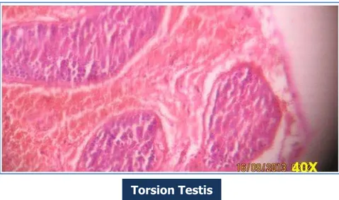
Torsion Testis: 16 cases of torsion testis received, grossly were enlarged, haemorrhagic & soft with histopathology showing intense congestion, extravasation of blood in to interstitial tissue of testis and epididymis, haemorrhagic infarction.
Chronic Non Specific Epididymo-Orchitis: 24 cases (30- 60 yrs) studied with histopathology showing, non-specific inflammation, necrosis, fibrous scarring, leydig cells are spared.
Graulomatous Epididymo-Orchitis: 5 cases of tuberculous etiology received had isolated involvement of testis and epididymis. Grossly testes was enlarged. C/S showed G/W areas -? Necrosis replacing entire testis. String sign negative. Histopathology showed, numerous caseating granulomas in the interstitium of testis, epididymis & spermatic cord, necrosis, fibrosis of tunica.
ORIGINAL ARTICLE
Syphilitic Orchitis: Only one case of 45 years observed which was a known case of syphilis. Histopathology showed diffuse interstitial inflammation, edema, lymphocyte and plasma cell infiltration.
Filarial Orchitis: One case of 60 yrs observed, grossly showing nodular firm mass in the testis involving peritesticular tissue and histopathology showed granulomatous inflammation in the stroma with cross sectional of the gravid filarial worm.
Granulomatous Orchitis
ORIGINAL ARTICLE
PYOCELE: 20 cases over a wide age range of 20 to 80 years observed histopathologically showing acute suppurative inflammation of the testis and epididymis, dense infiltration by neutrophils.
Atrophic Testis: Among 6 cases of atrophic testis observed, 3 were cases of cryptorchidism and 3 were cases of end stage chronic non-specific epididymo-orchitis. Grossly they were small, brown & firm and histopathologically showed seminiferous tubules as dense cords of hyaline connective tissue, prominent basement membranes, increase in interstitial stroma with prominent leydig cells
Maturation Arrest: 10 cases of maturation arrest observed of which 4 were cases of cryptorchidism and 6 were cases of chronic non-specific epididymo-orchitis. Histopathology showed maturation of germ cell was arrested at various levels from spermatogonia to spermatids.
Filarial Orchitis
ORIGINAL ARTICLE
Normal histology:
25 cases of normal histology observed.
16 cases of cryptorchidism.
All cases of bilateral orchidectomy for carcinoma prostate.
NEOPLASTIC LESIONS (7→5.8%):
Classic Seminoma: Two cases of seminoma of age 38yrs and 45yrs showed grossly enlarged testis measuring 10x6x4 cms & 9x7x4 cms respectively. C/s Solid homogenous grey white lobulated mass without any areas of hemorrhage and necrosis. No involvement of tunica albugenia epididymis or spermatic cord seen.
Histopathology in both cases showed sheets of uniform cells divided in to lobules by delicate fibrous septae infiltrated by lymphocytes the individual cells were large round to polygonal, with clear cytoplasm and a large central nucleus with one or two prominent nucleoli. Mitosis were rare.
ORIGINAL ARTICLE
Spermatocytic Seminoma:
One case of age 80 yrs, grossly-tumor was large-10X8X6 cms with C/s-solid pale, grey white soft mass with no involvement of tunica albugenia, epididymis and spermatic cord. Histopathology showed mixed population of 3 cell types-Medium sized cells (15-18m) with eosinophilic cytoplasm and round nucleus, smaller cells (6-8m) with a narrow rim of eosinophilic cytoplasm resembling secondary spermatocytes and scattered multi nucleated giant cells (50-100m).
Yolk Sac Tumor:
One case of age 1yr, grossly testis measured 6X5X4 cms on C/s-tumor was solid, grey white and yellow brown with gelatinous appearance and haemorragic areas. Histopathology showed lace like (Spider web) network of medium sized cuboidal cells with vacuolated cytoplasm, areas of papillary structures and endodermal sinus like structures called schiller-duval bodies.
Spermatocytic Seminoma
ORIGINAL ARTICLE
MIXED GERM CELL TUMOR:
One case of age 26 yrs, with combination of yolk sac tumor and mature teratoma. Grossly tumor was grey white yellow with areas of haemorrhage, cystic change and cartilage like areas.
NON - HODGKINS LYMPHOMA:
2 cases of age 65 yrs and 72 yrs. grossly testes were enlarged, C/s showed homogenous grey white masses in both cases. Histopathology showed features of small cell lymphoma in one case and diffuse large B cell lymphoma in another case.
SLL-showed total loss of normal architecture of testis, replaced by diffuse sheets of monotonous population of cells. Individual cells are round with scanty cytoplasm & some with prominent nucleoli with areas of necrosis. There is also infiltration of the peritesticular tissue with tumor tissue. There was no lymphatic or vascular invasion.
Non- Hodgkins Lymphoma (SLL)
ORIGINAL ARTICLE
DISCUSSION: Out of 119 orchidectomy specimens, 7 are neoplastic lesions. Out of 7 neoplastic lesions 5 are germ cell tumors i.e. 70%. This finding is in concordance with findings of other authors.1,2,3
Out of 5 germ cell tumours, 4 were (i.e., 80%) of one histological type and one was of more than one histological type. This finding is almost similar to the study of chitala A and Vadera V (1992) in which out of 154 germ cell tumors, 142 i.e. 92% were of one histological type.
In tumors with one histological type seminoma was the commonest in our study. Seminomas account for 50% of testicular germ cell tumors and more than 90% of these are classic seminomas.4
In our study, we had one case of spermatocytic seminoma. It is a rare germ cell tumors, also called spermatocytoma, first distinguished from classic seminoma by Masson (1946). WHO has updated its classification in 2004, but spermatocytic seminoma remains classified together with other testicular GCT (Eble et al, 2004). In differential diagnosis, it has to be distinguished from classical seminoma, pure embryonal carcinoma and testicular lymphoma. (Eble, 1994, Looijenga et al, 2007 & Lim et al 2011)
We had only one case of yolk sac tumor.
In our study, we had one case of mixed germ cell tumour with combination of yolk sac tumour and mature teratoma. The current incidence of mixed germ cell tumors in US is 6 per 100, 000. For unclear reasons, there has been a worldwide increase in the incidence of these tumors.5
Apart from germ cell tumors, we had 2 cases of testicular Non Hodgkin Lymphoma – 1 case of DLCL, 1 case of SLL. According to komal Bhatia et al study6 TNHL is a rare disease and
accounts for 1% of all NHLs, 2% of all extra nodal lymphomas and 1- 7% of all testicular neoplasms.
According to working formulations of the U.S. national cancer institute, most common (68%) of TNHL is intermediate grade, diffuse large B cell subtype followed by high grade diffuse small non-cleaved subtype (30%).7
Similar to other studies we found 1 case of primary testicular diffuse large B cell lymphoma of age 65yrs, presented as unilateral (left) testicular mass. In our study the age of DLBCL is 65yrs. But in 3 previous studies,8,9,10 the median ages were reported as 67, 69 and
66yrs respectively. But recent observation noted a shift of mean age to younger age group.11,12
In our study, among non-neoplastic lesions observed, chronic non-specific epididymo-orchitis is the most common inflammatory lesion (24cases) of the testis noted. Age range in our study is from 30 – 60yrs. Unlike in our study, highest incidence was between 20 -29yrs of age in one study of U.S. Army soldiers.13
We had 5 cases of caseating Granulomatous epididymo-orchitis. Genital TB accounts for 18% of cases of tuberculosis in India.14 Genitor urinary TB is the second most common form of
extra pulmonary TB after lymphnode involvement.15 Isolated epididymo-orchitis may produce
diagnostic difficulty while excluding a possible testicular neoplasm.16
ORIGINAL ARTICLE
scrotal mass as in our study. It is important for urologists to recognise the varied presentations of TBEO and to be familiar with its resemblance to malignancy. Genitor-urinary TB is a chronic infectious disease found predominantly in developing countries.18
We had only 1 case of syphilitic orchitis of in a known syphilitic tertiary patient aged 45 years. Syphilitic orchitis is a rare presentation of testicular swelling. A comprehensive literature search revealed only 11 confirmed cases in the past 59yrs19. Histopathology showed difuse
interstitial inflammation, edema, lymphocytes and plasma cell infiltrate. In a case report of syphilitic orchitis by TeraoT et al20 high orchidectomy was done to R/O testicular tumor.
Pathologically Granulomatous inflammation with lympho-plasmacytic infiltrate and endarteritis obliterance of small arterioles was seen in the specimen.
We had one case of filarial orchitis in 60yrs patient. While filarial orchitis is rare, yet reported manifestation.21 Tissue eosinophils is a useful diagnostic hint.22 However reaching a
preoperative diagnosis of testicular filariasis as seen in our case can be difficult. TB is an important differential diagnosis in addition to diverse neoplastic and non-neoplastic lesions of para-testicular region. Since the disease stimulates clinical malignancy, it is often the cause of unilateral orchidectomy. In our study grossly, there was a firm nodular mass in the testis involving the paratesticular tissue and histopathology showed Granulomatous inflammation in the stroma with cross-section of the gravid filarial worm.
Cryptorcchidism is the only congenital anomaly of testis observed in our study and all 23 cases received were unilateral, maximum age incidence being between 10-20yrs. grossly, 80% were normal, 20% were small and brown. Histopathologically 17 cases showed normal histology, 3 cases showed maturation arrest at different levels and 3 cases showed features of atrophic testis. According to Arfan Koni et al study of 51 men with inguinal unilateral undescended testis23
also there was wide range of histopathological changes but morphologically intra-tubular germ cell neoplasm was not seen in any patient similar to our study. Cryptorchidism is the main risk factor for testicular cancer, which is currently the most frequent cancer in young men. (Scores 1964, chilves et al 1984 etc). Several authors now believe that any form of cryptorchidism at birth, regardless of the outcome should be considered a risk factor for testicular cancer (Bererkowitz et al 1993).
CONCLUSION:
1. The results of the study show a relatively low frequency of testicular tumours in comparison with non-neoplastic lesions in patients treated at Gandhi hospital.
2. Generally any testicular mass in young patients is treated as malignancy, but infectious lesions like tuberculous and filarial epididymoorchitis should be considered especially in tropical countries before orchidectomy.
REFERENCES:
1. Dixon FJ. Moose RA. Tumors of the male sex organs – Atlas of tumor Path, fascicles 31b and 32, Washington DC, Armed forces institute of Path, 1952, 50-52.
ORIGINAL ARTICLE
3. Deotra A, Mathur DR, Vyas MC. A 18 yr study of testicular tumors in Jodhpur, Western Rajasthan. J Postgard Med 1994, 40, 68.
4. Nazeer T, Rojy, AmatoRJ, Park Yw, Ordone NG, Avala AG. Oncol Rep 1998 Nov-Dec; 5 (6) 1425-9.
5. Cotran, Ramzi S et al. The Male Genital Tract: in Robbins Pathologic Basis of Disease, 6th
ed, W.B. Sauders co, phil 1999.
6. Sao Paulo Med.J.Vol 125 no.5 sept – 2007.
7. Shahab N, Doll DC. Testicular lymphoma semin oncol. 1999, 26 (3); 259-69.
8. Ahmad M, Khan AH, Mansoor A, et al. NHL with primary manifestation in gonads – a clinicopathological study. J Pak Med Assoc 1994; 44: 86-88.
9. Lagrange JL, Ramaiah A, Theodore CH, et al, NHL of the testis: a retrospective study of 84 patients treated in the French anti-cancer centres. Ann oncol 2001; 12: 1313-1319.
10.Seymour JF, Solomon B, Wolf MM, et al, primary large cell NHL of the testis: a retrospective analysis of patterns of failure and prognostic factors. Clin Lymphoma 2001; 2: 109-115. 11.Buzelin F, Karan G, Moreau A, et al, Testicular tumor and the acquired immunodeficiency
syndrome. Eur urol 1994; 26: 71- 76.
12.Sokovich RS, Bormes TP, Mekiel CF. AIDS presenting as testicular lymphoma. J. Urol 1992, 147: 1110-1111.
13.Mittemayer BT, Lennox KW, Borski AA. Epididymitis – a review of 610 cases, J. Urol.1996, 95 (3); 390-392.
14.Das P, Abhuja A, Datta Gupta S (2008) – Incidence, etiopathogenesis and pathological aspects of genitourinary tuberculosis in India: A journey restricted. Indian J. Urol24: 356-361.
15.Kapoor R, Ansari MS, Madhavi A, Gulia A (2008) Clinical presentation and diagnostic approach in cases of genitourinary TB. Indian J urol 24: 401-405.
16.Shah H, Shahk, Dixit R, Shah KV (2004). Isolated TBEO. Indian J Tubercl 51: 159-162 17.Southeast Asian J Trop Med Public health. Vol 43, no.4, July 2012.
18.Goke et al, 2002, kulchavenya & khomyako 2006; Zarrabi and Heyns, 2007; Hsu et al 2010. 19.Inf. J. STD AIDS 2011 Sep; 22 (9) 531-3 do 4: 1258/ij ser 2009.00 9442.
20.Himyokika kiyo. 1993 oct; 39 (10): 973-6.
21.Chitale A, Vadera V; In pathology of urinary and male genital system. Edited by Chitale A, Vadera V., 1992, 195-225.
22.Tuberculosis and parasitic diseases of the genitor-urinary system. In Campbells Urology. Volume 1. 7th ed, edited by Walsh Patrick, Retik B, Vaughav ED Jr, Wein AJ. Philadelphia,
PA; Saunders; 751-766.
ORIGINAL ARTICLE
4. Professor & HOD, Department of Pathology, Gandhi Medical College, Secunderbad.
NAME ADDRESS EMAIL ID OF THE CORRESPONDING AUTHOR:
Dr. T. Sundari Devi,
H. No. 2-4-1000/2, Plot. No.11, Road.No. 3/A, Samathapuri colony, New Nagole, Hyderabad.
E-mail: [email protected]
Date of Submission: 19/06/2015. Date of Peer Review: 20/06/2015. Date of Acceptance: 22/06/2015. Date of Publishing: 26/06/2015.
AUTHORS:
1. T. Sundari Devi 2. B. Vijaya Nirmala 3. N. Srivani
4. O. Shravan Kumar
PARTICULARS OF CONTRIBUTORS:
1. Assistant Professor, Department of Pathology, Gandhi Medical College, Secunderbad.
2. Assistant Professor, Department of Pathology, Gandhi Medical College, Secunderbad.