)274(
COPYRIGHT © 2015 BY THE ARCHIVES OF BONE AND JOINT SURGERY
Arch Bone Jt Surg. 2015; 3(4):274-279. http://abjs.mums.ac.ir
the online version of this article abjs.mums.ac.ir
Reza Firoozabadi, MD; Paul Stafford, MD; Milton Routt, MD Research performed at Harborview Medical Center, Seattle, WA USA
Introduction
T
he ilioinguinal surgical exposure provides access to the anterior pelvis and facilitates acetabular fracture reduction and fixation (1-3). To safely perform the approach and to prevent post-operative inguinal hernias, one must thoroughly understand the inguinal anatomy.In addition to understanding the normal anterior
pelvic region anatomy, the surgeon must be aware of inguinal canal and spermatic cord abnormalities (4). Potential aberrant findings include direct and indirect inguinal hernias as well as spermatic cord lesions (4). These abnormalities may be either related to the traumatic injury or be long standing, and can increase the complexity of the exposure and/or closure. Also, inguinal canal abnormalities often require dissection
Corresponding Author: Reza Firoozabadi, Department of
Orthopaedic Surgery and Sports Medicine, Harborview Medical Center, 325 Ninth Avenue, Box 359798, Seattle, WA USA Email: [email protected]
RESEARCH ARTICLE
Received: 4 June 2015 Accepted: 12 July 2015
Inguinal Abnormalities in
Male Patients with Acetabular Fractures Treated
Using an Ilioinguinal Exposure
Abstract
Background: Surgeons performing an ilioinguinal exposure for acetabular fracture surgery need to be aware of
aberrant indings such as inguinal hernias and spermatic cord lesions. The purpose of this study is to report these occurrences in a clinical series of adult males undergoing acetabular fracture ixation and a series of adult male cadavers. The secondary aim is to characterize these abnormalities to aid surgeons in detecting these abnormalities preoperatively and coordinating a surgical plan with a general surgeon.
Methods: Clinical study- Retrospective review of treated acetabular fractures through an ilioinguinal approach. Incidence of inguinal canal and spermatic cord abnormalities requiring general surgery consultation were identiied. Corresponding CT scans were reviewed and radiographic characteristics of the spermatic cord abnormalities and/ or hernias were noted. Cadaveric study- 18 male cadavers dissected bilaterally using an ilioinguinal exposure. The inguinal canal and the contents of the spermatic cord were identiied and characterized.
Results: Clinical Study- 5.7% (5/87) of patients had spermatic cord lesion and/or inguinal hernia requiring general surgical intervention. Preoperative pelvic CT scan review identiied abnormalities noted intraoperatively in four of the ive patients. Cord lipomas visualized as enlargements of the spermatic cord with homogeneous density. Hernias visualized as enlarged spermatic cords with heterogeneous density.
Cadaver Study- 31% (11/36) of cadavers studied had spermatic cord and/or inguinal canal abnormalities. Average cord diameter in those with abnormalities was 24.9 mm (15-28) compared to 16 mm (11-22) in normal cords, which was statistically signiicant.
Discussion: The clinical and cadaveric indings emphasize the importance of understanding inguinal abnormalities and the value of detecting them preoperatively. The preoperative pelvic CT scans were highly sensitive in detecting inguinal abnormalities.
and repair by a general surgeon. The orthopedic surgeon must detect these inguinal findings preoperatively and coordinate a surgical plan with the general surgery team. Several studies have discussed inguinal canal and spermatic cord abnormalities (5-10). However, we are unaware of any previous reports discussing these abnormalities in relation to acetabular fracture management. The primary aim of this study is to report these occurrences in a clinical series of adult males undergoing acetabular fracture fixation and in a series of adult male cadavers. The secondary aim is to characterize these abnormalities to aid surgeons in detecting these abnormalities preoperatively so that a coordinated plan can be made together with the appropriate consultants.
Material and Methods
Cadaveric study
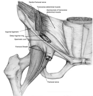
Eighteen embalmed male cadavers were dissected bilaterally (36 sides). Available demographic data included subject age, height, and weight from post-mortem data sheets. The dissections were performed using an ilioinguinal exposure according to Letournel and include the removal of the soft tissues anterior to the rectus sheath and external oblique aponeurosis (1, 11). The spermatic cord and the genital branch of the genitofemoral nerve as they exit the external (superficial) inguinal ring were dissected and identified [Figure 1a]. The ilioinguinal nerve which most often pierces through Figure 1a. Inguinal Anatomy Superficial Layer. The external oblique muscle and aponeurosis make up the most outer layer of the abdominal wall. The external (superficial) inguinal ring is the medial opening in this layer. The ilioinguinal nerve course is variable. It often pierces the external oblique aponeurosis cephalad to the superficial ring but sometimes exits through the external inguinal ring outside the spermatic cord. The genitofemoral nerve exits the superficial ring along with the spermatic cord.
Figure 1b. Inguinal Anatomy Middle Layer. The internal oblique muscle is the “floor” of the inguinal canal and lies directly below the external oblique aponeurosis. The three sensory nerves of the groin are seen in this layer – iliohypogastirc, ilioinguinal, and the genital branch of the genitofemoral.
INGUINAL ABNORMALITIES IN ACETABULAR FRACTURES THE ARCHIVES OF BONE AND JOINT SURGERY. ABJS.MUMS.AC.IR
VOLUME 3. NUMBER 4. OCTOBER 2015
)276(
the external oblique aponeurosis just cephalad to the superficial inguinal ring was exposed. The external oblique aponeurosis was incised in line with the inguinal ligament and retracted proximally and distally to open the inguinal canal [Figure 1b; 1c]. When any abnormality was noted, the incision was extended medially and distally through the external spermatic fascia to fully expose the contents of the spermatic cord. Spermatic cord diameter was measured uniformly, and any local masses were noted. Bowel hernias were identified as “direct” or “indirect” by dissecting the hernia sac and identifying their point of origin into the inguinal canal.
Clinical study
A retrospective review of prospectively gathered data at a Level I trauma center was performed to identify patients who underwent acetabular fracture operative reduction and then fixation over a consecutive three year period, utilizing an ilioinguinal approach. After obtaining IRB approval, the operative studies including
standardized pelvic computed tomography and the individual operative notes were reviewed to determine the incidence of inguinal canal and spermatic cord abnormalities requiring general surgical consultation. The associated general surgery operative notes were used to confirm the etiology of the abnormality and details of the surgical intervention. Radiographic characteristics of the spermatic cord abnormalities and/or hernias were noted from the scans.
Results
Cadaver Study
Average specimen age was 80 years old (range 22-100), average height of 169 cm (range 152-191) and average weight of 64 kg (range 34-114). Eleven of 36 (31%) had spermatic cord and/or inguinal canal abnormalities. This included one cadaver with bilateral indirect inguinal hernias, three cadavers with bilateral spermatic cord lipomas, and three cadavers with unilateral spermatic cord lipomas. The average cord diameter in
Table 1. Acetabular Fractures and Inguinal Abnormalities
Age Abnormality CT Findings Surgical Details
39 Indirect Hernia Not detected Hernia reduction, mesh reconstruction
56 Direct Hernia, Cord Lipoma Enlarged cord, homogeneous density Lipoma resection, mesh reconstruction
41 Cord Lipoma Enlarged cord, homogeneous density Lipoma resection, mesh reconstruction
49 Indirect Hernia Enlarged cord, heterogeneous density Hernia reduction, repair of inguinal canal
55 Direct Hernia, Cord Lipoma Enlarged cord, homogeneous density Lipoma resection, repair of inguinal canal
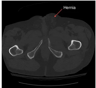
Figure 2a. Cord Lipoma. On the preoperative CT scan, the cord lipoma is hypodense and homogeneous, suggesting a fatty etiology. The affected cord has a notably larger diameter than that of the contralateral normal cord.
those with abnormalities was 24.9 mm (range 15 to 28). The average cord diameter in normal cords was 16 mm (range 11 to 22). The difference between normal and abnormal cord diameters was statistically significant utilizing a Student’s t-test (P=0.00001).
Clinical Study
Two hundred seventy-seven displaced acetabular fractures were identified over the three year period. Of the 120 fractures treated through an ilioinguinal exposure, 87 were males with an average age of 44 years (range 14 to 70). Five of these 87 male patients (5.7%) had a hernia and/or spermatic cord lesion requiring general surgical intervention under the same anesthesia through the same exposure [Table 1]. Two of the five had indirect inguinal hernias. After reduction of the indirect hernia, one of the patients underwent repair of the inguinal canal while the other required mesh reconstruction. One of the five patients had an isolated cord lipoma without associated bowel herniation. The lipoma was resected and the inguinal canal required mesh reconstruction. The remaining two patients had both direct inguinal hernias and cord lipomas. Both patients had resection of their lipomas. One patient had a primary repair of the inguinal canal while the other required a mesh augmentation.
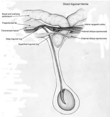
The preoperative pelvic CT scans of all five patients were reviewed in detail, and the abnormality was easily Figure 3. Indirect Inguinal Hernia. By definition, the hernia enters the inguinal canal through the internal (deep) inguinal ring, lateral to the epigastric vessels. The bowel protrudes through the internal ring, into the inguinal canal, and given time, may advance through the external (superficial) inguinal ring and into the scrotum.
identified on four of the five patients [Table 1]. All lipomas appeared as spermatic cord enlargements with homogeneous density. The homogeneous density on the CT scan was consistent with the intraoperative finding of fat within the abnormal spermatic cord [Figure 2a]. All hernias appeared as enlargements of the spermatic cord with a heterogeneous density. This was consistent with the intraoperative findings of a bowel sac and normal cord structures within the spermatic cord [Figure 2b].
The one instance not detected on the preoperative CT scan was well documented in the preoperative history. The 56 year-old male reported a history of inguinal hernia with episodes of incarceration which he manually reduced. The hernia was confirmed and repaired during the ilioinguinal approach.
Discussion
The surgeon performing an ilioinguinal approach must understand normal inguinal anatomy and be aware of abnormalities including spermatic cord lipomas and inguinal hernias as well as other potential soft tissue abnormalities. Both cord lipomas and hernias have been described in the general surgery literature (5-7, 9, 10, 12, 13). However, we are unaware of any reports describing these abnormalities in association with acetabular fractures and their operative management, particularly the anterior ilioinguinal surgical exposure fixation.
INGUINAL ABNORMALITIES IN ACETABULAR FRACTURES THE ARCHIVES OF BONE AND JOINT SURGERY. ABJS.MUMS.AC.IR
VOLUME 3. NUMBER 4. OCTOBER 2015
)278(
vessels, and inferiorly by the inguinal ligament. Indirect hernias enter the inguinal canal through the internal (deep) inguinal ring, lateral to the epigastric vessels (Remote from Hasselbach’s Triangle) [Figure 3]. Direct inguinal hernias enter the inguinal canal by breaching through a weakened area of the inguinal floor (transverses abdominus muscle), medial to the inferior epigastric vessels (within Hasselbach’s Triangle) [Figure 4] (4, 14). Both types may present as an inguinal or groin mass and/or bulge in the spermatic cord.
Spermatic cord lipomas may also present as enlargement of the spermatic cord. Although these abnormalities are loosely labeled “lipomas”, they are not true lipomas. Instead, cord lipomas are actually retroperitoneal fat that has entered the inguinal canal (9). The fat protrudes through the internal (deep) inguinal ring, outside the internal spermatic fascia and lateral to the cord. Therefore, by definition, they are “indirect”. They may occur in isolation or in association with a direct or indirect bowel hernia. It is important to note that their initial presentation may be indistinguishable from a bowel herniation. The spermatic cord must be opened and explored to identify the etiology of the mass.
Several previous studies report the occurrence and incidence of cord abnormalities (5, 8-10, 14). Heller found a discrete mass of fat (contiguous with the preperitoneal fat through the deep inguinal ring) within the inguinal canal in 75% (27 of 36) of male cadavers. Sixteen percent of these (6 cadavers) extended beyond the external (superficial) inguinal ring (7). Lilly found spermatic cord lipomas in 22.5% of patients undergoing inguinal hernia repairs (9). Bissada reported unusual causes of spermatic cord enlargement including three cases of sarcoma (5). These studies emphasize the significant incidence of hernias and cord lipomas in the adult male population.
The 31% incidence of spermatic cord and/or inguinal canal abnormalities in our cadaver study was higher than the 16% reported by Heller (7). While two of our twelve abnormalities consisted of indirect inguinal hernias, Heller observed no cases of inguinal hernia. Although the median age between the two groups was distinctly different (80 years vs. 56 years), prior clinical studies do not support age as a factor in the development of inguinal abnormalities (12, 13). Heller only noted abnormalities that extended beyond the external ring while we included all abnormalities with extension into the inguinal canal. This alone may explain the difference (16% vs. 31%) in our incidence of abnormalities.
The difference in incidence between our cadaveric study and clinical series is pronounced (31% vs. 5.7%). We examined the age, height, and weight for any difference between the two groups. The median age in our cadaver study was 80 years compared to 44 years in our clinical series. However as mentioned previously, Carbonell did not find age to be a causative factor in the development of inguinal abnormalities. Instead, he reported that patients with indirect inguinal hernias were significantly heavier, taller, and expended more physical effort than patients without abnormalities (12). A history suggesting strenuous physical activities or heavy manual labor was
not available from either the cadaver or clinical group. The most likely factor explaining the difference between our clinical and cadaveric study is the difference in our definition of “inguinal abnormalities” between the two groups. In our cadaveric study, we aggressively investigated minor asymmetries in cord diameter. This practice identified patients as having “inguinal abnormalities” when miniature lipomas and minor disruptions of the inguinal anatomy were present. However, this aggressive approach is not warranted in clinical practice, and the lower incidence of abnormalities in our clinical series may reflect this difference. In our clinical series, only inguinal abnormalities requiring general surgical intervention were included.
This study has a number of limitations inherent in a retrospective study evaluating prospective data. Most important, medical records and operatives reports were utilized to identify patients with inguinal abnormalities. Theoretically it is possible in a retrospective study that some patients with inguinal abnormalities were not discovered due to inadequate documentation. Furthermore, while cord diameter was recorded we did not record the size of the abnormalities found in the cadaveric or clinical setting. Therefore, caution should be used in drawing conclusions about the incidence of abnormalities. However the radiographic characterization of these abnormalities should not be effected by these limitations.
The clinical and cadaveric findings emphasize the importance of understanding inguinal abnormalities and the value of detecting them preoperatively. The orthopedic surgeon must ask patients with acetabular fractures about prior inguinal procedures and recent inguinal complaints. Physical exam may reveal an inguinal “bulge” and/or a mass protruding into the testes. The preoperative pelvic CT scans can be useful in detecting inguinal abnormalities. Cord lipomas are visualized as enlargements of the spermatic cord with homogeneous density. Hernias are readily visualized as enlarged spermatic cords with heterogeneous density. In summary, preoperative pelvic CT scans should be carefully studied for both osseous and inguinal abnormalities, which could be treated during the same anesthesia using the same exposure. When abnormalities are detected, the orthopedic surgeon must coordinate the operative plan with a general surgeon.
Acknowledgments
The authors would like to thank Samantha Kickingbird for her illustrations and Theresa Bergholz for editing assistance.
Reza Firoozabadi MD
Department of Orthopaedic Surgery and Sports Medicine, Harborview Medical Center, Seattle, WA USA
Paul Stafford MD
Orthopedic and Trauma Service of Oklahoma, Tulsa, OK USA
Milton Routt MD
References
8. Lilly MC, Arregui ME. Ultrasound of the inguinal floor for evaluation of hernias. Surg Endosc. 2002; 16(4):659-62.
9. Lilly MC, Arregui ME. Lipomas of the cord and round ligament. Ann Surg. 2002; 235(4):586-90.
10. Nasr A, Tormey S, Walsh TN. Lipoma of the cord and round ligament: an overlooked diagnosis? Hernia. 2005; 9(3):245-7.
11. Letournel E, Judet R. Fractures of the Acetabulum. 2nd ed. Berlin: Springer Science & Business Media; 1993.
12. Carbonell JF, Sanchez JL, Peris RT, Ivorra JC, Del Bano MJ, Sanchez CS, et al. Risk factors associated with inguinal hernias: a case control study. Eur J Surg. 1993; 159(9):481-6.
13. van Wessem KJ, Simons MP, Plaisier PW, Lange JF. The etiology of indirect hernias: congenital and/or acquired? Hernia. 2003; 7(2):76-9.
14. Irwin T, McCoubrey A. Adult groin hernias. Surgery. 2012; 30(2):290-5.
1. Letournel E. The treatment of acetabular fractures through the ilioinguinal approach. Clin Orthop Relat Res. 1993; 292(292):62-76.
2. Routt MJ, Simonian PT, Swiontkowski M. Stabilization of pelvic ring disruptions. Orthop Clin North Am. 1997; 28(3):369-88.
3. Routt ML Jr, Swiontkowski MF. Operative treatment of complex acetabular fractures. Combined anterior and posterior exposures during the same procedure. J Bone Joint Surg Am. 1990; 72(6):897-904.
4. Gilbert A. An anatomic and functional classification for the diagnosis and treatment of inguinal hernia. Am J Surg. 1989; 157(3):331-3.
5. Bissada NK, Redman JF. Unusual masses in the spermatic cord: report of six cases and review of the literature. South Med J. 1976; 69(11):1410-2. 6. Carilli S, Alper A, Emre A. Inguinal cord lipomas.
Hernia. 2004; 8(3):252-4.