193 Radiol Bras. 2018 Mai/Jun;51(3):193–199
Fonseca EKUN, Peixoto MR, Cavalcante Jr FA, Rahal Jr A, Francisco Neto MJ, Funari MBG. Ultrasound evaluation of inguinoscrotal pain: an imaging-based review for the ultrasonographer. Radiol Bras. 2018 Mai/Jun;51(3):193–199.
Abstract
Resumo
Emergencies involving the inguinal region and scrotum are common and can be caused by a plethora of different causes. In most cases, such conditions have nonspecific symptoms and are quite painful. Some inguinoscrotal conditions have high complication rates. Early and accurate diagnosis is therefore imperative. Ultrasound is the method of choice for the initial evaluation of this vast range of conditions, because it is a rapid, ionizing radiation-free, low-cost method. Despite the practicality and accuracy of the method, which make it ideal for use in emergency care, the examiner should be experienced and should be familiarized with the ultrasound findings of the most common inguinoscrotal diseases. On the basis of that knowledge, the examiner should also be able to make an accurate, direct, precise report, helping the emergency room physician make decisions regarding the proper (clinical or surgical) management of each case. Here, we review most of the inguinoscrotal conditions, focusing on the imaging findings and discussing the critical points for the appropriate characterization of each condition.
Keywords: Hernia, inguinal; Inguinal canal; Orchitis; Fournier gangrene; Ultrasonography.
As emergências envolvendo a região inguinal e o escroto são frequentes, derivadas de diferentes causas, e na maioria das vezes apresentam-se clinicamente de modo não específico e bastante dolorosas. Algumas destas condições apresentam elevado poten-cial de gravidade, sendo imperioso um diagnóstico rápido e preciso. A ultrassonografia é, indiscutivelmente, o método de escolha na avaliação inicial de todo o vasto leque de doenças nessas regiões, sendo rápido, de baixo custo e sem uso de radiação. Além da praticidade e acurácia do método, ideal para a prática em âmbito de pronto-atendimento, a experiência do examinador, o qual deve estar familiarizado com os principais achados de imagem, é fundamental para a precisão diagnóstica. Some-se a isto a neces-sidade de um relatório claro e assertivo, auxiliando o médico emergencista na terapêutica apropriada a cada caso, seja clínica ou cirúrgica. Procuramos trazer uma revisão baseada nos achados de imagem das principais afecções dolorosas inguinais e escrotais, discutindo os pontos-chaves para sua adequada caracterização.
Unitermos: Hérnia inguinal; Canal inguinal; Orquite; Gangrena de Fournier; Ultrassonografia.
Study conducted in the Imaging Department of the Hospital Israelita Albert Einstein, São Paulo, SP, Brazil.
1. MD, Resident in the Imaging Department of the Hospital Israelita Albert
Einstein, São Paulo, SP, Brazil.
2. MD, Radiologist in the Imaging Department of the Hospital Israelita Albert
Einstein, São Paulo, SP, Brazil.
3. MD, PhD, Radiologist and Coordinator of the Ultrasound Group in the Imaging
Department of the Hospital Israelita Albert Einstein, São Paulo, SP, Brazil.
4. MD, PhD, Radiologist and Coordinator of the Imaging Department of the
Hos-pital Israelita Albert Einstein, São Paulo, SP, Brazil.
Mailing address: Dr. Antonio Rahal Júnior. Departamento de Imagem – Hospital
Israelita Albert Einstein. Avenida Albert Einstein, 627, Morumbi. São Paulo, SP, Bra -zil, 05652-901. E-mail: antoniorahal@yahoo.com.br.
Received September 20, 2016. Accepted after revision November 3, 2016.
is essential for differentiating between more severe con-ditions and less severe ones, as well as for determining which patients can be released with medication and which ones must be hospitalized or require emergency surgical intervention. Making a rapid and accurate diagnosis can also allow early intervention for complications such as the progression from testicular torsion to permanent injury and atrophy.
The objective of this study was to discuss the tech-nique for ultrasound examination of the inguinal region and scrotum, as well as to identify the most common causes of inguinoscrotal pain seen in the emergency
room. We also list the specific imaging characteristics that
should be known to ultrasound examiners.
TECHNIQUE
The structures of the inguinal region are superficial
and can be evaluated well with linear transducers at 10 MHz or at lower frequencies (7 MHz) in more obese patients. INTRODUCTION
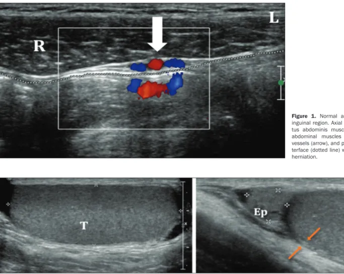
Figure 1 shows the normal anatomy of the inguinal region in the axial plane. The Valsalva maneuver is essential and a critical component of the examination of the region, al-lowing dynamic evaluation, especially in cases of suspected hernia, because hernias can disappear completely when
the patient is at rest, making them difficult to detect(1,2).
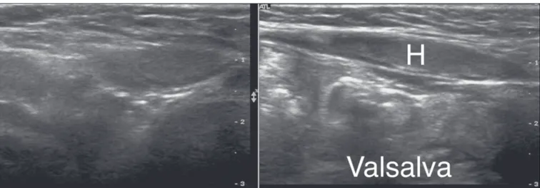
The normal aspect of the scrotum is shown in Figure 2.
IMAGING FINDINGS Hernias
Hernias occur at the weakest points in the abdominal wall, through which the vessels can push and the testes can
migrate. They are classified as indirect or direct, depending
on their origin in relation to the inferior epigastric vessels. Indirect inguinal hernias – Indirect inguinal hernias are seen protruding anteriorly toward the transducer, origi-nating from an area lateral to the inferior epigastric vessels (Figure 3). They are congenital and are more common in men, in whom their occurrence is due to the persistence of a patent processus vaginalis. In women, the cause is the delayed closure or non-closure of the canal of Nuck(1–3).
Figure 1. Normal anatomy of the inguinal region. Axial plane. The rec-tus abdominis muscle (R), oblique abdominal muscles (L), epigastric vessels (arrow), and peritoneal fat in-terface (dotted line) with no signs of herniation.
Figure 2. Normal anatomy of the scrotum. The testes (T) show homogeneous granular echogenicity and the testicular mediastinum is seen as a hyperechogenic linear band at the center of each testis. The tunica albuginea appears as a hyperechogenic line around the testicles (demarcated between the arrows), usually with small amount of anechoic fluid between its layers. The head of the epididymis (Ep) can be clearly seen in the sagittal plane, resting on the testicle and with similar echogenicity.
Direct inguinal hernias – Direct inguinal hernias originate medial to the inferior epigastric vessels, in Has-selbach’s triangle, which can be evaluated in images of the area superior to the inguinal canal (Figure 4). They are usually acquired, denoting weakness of the transverse fas-cia. Incarcerated and strangulated hernias are less com-mon because the width of their neck is usually greater than is their depth.
Femoral hernias – Femoral hernias also belong to the inguinal group of hernias, due to the proximity of their sites of presentation. More common in females, femoral hernias pass through the femoral canal, therefore being located medial to the femoral vein, and insinuate them-selves into the superomedial portion of the thigh(3).
The vast majority of hernias detected by ultrasound do not contain intestinal loops, comprising only adipose tissue. Incarcerated hernias are those that are not reduc-ible. Possible complications of the incarceration of intes-tinal loops in hernias include complete or partial obstruc-tion and strangulaobstruc-tion. Incarcerated hernias with vascular
frequently do not present vascularity on the Doppler flow
study, some cases showing parietal thickening and loss of peristalsis if they contain intestinal loops(1,2). In such
cases, emergency surgery is indicated.
Spermatic cord torsion
Cases of acute scrotum should, until proven other-wise, be considered attributable to spermatic cord torsion. Such torsion, which can be extravaginal or intravaginal, ac-counts for a third of all cases of acute scrotum.
Extravagi-nal torsion affects newborns in the first days of life, during the final stage of fixation of the testis. Intravaginal torsion
is more common, occurring in older children and adults, its incidence peaking at puberty. Failure of the tunica
albu-ginea during fixation results in the so-called “bell clapper”
deformity, which often occurs in both testes, requiring bi-lateral orchidopexy to avoid contrabi-lateral torsion.
The testis can be elevated and fixed, with the epididy -mis in a medial position. Ultrasound in B mode can reveal entanglement of the spermatic cord, similar to a whirl. The persistence of testicular echogenicity indicates that the tes-tis remains viable, and it is imperative that the surgery be
performed immediately. Color Doppler ultrasound of the
scrotum confirms the diagnosis of torsion. In cases of tor -sion that is partial (less than 360°), spectral Doppler ultra-sound can be of great value in identifying lower diastolic
flow in the affected testis than in the contralateral testis, denoting resistance to blood flow in the former. In addition
to being minimally invasive and affordable, spectral Dop-pler ultrasound shows the anatomy of the spermatic cord
and the blood flow, which in cases of twisting is reduced or
absent(4), as shown in Figure 5.
Torsion of the appendix testis
The appendix testis can also be the cause of acute testicular pain, mimicking testicular torsion. The charac-teristic blue dot sign is seen in only 20% of patients with torsion of the appendix testis, and ultrasound is funda-mental in making the differential diagnosis. When the ap-pendix testis is twisted, it will appear enlarged, with vari-able echogenicity, and asymmetry with the contralateral appendix testis is a good comparative parameter.
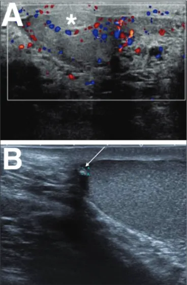
A Doppler flow study will show little or no vascularity
in the twisted appendix, with possible hyperemia of
adja-Figure 3. Indirect inguinal hernia. Hernia sac (H) penetrating the scro-tum through the inguinal canal. (T, testis).
cent tissues. More importantly, there will be no changes in the ipsilateral testis (Figure 6), which rules out the dif-ferential diagnosis of spermatic cord torsion(5). Making
the differential diagnosis with spermatic cord torsion is an important part of the management of these cases: the torsion of the appendix testis can be managed with symp-tomatic treatment, whereas spermatic cord torsion is a urological emergency, requiring immediate surgery to save the affected testis. In chronic cases, the appendix testis
can detach and calcify, generating a freely mobile calcified
body known as a scrotolith or scrotal pearl.
Epididymitis
The most common cause of acute scrotal pain in men is epididymitis, the incidence of which peaks between the
fifth and sixth decades of life. The pain is initially insidi -ous and increases after 24–48 h. Epididymitis often re-sults from an infection in the lower urinary tract, the most common etiological agent being Escherichia coli. In young men, epididymitis is typically a sexually acquired disease occurring from the second to the fourth decades of life, the main causative pathogens being Chlamydia trachoma -tis and Neisseria gonorrhoeae.
In acute epididymitis, ultrasound shows thickening and enlargement of the epididymis, initially affecting the caudal portion but potentially affecting the entire organ. The echogenicity is often diminished, and the echotex-ture is heterogeneous. Reactive hydrocele and cutaneous thickening are common. The effects can extend to the tes-tis, which will produce a hypoechoic area with increased
vascularity on a Doppler flow study. In some cases, notably
in patients with the mumps, orchitis can occur in the ab-sence of epididymitis.
In cases of uncomplicated orchitis, spectral Doppler
ultrasound can reveal increased diastolic flow. If orchitis
is not treated promptly, the entire testis can be affected, becoming hypoechoic and enlarged. Testicular edema can
cause a secondary increase in pressure, with a consequent increase in the risk of venous infarction and hemorrhage.
In such cases, a reduction in the diastolic flow seen on
spectral Doppler ultrasound serves as a warning sign,
de-noting resistance to venous blood flow and the possibility
of testicular infarction. In addition to infarction, the com-plications of epididymitis include abscess and pyocele(4),
as depicted in Figures 7 and 8.
Thrombosis of the pampiniform plexus
Spontaneous thrombosis of the pampiniform plexus is
rare and difficult to diagnose, with a clinical profile simi -lar to those of other causes of scrotal pain. It is typically associated with intense physical exertion, which leads to an increase in intra-abdominal pressure and a reduction in
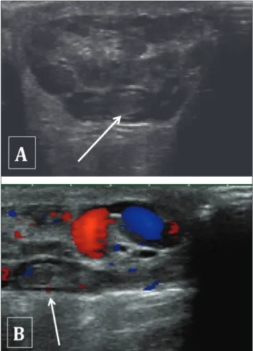
the venous return. The ultrasound findings are similar to
those of varicocele—that is, the vessels of the pampiniform plexus are dilated, with a caliber > 3 mm—although with echoic material characteristic of intraluminal thrombi(6), as
can be seen in Figure 9.
Figure 5. Hypoechoic, heterogeneous, rounded testis. Absence of intratesticu-lar flow on the Doppler flow study.
Figure 6. A: Torsion of the appendix testis. Note the enlargement of the appen-dix testis (asterisk), showing no flow on the Doppler flow study. The adjacent testis presents only reactive hyperemia, without alteration of its echotexture.
Trauma
Another common cause of acute testicular pain is
trauma. Although the clinical history is often sufficient for
making the diagnosis, ultrasound plays a central role in the evaluation of suspected testicular rupture and is indi-cated if there is a break in the continuity of the testicular surface, which is demarcated by the tunica albuginea. In all cases of blunt force testicular trauma, ultrasound is indicated(7). If the tunica albuginea is not intact, surgery
should be performed promptly in order to save the organ. The same applies if there is extruded material or loss of testicular homogeneity(8,9), as depicted in Figure 10.
Ul-trasound is also crucial in the follow-up of cases of trauma with hematocele, because blood collections can lead to a pressure increase within the tunica, generating ischemic injury and subsequent testicular atrophy(7,10), as well as
increasing the risk of subsequent infection.
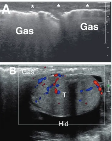
Fournier’s gangrene
Necrotizing infection of the fasciae of the perineum, commonly with gas-producing organisms, is known as Fournier’s gangrene. Ultrasound can reveal involvement of the soft tissues of the scrotum, showing hyperechoic
Figure 9. Partial thrombosis of the pampiniform plexus. Note the hypoechoic material in the vascular lumen and the absence of vascularization on the Dop-pler flow study (arrows).
Figure 7. Heterogeneous testis and epididymis, both showing enlargement and hypervascularity on the Doppler flow study. Note the small reactive hydrocele (arrow). (Ep, epididymis; LT, left testis; L, left).
L
areas with acoustic shadowing indicative of gas. Although the blood supply to the walls and adjacent soft tissues of the scrotum is provided by branches of the pudendal arteries, the testes are supplied by direct branches of the aorta, explaining the fact that Fournier’s gangrene often spares the testes(11,12). When there is testicular
involve-ment, the infectious focus is usually retroperitoneal or intra-abdominal(11), as shown in Figure 11.
borders, with or without a surrounding halo. The interior of such an abscess is necrotic, with no vascularity seen on
Doppler flow studies, whereas the periphery shows hyper -vascularity(10), as can be seen in Figure 12.
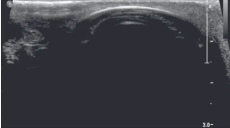
Testicular prostheses
Testicular prostheses appear as anechoic ovoid struc-tures (Figure 13), which should be recognized by the ul-trasound technician in order to avoid possible confusion.
CONCLUSION
Ultrasound is the method of choice for the initial evalu-ation of the inguinal region, its main advantages being its low cost, good accuracy, and wide availability. It is impor-tant to use B mode ultrasound imaging, together with color and spectral Doppler ultrasound, and to make a comparison with the contralateral side, the correlation with the clinical
Figure 10. Testicular trauma. Post-trauma ultrasound of the testis showing a fracture line in the tunica albuginea (arrows). Note the diffusely heterogeneous echotexture, with hypoechoic foci of permeation, indicative of intratesticular hematoma.
Figure 11. A: Pronounced thickening and heterogeneity of the scrotal wall and soft tissues adjacent to the testes (asterisks) extending to the perineum and containing areas of gas. The transition between the normal and thickened sub-cutaneous tissue is clearly seen in the image of the left testis. B: The testis (T) was preserved, with only a small reactive hydrocele (Hid).
Figure 12. Markedly thickened and hyperechoic scrotal wall, indicating an in-flammatory/infectious process, with a heterogeneous liquid collection (prob -able abscess) near the base of the penis.
MIDDLE LINE
history and physical examination findings being fundamen -tal. The examiner should be familiar with the imaging as-pects of the different conditions affecting this region, mak-ing an accurate, concise report, facilitatmak-ing the decision-making process and consequently increasing the chance of therapeutic success.
REFERENCES
1. Shadbolt CL, Heinze SB, Dietrich RB. Imaging of groin masses: in-guinal anatomy and pathologic conditions revisited. Radiographics. 2001;21 Spec No:S261–71.
Ultrasound Med. 2007;26:1649–55.
7. Resende DAQP, Souza LRMF, Monteiro IO, et al. Scrotal collec -tions: pictorial essay correlating sonographic with magnetic
reso-nance imaging findings. Radiol Bras. 2014;47:43–8.
8. Bhatt S, Dogra VS. Role of US in testicular and scrotal trauma. Radiographics. 2008;28:1617–30.
9. Morey AF, Brandes S, Dugi DD 3rd, et al. Urotrauma: AUA guide-line. J Urol. 2014;192:327–35.
10. Vital RJ, Mattos LA, Souza LRMF, et al. Sonographic findings in
non-neoplastic testicular lesions. Radiol Bras. 2007;40:61–7. 11. Levenson RB, Singh AK, Novelline RA. Fournier gangrene: role of
imaging. Radiographics. 2008;28:519–28.
12. Grayson DE, Abbott RM, Levy AD, et al. Emphysematous infec-tions of the abdomen and pelvis: a pictorial review. Radiographics. 2002;22:543–61.
13. Dogra VS, Gottlieb RH, Oka M, et al. Sonography of the scrotum.
Radiology. 2003;227:18–36.
Figure 13. Some patients undergoing orchiectomy may opt for testicular pros -thesis placement. Although the existence of a testicular pros-thesis can be dis-covered during the anamnesis, that information is not always available. There-fore, the ultrasound technician must be able to recognize such prostheses, which present as anechoic ovoid structures.