Campus de Araçatuba
PAULA FERNANDA KRELING DOMINGUES
Avaliação do potencial antimicrobiano e citotoxicidade de
fragmentos peptídicos catiônicos isolados ou combinados na
prevenção da cárie dentária
Araçatuba - SP 2016
Campus de Araçatuba
PAULA FERNANDA KRELING DOMINGUES
Avaliação do potencial antimicrobiano e citotoxicidade de
fragmentos peptídicos catiônicos isolados ou combinados na
prevenção da cárie dentária
Tese apresentado à Faculdade de Odontologia da Universidade Estadual Paulista “Júlio de Mesquita Filho”, Campus de Araçatuba, como parte dos requisitos para a obtenção de título de Doutor em Ciência Odontológica - Área de Concentração: Saúde Bucal da Criança.
Orientadora: Profa. Dra. Cristiane Duque
Coorientador: Prof. Titular Célio Percinoto
Araçatuba - SP 2016
Catalogação-na-Publicação
Diretoria Técnica de Biblioteca e Documentação – FOA / UNESP
Domingues, Paula Fernanda Kreling.
D671a Avaliação do potencial antimicrobiano e citotoxicidade de fragmentos peptídicos catiônicos isolados ou combinados na prevenção da cárie dentária / Paula Fernanda Kreling Domin- gues - Araçatuba, 2016
72 f. : il. ; tab. + 1 CD-ROM
Tese (Doutorado) – Universidade Estadual Paulista, Faculdade de Odontologia de Araçatuba
Orientadora: Profa. Cristiane Duque Coorientador: Prof. Célio Percinoto
1. Cárie dentária 2. Peptídeos 3. Testes de sensibilidade microbiana 4. Células cultivadas I. Título
Black D27
Paula Fernanda Kreling Domingues
Nascimento
11.02.1984
Londrina
PR
Filiação
Luiz Valdemar Kreling
Maria Cristina Beffa Kreling
2004
2008
Curso de Graduação em Odontologia pela Universidade Estadual
de Londrina – UEL
2009 – 2011
Curso de residência em Odontologia para Bebês – Bebê
clínica/Núcleo de Odontologia para Bebês – Universidade
Estadual de Londrina – UEL
2011
2013
Curso de pós
graduação em Odontologia Clínica – Nível de
mestrado pela – Universidade Estadual de Londrina – UEL
2013 – 2016
Curso de pós
graduação em Ciência Odontológica – Área de
Concentração: Saúde Bucal da Criança
Nível de dutorado pela
Faculdade de Odontologia de Araçatuba
UNESP
Associações
CROPR – Conselho Regional de Odontologia do Paraná
SBPqO
Sociedade Brasileira de Pesquisa Odontológica
À Deus
Meu criador
Sempre em primeiro lugar
À minha família
Meu marido e filho, amores da minha vida
Aos meus queridos pais
À Deus
Pela Sua presença em minha vida;
Por proteger meus caminhos e minha família;
Pelo Seu amor e misericórdia perante os meus erros;
“Guarda
me, ó Deus, pois em ti tenho segurança! Eu disse a Deus, o SENHOR: Tú és o meu
Senhor, tudo o que tenho de bom vem de ti.” Salmo 16, 1:2
Ao meu marido Vagner
Pelo amor, respeito, apoio, amizade. Pela paciência, por ter sido tão generoso nesses anos de
idas e vindas, tendo respeitado minha ausência em casa para cumprimento deste trabalho.
Por interromper seus projetos pessoais para viver um sonho meu e se aventurar comigo num
pais estrangeiro, num frio intenso e nas dificuldades que passamos.
Eu não teria conseguido sem seu apoio e confiança.
Amo
te para sempre.
Ao meu filho Antônio
Meu melhor projeto;
Minha vida;
Meu tudo;
Amor de nossas vidas, amamos você, filho.
Aos meus pais Luiz e Cristina
Por nos ensinar os mais importantes valores de vida, nos tornando pessoas honestas,
trabalhadoras e respeitosas.
Por terem aberto mão de tantos sonhos para que tivessemos os melhores estudos.
Obrigada por tudo.
Aos meus irmãos Thaís e Aluisio
Por me apoiarem em minhas decisões, por estarem sempre ao meu lado.
Aos meus sogros Sérgio e Edney
Pelo amor e carinho;
Por terem me dado meu melhor presente;
Por me apoiarem sempre.
Aos meus cunhados e cunhadas
Pelos momentos de alegria;
Por serem tão presentes e cuidadosos.
Aos meus queridos sobrinhos
Vocês são a alegria em nossas vidas.
Amo todos vocês.
À Faculdade de Odontologia de
Araçatuba - Unesp
À minha orientadora
Prof. Dra. Cristiane Duque
Por ter me aceito e acreditado em meu potencial. Por toda paciência e entendimento das
minhas voltas pra casa. Por ter me dado a chance e se empenhado para que eu pudesse
estudar fora do país. Por toda dedicação, tempo investido em seus alunos, sempre pronta a
nos ajudar. Obrigada.
Ao meu coorientador
Prof. Dr. Célio Percinoto
Pelos ensinamentos clínicos, conselhos e disponibilidade sempre que precisei.
À minha amiga Loiane
Pela amizade, acolhimento, por sua dedicação.
Minha amiga, você sabe que não teria conseguido sem você!
À minha amiga Kelly
Companheira de viajem e de moradia;
Sua presença trouxe ânimo para finalizar meus trabalhos.
Aos meus amigos Paranaenses
À todos os meus colegas de
Pós-Graduação
Pela amizade, convívio e experiências trocadas;
Pela ajuda recebida.
Aos docentes da disciplina de Saúde
Bucal da Criança da FOA - Unesp
Prof. Dr. Alberto Carlos Botazzo Delbem, Prof. Dr. Célio Percinoto, Profª. Drª. Cristiane
Duque, Prof. Dr. Juliano Pelim Pessan, Prof. Dr. Robson Frederico Cunha, Profª. Drª. Sandra
M. H C. Avila de Aguiar
Pelos ensinamentos transmitidos e contribuição na minha formação profissional.
Ao curso de pós-graduação em Ciência
Odontológica da FOA
Na pessoa do coordenador Prof. Dr. Alberto Carlos Botazzo Delbem.
Aos funcionários do Departamento de
Odontopediatria
Ricardo, Mário e Luisinho.
Aos funcionários da biblioteca da FOA
Ana Cláudia, Luzia, Ivone, Cláudio, Maria Cláudia, Luiz, Denise e Izamar.
Pela atenção e disponibilidade.
Aos funcionários da sessão de Pós
Graduação da FOA
Valéria, Cristiane e Lilian.
Pelo profissionalismo e disponibilidade.
À coordenação de aperfeiçoamento de
pessoal de nível superior – Capes
Pela concessão da bolsa de estudo.
À Fundação de Amparo e Pesquisa do
Estado de São Paulo – Fapesp
Pela concessão da bolsa de estudo.
Aos professores da Universidade Estadual
de Londrina
Pela base ensinada a mim.
Pelo empenho e dedicação para que pudesse chegar até aqui.
KRELING, PF. Avaliação do potencial antimicrobiano e citotoxicidade de fragmentos peptídicos catiônicos isolados ou combinados na prevenção da cárie dentária. 2016. 74p. Tese (Doutorado em Ciência Odontológica, Área de Saúde Bucal da Criança), Faculdade de Odontologia de Araçatuba, Universidade Estadual Paulista “Júlio de Mesquita Filho”, Araçatuba, 2016.
RESUMO
O sistema imune tem diversas formas de defesa contra microrganismos patogênicos. As membranas da mucosa são uma fonte de peptídeos catiônicos antimicrobianos contra uma ampla variedade de bactérias, fungos e vírus encapsulados. O objetivo deste estudo foi avaliar a citotoxicidade e atividade antimicrobiana em condições planctônicas e de biofilme de fragmentos derivados de peptídeos catiônicos (PC): LL-37 (originário de hCAP-18), D6-17 e D1-23 (originários de ortólogo da β-defensina-3 humana) contra bactérias cariogênicas. Para análise citotóxica, duas linhagens de células epiteliais foram expostas a diluições seriadas de fragmentos de PC. Ensaios de MTT e coloração de DAPI foram realizados para avaliar o metabolismo e a morfologia celular, respectivamente. A concentração inibitória mínima (CIM) e a concentração bactericida mínima (CBM) foram determinadas para fragmentos de PC e controle (digluconato de clorexidina - CHX) contra Streptococcus mutans (Sm), S. mitis, S. oralis, S. salivarius, S. sanguinis, Lactobacillus acidophilus, L. casei, L. rhamnosus, L. brevis, L.
Actinomyces. D6-17 mostrou atividade bactericida apenas contra S. mutans, L. brevis e
L. fermentum. Combinações de fragmentos de PC não mostraram efeito sinérgico contra S. mutans. D1-23 (10x CBM) apresentou atividade contra biofilme de S. mutans
superior a CHX. A microscopia confocal mostrou alta taxa de células mortas em relação a células vivas para D1-23 e CHX quando comparado ao grupo controle (meio de cultura). D1-23 também diminuiu a espessura do biofilme em relação ao grupo controle. Conclui-se que D1-23 mostrou relevante atividade antimicrobiana/antibiofilme contra bactérias cariogênicas e baixa toxicidade em células epiteliais.
KRELING, PF. Cytotoxicity and microbiological effect of cationic peptide fragments isolated or combined for dental caries prevention. 2016. 74p. Tese (Doutorado em Ciência Odontológica, Área de Saúde Bucal da Criança), Faculdade de Odontologia de Araçatuba, Universidade Estadual Paulista “Júlio de Mesquita Filho”, Araçatuba, 2016.
ABSTRACT
The immune system has several forms of defense against pathogenic microorganisms. The mucous membranes are a source of potent antimicrobial cationic peptides against a broad range of bacteria, fungi and enveloped viruses. The aim of study was evaluated the cytotoxicity and antimicrobial activity under planktonic and biofilm conditions of fragments derived from cationic peptides (CP): LL-37 (from hCAP-18), D6-17 and D1-23 (from β-defensin-3 derivative) against cariogenic bacteria. For cytotoxicity analysis, two lines of epithelial cells were exposed to serial dilutions of the CP fragments. MTT assays and DAPI staining were performed to evaluate cell metabolism and morphology, respectively. Minimum inhibitory concentration (MIC) and minimal bactericidal concentration (MBC) were determined for the CP fragments and control (Chlorhexidine digluconate-CHX) against Streptococcus mutans (Sm), S. mitis, S. oralis, S. salivarius, S. sanguinis, Lactobacillus acidophilus, L. casei, L. rhamnosus, L. brevis, L.
fermentum and Actinomyces israelii. Fractional inhibitory concentration (FIC) was
obtained for the combinations of CP fragments on S. mutans. Biofilm assays were
conducted with CHX and the best antimicrobial CP fragment against S. mutans strains.
Confocal Laser Scanning Microscopy (CLSM) was used to analyze the Live/Dead cells and biofilm thickness. The results indicated that D6-17 did not affect the metabolism of either cell line. D1-23, LL-37 and CHX were not toxic for both cells, in concentrations below 0.2, 0.02 and 0.01mM, respectively. DAPI-staining cells demonstrated nuclei fragmentation for LL-37 group and cells with the aspect of apoptosis in the CHX group. Cells treated with D1-23 and D6-17 presented morphology similar to the control group (culture medium). D1-23 presented the best bactericidal activity against S. mutans, S. mitis and S. salivarius. LL-37 had a better effect against Lactobacillus and Actinomyces
compared to CHX. CLSM analysis showed that D1-23 and CHX groups presented higher quantification of dead cells when compared to control (culture medium). The biofilm thickness were lower in the D1-23 group compared to CHX and control groups. We concluded that D1-23 showed a remarkable antimicrobial/anti-biofilm effect against cariogenic bacteria and low toxicity for epithelial cells.
Lista de Figuras
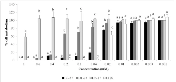
Figure 1. Mean (bars = standard deviation) of percentage of HaCat cell metabolism (MTT assay) after CHX and peptide fragments exposure.
Note: a Different lower letters show statistical difference among the groups,
considering each concentration separately, according to ANOVA and Tukey tests.
Figure 2. Mean (bars = standard deviation) of percentage of OBA-9 cell metabolism (MTT assay) after CHX and peptide fragments exposure.
Note: a Different lower letters show statistical difference among the groups,
considering each concentration separately, according to ANOVA and Tukey tests.
Figure 3. Fluorescence microscopic images (x200) of HaCaT cells treated with peptide fragments (LL-37, D1-23 and D6-17) and CHX for 24h. Red narrows shows higher intensity DAPI-staining cells and the presence of fragmented nuclei observed for LL-37 group. White narrows shows cells with aspect of apoptosis in the CHX group. Control = culture medium (DMEM).
Figure 4. Box-whisker plots of the activity of D1-23 (2X MBC and 10X MBC) and CHX (2xMBC and 10xMBC) against biofilm of S. mutans ATCC and clinical strains (CS1 and CS2). Different capital letters show statistical difference among the groups, considering each strain separately, according to Mann-Whitney tests.
Note: Bars indicate minimum and maximum values. Boxes indicate lower and upper quartiles. Line in the middle of boxes is median.
Figure 5. Confocal microscopy (CLSM) of S. mutans biofilms on enamel blocks after
exposure to D1-23 and CHX. A. Representative images (x63 immersion) of dead cells and live cells and merge images of S. mutans biofilms. D1-23 and CHX showed higher quantification of dead cells (red points) when compared to control (no treatment) which presented strong predominance of live cells (green points). B. Means (bars-standard deviations) of percentage of dead cells obtained after CLSM analysis of S. mutans biofilm. Note: aDifferent lower case letter show statistical differences among
the groups, according to ANOVA and Tukey tests (p<0.05).
Figure 6. Biofilm thickness. A. Representative 3D images obtained by CLSM of S. mutans biofilms after D1-23 and CHX exposure for 24h. B. Means (bars-standard deviations) of biofilm thickness obtained after CLSM analysis of S. mutans biofilm. Note: aDifferent lower case letter show statistical differences among the groups,
Lista de Tabelas
Table 1. Values of MIC and (MBC)† in mM obtained by cationic peptide fragments
against oral bacteria.
Note: † MIC – Minimum Inhibitory Concentration; MBC – Minimal Bactericidal Concentration
Table 2. Values of FIC obtained by combinations of fragments of cationic peptides against S. mutans (ATCC 25175).
Sumário
Introdução Geral ... 18
Artigo ... 23 Abstract ... 25 1. Introduction... 26 2. Materials and methods ... 27 3. Results ... 33 4. Discussion ... 35 5. Conclusion ... 38 References ... 39 Tables and Figures ... 44
Referências Bibliográficas Gerais ... 53
Anexos ... 58 Anexo A ... 59
19
A cárie é uma doença infecciosa, causada por ácidos provenientes da fermentação microbiana dos carboidratos da dieta que, com o tempo, causam a desmineralização dos tecidos duros do dente (Fejerskov e Kidd, 2005). Apesar de haver um declínio de sua prevalência no mundo, a cárie dentária continua sendo a doença infecciosa crônica mais comum em crianças, tanto em países em desenvolvimento, quanto nos desenvolvidos (Vadiakas, 2008; Colak et al., 2013). Se não tratada, pode trazer diversos problemas para as crianças, incluido dor (Lygidakis et al., 1998), diminuição na alimentação, maloclusão e até mesmo problemas em seu desenvolvimento (Isong et al., 2014). Quando atinge crianças muito jovens, a partir da erupção dos primeiros dentes decíduos é denominada cárie precoce da infância – CPI (Drury et al., 1999). No Brasil, a CPI apresenta-se como grave problema de saúde pública, com prevalência de 26,5% em crianças com 5 anos de idade (SBBrasil, 2010). Para facilitar o diagnóstico clínico e a padronização terminológica, foi estabelecido que a CPI é caracterizada pela presença de uma ou mais superfícies dentárias cariadas, cavitadas ou não, perdidas ou restauradas, em menores de 6 anos (American Academic of Pediatric Dentistry, 2009). Devido à rápida destruição dentária em curto período de tempo, tem sido sugerida a associação entre os seguintes fatores na etiologia da CPI: ingestão frequente de dieta rica em carboidratos fermentáveis como a sacarose, altas contagens de estreptococos mutans e maior vulnerabilidade imunológica da criança (Mattos-Graner et al., 1998; Mohebbi et al., 2008). Por isso, medidas preventivas e buscas por alternativas que venham amenizar esse quadro são fundamentais para a saúde e bem estar das crianças (Isong et al., 2014).
O grupo bacteriano considerado mais cariogênico é o dos estreptococos mutans (SM), especialmente Streptococcus mutans (Mattos-Graner et al., 2001; van
Houte et al., 1991). Embora a associação entre SM e CPI pareça convincente, grande percentual das crianças colonizadas por essa espécie bacteriana não manifestam a doença (Mattos-Graner et al., 1998; Ramos-Gomes et al., 2002). Estudos têm mostrado que características genotípicas e fenotípicas do S. mutans podem determinar o
estabelecimento da espécie no biofilme dental e sua cariogenicidade (Duque et al. 2009; Saxena et al., 2008; Lemos et al., 2005). Assim, outras espécies acidogênicas e acidúricas, incluindo estreptococos não mutans, Actinomyces estão envolvidas com o
20
Nyvad (2011) concluíram que muitos microrganismos são igualmente acidogênicos e acidúricos, incluindo espécies de Actinomyces e Lactobacillus, ambos com importante papel no desenvolvimento do processo carioso. Lactobacillus estão principalmente envolvidos na progressão da cárie dentária, uma vez que tem sido encontrado em lesões cariosas avançadas em dentina (Becker et al., 2002). Actinomyces é considerado um colonizador inicial, com importante papel no desenvolvimento do biofilme dental (Arai et al., 2015) contribuindo para a colonização de outras espécies bacterianas (Palmer et al., 2003), além disso, também está ligado ao aparecimento da lesão de cárie na superfície radicular (van Houte et al., 1994).
Para auxiliar na defesa contra microbiota patogênica o sistema imunológico humano conta com diversas formas de defesa. As mucosas, além de apresentarem a função de barreira física contra a entrada de organismos estranhos, são fontes de potentes peptídeos catiônicos antimicrobianos (PCAM). Estes apresentam ação contra uma ampla variedade de bactérias, fungos e vírus encapsulados e promovem modulação da resposta imune do hospedeiro, mantendo a microbiota normal em estado estável em diferentes nichos, como a pele, os intestinos e a cavidade bucal (Mccormick e Weinberg, 2010; Wiesner e Vilcinskas, 2010). Entre os principais PCAM presentes na saliva e também no fluido crevicular estão as defensinas e catelicidinas.
As defensinas são peptídeos pequenos, de 15 a 45 aminoácidos, que dependendo do padrão de pareamento de seus resíduos de cisteína, são subdivididas em duas principais subfamílias: α e β-defensinas, ambas apresentam função imunomoduladora, modificando a migração e maturação celular, induzindo citocinas e a liberação de histamina e prostaglandina A2 de mastócitos (Abiko et al., 2003; Mccormick e Weinberg, 2010). Além disso, esses peptídeos vêm demonstrando potente atividade antimicrobiana contra um amplo espectro de patógenos, incluindo os bucais (Ouhara et al., 2005). As α-defensinas são produzidas principalmente pelos neutrófilos, peptídeo neutrofílico humano (HNP-1 a 4), e células de Paneth, defensinas humanas 5 e 6 (HD5 and HD6) (Jones e Bevin, 1992; Chairatana et al., 2016; Nakamura et al., 2016). As β-defensinas (hBDs-1 a 3) são produzidas predominantemente no epitélio (Dale e Fredericks, 2005), superfície da mucosa e trato reprodutivo, têm demostrado um forte espectro antimicrobiano in vitro, além de ser quimioatrativos
21
2014). Reynolds et al. (2010) avaliaram a atividade antibacteriana de fragmentos de β -defensina 3 e encontraram que a metade N-terminal do aminoácido 23 (D1-23) do Defb14-1CV (ortólogo de rato da β-defensina 3 humana) é um potente agente antimicrobiano.
A catelicidina (hCAP-18) é um peptídeo catiônico α-helical sem cisteína (Bals; Wilson, 2003). É o único desse gênero encontrado em seres humanos e foi primeiramente identificado através do isolamento de granulos de neutrólilos. hCAP-18 é produzida também por células epiteliais do pulmão, intestino, cavidade bucal e trato urogenital, sendo encontrada no plasma seminal e plasma sanguíneo. Após a secreção, ocorre a quebra de hCAP-18 pela ação de proteases em um peptídeo de cadeia longa LL-37, além de outros peptídeos menores. LL-37 é peptídeo catiônico com 37 resíduos, componente α-helicoidal do hCAP-18 (Low et al., 1999). É um modulador do sistema imuno inato, envolvendo funções como antividade antibacteriana, incluindo cepas bucais do grupo estreptococos (S. mutans, S. sobrinus, S. mitis e outras) (Ouhara et al., 2005). A função antibacteriana do LL-37 tem sido atribuída a sua capacidade de formar poros na membrana, além de, em altas concentrações, é citotóxica para células eucariotas (de Yang et al., 2000; Scott et al., 2002; Yang et al., 2004).
Os PCAM na saliva podem contribuir para a manutenção da saúde bucal geral e ter um papel junto à primeira linha do organismo contra as infecções (Mccormick e Weinberg, 2010; Wiesner e Vilcinskas, 2010; Zhang; Cherryholmes e Shively, 2008) uma vez que têem sido considerados contribuidores na saúde da mucosa oral, o que, provavelmente pode ser um fator biológico que influencia a suscetibilidade cariogênica (Dale et al., 2006). Alguns estudos relacionaram a presença de PCAM e CPI (Tao et al., 2005; Davidopoulou et al., 2012; Colombo et al., 2016). Tao et al., 2005 observaram
22
antimicrobiana desses peptídeos sobre microrganismos cariogênicos, estudo verificou que cepas de S. mutans isoladas de crianças com cárie ativa mostraram maior resistência a HNP-1-2, HBD-2-3 e LL-37 em concentrações variadas quando comparadas às crianças livres de cárie, demonstrando que essas cepas apresentam uma vantagem ecológica para a colonização mais efetiva do biofilme, aumentando o risco à doença cárie (Phattarataratip et al., 2011). Colombo et al. (2016) encontraram correlações positivas entre hBD-2, hBD-3 e LL-37, e entre esses, LL-37 foi o mais associado aos níveis de cárie. Assim, os PCAM poderiam ser uma alternativa aos antimicrobianos convencionais por serem alvos seletivos de células procariotas, diminundo a probabilidade de resistência microbiana (Zasloff, 2009).
24
Cytotoxicity and effect of cationic peptide fragments against cariogenic bacteria under planktonic and biofilm conditions
Paula Fernanda Krelinga, Kelly Limi Aidaa, Loiane Massunarib, Karina Sampaio Caiaffab,
Célio Percinotoa, Telma Blanca Lombardo Bedranc, Denise Madalena Palomari
Spolidorioc, Eduardo Maffud Cillid, Cristiane Duquea*
aDepartment of Pediatric Dentistry and Public Health, Araçatuba Dental School, UNESP
- Univ Estadual Paulista, Araçatuba, São Paulo, Brazil
bDepartment of Endodontics, Araçatuba Dental School, UNESP - Univ Estadual Paulista,
Araçatuba, São Paulo, Brazil
cDepartment of Physiology and Pathology, Araraquara Dental School, UNESP - Univ
Estadual Paulista, Araraquara, São Paulo, Brazil
dDepartment of Biochemistry and Chemical Technology, Institute of Chemistry, UNESP
- Univ Estadual Paulista, Araraquara, São Paulo, Brazil
*Corresponding author: Cristiane Duque
Department of Pediatric Dentistry and Public Health, Araçatuba Dental School, UNESP - Univ Estadual Paulista
Address: R. José Bonifácio, 1193, CEP: 16015-050, Araçatuba-SP, Brazil Tel: (+55) 1836363315
E-mail: cristianeduque@yahoo.com.br, cduque@foa.unesp.br
25
Abstract
This study evaluated the cytotoxicity and effect of fragments derived from oral cationic peptides (CP): LL-37, D6-17 and D1-23 against cariogenic bacteria under planktonic and biofilm conditions. For cytotoxicity analysis, two lines of epithelial cells were used. Minimum inhibitory concentration and minimal bactericidal concentration were determined for the CP fragments and control (chlorhexidine-CHX) against cariogenic bacteria. Fractional inhibitory concentration was obtained for the combinations of CP fragments on Streptococcus mutans. Biofilm assays were conducted with the best
antimicrobial CP fragment against S. mutans. The results indicated that D6-17 was not
cytotoxic. D1-23, LL-37 and CHX were not cytotoxic in low concentrations. D1-23 presented the best bactericidal activity against S. mutans, S. mitis and S. salivarius.
Combinations of CP fragments did not show synergic effect. D1-23 presented higher
activity against S. mutans biofilm than CHX. It was concluded that D1-23 showed a remarkable effect against cariogenic bacteria and low cytotoxicity.
26
1. Introduction
Early childhood caries (ECC) represents the most common chronic disease in childhood with a prevalence of around 26% in Brazil (SB Brasil 2010) and 23% in the USA (Dye et al. 2015) among 5-6 year old children, and can be observed in toddlers as young as 12 months of age (SB Brasil 2010; Dye et al. 2015). ECC can progress and lead to severe destruction of primary teeth, causing infection, pain, chewing and speech difficulties, physiological trauma and early dental loss (Losso et al. 2009). Besides the negative effects on health, quality of life and high treatment costs, children who present ECC remain at a high risk for future caries recurrences and under continuous dental interventions, such as topical fluoride/antimicrobial applications (O’Sullivan and Tinanoff 1996).
By virtue of rapid tooth destruction in a short period of time, the association between the following factors has been suggested in the etiology of ECC: frequent intake of a diet rich in fermentable carbohydrates such as sucrose, high microorganism count and immunological vulnerability (Mattos-Graner et al. 1998, 2001). The bacterial group considered most cariogenic is mutans streptococci, especially Streptococcus mutans, one of the primary bacterial colonizers of dental enamel and less frequently Streptococcus sobrinus (Mattos-Graner et al. 2001, 2014; van Houte et al. 1991).
However, other acidogenic and aciduric species, such as Lactobacillus and Actinomyces, are involved in the initiation of carious lesions (Sansone et al. 1993; van
Houte et al. 1996).
27
S. mutans, S. sobrinus, Fusobacterium nucleatum and Porphyromonas gingivalis
(Ouhara et al. 2005). The human cationic peptide (hCAP-18) is the only cathelicidin identified in humans, produced by epithelial cells from the lungs, gut, urogenital tract and oral cavity. After secretion, hCAP-18 is broken, by protease activity, to a small peptide called LL-37. This peptide fragment is a multifunctional immune modulator with antibacterial function and the ability to stimulate angiogenesis, skin healing and chemotaxis of inflammatory cells (Mccormick and Weinberg 2010; Wiesner and Vilcinskas 2010).
Limited research has been conducted to determine the effectiveness of synthetic or natural chemotherapeutic agents, individually or in combination, to prevent or reduce the incidence of ECC (Horowitz 1998). There has been recent interest in the use of peptides for the prevention of dental caries (Bernegossi et al. 2015; da Silva et al. 2013). Although AMCP have been pointed out as a new class of antibiotics, the long length of their amino acid chain or chemical linkages make their production as a therapeutic agent difficult. These peptides could serve as a template for the design of effective antibiotics for oral application, potentially reducing the cost of production and optimizing their antimicrobial properties (Batoni et al. 2011). Synthetic analogues of AMCP have reached clinical trials to be indicated for patients, as reported for defensin mimetic PMX-30063 and histatin-5 P113 (Gordon et al. 2005). The aim of this study was to evaluate the cytotoxicity and the effect of fragments derived from oral cationic peptides (CP): LL-37 (from hCAP-18), D6-17 and D1-23 (from ortologue of β-defensin-3) against cariogenic bacteria under planktonic and biofilm conditions.
2. Materials and methods
2.1 Preparation of peptides and controls
The peptide fragments LL-37 (LLGDFFRKSKEKIGKEFKRIVQRIKDFLRNLVPRTES) derived from hCAP-18 (Ji et al. 2007); Def14-1CV (6-17) or D6-17 (LRKFFARIRGGR) and
Defb14-1CV (1-23) or D1-23 (FLPKTLRKFFARIRGGRAAVLNA) derived from Defb14, the mouse
orthologue of human β-defensin-3 (Reynolds et al. 2010) were purchased from Invitrogen (Life Technologies, Carlsbad, CA, USA). Defb14-1Cv is the peptide with
six-28
cysteine motif. The synthetic peptides were resuspended in sterile deionized water at 20 mM and stored at -20˚C prior to their use. Chlorhexidine digluconate (CHX, Sigma Aldrich, St. Louis, MO, USA) was used as a control. All subsequent experiments were performed in triplicate, in three independent assays.
2.2 Cytotoxicity tests (Apêndice A) 2.2.1 Epithelial cell cultures
The following cell lines were tested: immortalized human gingival epithelial cell line OBA-9 and skin epidermal HaCaT. The OBA-9 cells were cultured in K-SFM serum-free medium (Life Technologies), containing insulin, epidermal growth factor, fibroblast growth factor and 100 µg mL-1 of penicillin G/streptomycin. The HaCaT cells were
cultured in Dulbecco’s modified Eagle’s medium – DMEM (Gibco BRL, Carlsbad, CA, USA) plus 10% fetal calf serum and 100µg mL-1 penicillin G/streptomycin. Both cell
lines were grown until they reached subconfluent density at 37oC in 5% CO
2 (Bedran et
al. 2014).
2.2.2 Stimulation of epithelial cells by peptide fragments
The epithelial cells were harvested following a trypsin treatment (5 min) (TrypLETM Express; Life Technologies Inc.) at 37°C. Proteases were then inactivated by adding 0.3 mg mL-1 of trypsin inhibitor and cells were harvested by centrifugation (500xg for 5
min), suspended in fresh medium, seeded in a 96-well microplate (200 µL/well, 1x10⁶
cells/mL) and incubated overnight at 37°C in a 5% CO2 atmosphere to allow cell
adhesion before stimulation. The cells were then stimulated with the peptide fragments (LL-37; D6-17; D1-23) and CHX at concentrations ranging from 1 to 0.001mM for 24 h at 37°C in 5% CO2 (Bedran et al. 2014).
2.2.3 Determination of cell viability
29
Aldrich). Next, the culture medium with the MTT solution was aspirated and replaced with 100 µL of acidified isopropanol solution. Two 50 µL aliquots of each well were transferred to 96-well plates. Cell viability was evaluated using spectrophotometry, being proportional to the absorbance measured at 570 nm wavelengths with an ELISA microplate reader (Bio-Rad Laboratories, Hercules, CA, USA). The means were calculated for the groups and transformed into percentages, which represented the inhibitory effect of the mitochondrial activity of the cells by peptides/CHX. The negative control (DMEM or K-SFM) was defined as having 100% cell metabolism (Bedran et al. 2014).
2.2.4. DAPI staining
Apoptotic nuclear morphology was observed using 4,6-Diamidino-2-phenylindole dihydrochoride (DAPI). HaCaT cells at a density of 2x 10⁵ cells/well were placed onto
24-well slides and treated with 0.1mM of each peptide fragment or CHX for 24h. Next, cells were washed with phosphate-buffered saline (PBS) and stained with DAPI solution, as described previously (Lai et al. 2011). After staining, the cells were examined and photographed using a fluorescence microscope (Leica, DM5500 B, Wetzer, Hesse, Germany).
2.3 Antimicrobial tests
2.3.1 Bacterial conditions
The following bacterial strains used in the present study were kindly provided by the Oswaldo Cruz Foundation (FIOCRUZ - Rio de Janeiro, São Paulo, Brazil): Streptococcus mutans (ATCC – 25175), Streptococcus mitis (ATCC 4945), Streptococcus oralis (IAL
-1676), Streptococcus sanguinis (ATCC 10557), Streptococcus salivarius (ATCC 7073), Lactobacillus acidophilus (ATCC 4356), Lactobacillus paracasei (ATCC 335), Lactobacillus rhamnosus (ATCC 9595), Lactobacillus brevis (ATCC 367), Lactobacillus fermentum (ATCC 9338) and Actinomyces israelii (ATCC 12102). Clinical S. mutans strains 1 and 2 (CS1 and CS2) were kindly provided by Dr. Renata Mattos-Graner and
30
Salivarius Agar Base (Difco Laboratories, Detroit, MI, USA) with 0.2 U mg mL-1
Bacitracin (Sigma-Aldrich) for S. mutans strains, Mitis Salivarius Agar (Difco Laboratories) for the other Streptococcus strains (Difco Laboratories), Rogosa Agar (Difco Laboratories) for Lactobacillus and Brain Heart Infusion Agar (Difco Laboratories) for Actinomyces and incubated at 37°C for 24 h in 5% CO2. Growth curve assays were
performed for each bacterium in order to determine the optical density (OD) at the mid-log phase with approximately 5-10x10⁸ CFU mL-1 to be used in the following
experiments. The absorbance was measured using a microplate reader (Eon Microplate Spectrophotometer, BioTek Instruments, Winooski, VT, USA) to assess cell density.
2.3.2 Determination of MIC and MBC (Apêndice B)
Minimal inhibitory concentration (MIC) and minimal bactericidal concentration (MBC) were determined by the broth microdilution method, in 96-well microtiter plates, following the criteria previously described by the Clinical Laboratory Standards Institute M7-A9 (CLSI 2012) for bacteria. Bacterial cell cultures at the mid-log phase were harvested by centrifugation (Hanil Combi centrifuge, 514R) for 10 min, at 3000xg, the supernatant was discarded and the pellet re-suspended in Mueller-Hinton broth (Difco Laboratories). The final concentration of bacterial suspension in the wells was 5-10x105 CFU mL-1. The fragments of cationic peptides were serially diluted in sterile
deionized water at concentrations ranging from 1 to 0.001 mM. Next, bacterial suspension was inoculated in each well. The plates were incubated at 37°C for 24 h in 5% CO2. Afterwards, 15 µL of 0.01% resazurin staining (Sigma-Aldrich) was applied to
each well and incubated for 4 h to determine cell viability (Hahnel et al. 2012). After that, wells corresponding to MIC and at least three previous wells were homogenized, serially diluted and plated on Mueller-Hinton agar to determine the MBC. The plates were incubated at 37°C for 24 h in 5% CO2. The number of colonies forming units/mL
(CFU mL-1) of bacteria was determined. The MBC was considered when the peptides
31
2.3.3 Determination of FIC
The combined effects of peptide fragments were evaluated by the fractional inhibitory concentration (FIC) index, using the checkerboard assays as previously described (Tong et al. 2011). Briefly, the rows of a 96-well microplate contained the same concentrations of one of the peptide fragments (or two peptides for triple combinations), diluted from 1 to 0.001 mM along the y-axis. The column contained the same concentration of another peptide fragment, diluted from 1 to 0.001 mM along the x-axis. After 24h of incubation at 37°C in 5% CO2, plates were stained with
resazurin for 4 h. The FIC index was calculated according to the equation: FIC index = FIC A (MIC of antimicrobial A in combination/MIC of A alone)+ FIC B (MIC of antimicrobial B in combination/MIC of B alone). The FIC values were interpreted as synergy if the values were ≤0.5, no interaction if the values were between 0.5 and 4.0 and antagonism if the values were >4.0.
2.3.4. Biofilm assays (Apêndice C)
Biofilm assays were conducted with the peptide fragment, which demonstrated the best bactericidal effect against S. mutans strains (D1-23). This part of the study was reviewed and approved by the Animal and Human Research Ethics Committee of Araçatuba Dental School, Universidade Estadual Paulista, Brazil (Protocols:198/2013 and #CAAE 13079213.4.0000.5420) (Anexo A). These assays were based on the study of Ccahuan-Vásquez and Cury (2010) with some modifications. Enamel blocks (2 mmx2mmx2 mm) from bovine incisor teeth were cut and sequentially polished and selected through measurement of surface free energy (Drop Shape Analyzer – DSA100, Krüss, GmbH, Hamburg, Germany)(Brambilla et al. 2012). The mean±standard deviation of free energy on the enamel blocks was 114±15 mM/m and they were carefully randomized and distributed into three groups (n=6): negative control (culture medium), D1-23 and CHX. The enamel blocks had been previously sterilized in water inside glass tubes at 121oC for 30 min and their sterility was tested before use (Amechi
et al. 1998) (Apêndice D). The enamel blocks were fixed with double sided tape to the bottom of sterile polystyrene 96-well microplates, with a U-shaped base, and pretreated with 200 µL of the stimulated saliva per well for 4 h at 37°C in 5% CO2
32
supernatant filtered through a 0.22 µm membrane filter (Corning Inc., Corning, USA). After the incubation time, the saliva was removed and 10 µL of each microorganism suspension (approximately 5-10x10⁶ CFU mL-1) was inoculated in each well containing
90 µL of BHI broth supplemented with 1% sucrose. The plates were incubated at 37° in a 5% CO2 atmosphere. After 48 h, the culture medium was removed and the wells
were washed with sterile saline (0.9% NaCl) for subsequent addition of 200 µL of D1-23, CHX and water. The concentrations used for these assays were 2 and 10 times higher than the MBC concentration. The microplates were incubated in the same conditions for 24 h. Specimens were carefully removed from the wells, washed in saline and individually transferred to microtubes containing 1mL of saline and sonicated at 7W for 30 s (Branson, Sonifier 50, Danbury, CT, USA) to detach cells from the biofilm formed on the enamel specimens (Ccahuana-Vásquez and Cury 2010). Aliquots of the suspension were diluted and inoculated in BHI Agar (Difco Laboratories). The plates were incubated for 48 h at 37o C, 5% CO
2. After this period,
bacterial colonies were counted and expressed in CFU mL-1.
2.3.5. Confocal Laser Scanning Microscopy (CLSM) (Apêndice E)
Biofilm assays for CLSM analysis were conducted with S. mutans ATCC testing the
33
order to analyze the Live/Dead cells ratios on enamel slices, all scans were reconstructed in a three-dimensional model by the same software. The quantification of red fluorescence ratio in relation to green-and-red fluorescence and biofilm thickness were determined by software Image J 1.48 (NIH, Bethesda, MA, USA) (Lee et al. 2013).
2.4 Statistical analysis
Data from cytotoxicity were submitted to the ANOVA/Tukey tests in order to compare the effects of the peptide fragments on epithelial cells, considering each concentration separately. Box-whisker plots were performed to represent the distribution of non-parametric data obtained in the biofilm assays and Mann-Whitney tests were applied to compare D1-23 with CHX for each S. mutans strain. CLSM data (quantification of
dead cells in relation to total cells and biofilm thickness) were converted in means/standard deviations and submitted to ANOVA and Tukey tests (p<0.05). SPSS 19.0 software (SPSS Inc., Chicago, IL, USA) was used to run the statistical analysis.
3. Results
3.1 Cytotoxicity tests
34
3.2 Antimicrobial activity (in planktonic conditions)
Table 1 shows the MIC and MBC values obtained for the cationic peptide fragments. CHX demonstrated the best antibacterial effect against all bacteria tested. Among the peptide fragments, D1-23 presented the best bactericidal activity against S. mutans
strains, S. mitis and S. salivarius with MIC values ranging from 0.003 mM to 0.1 mM and MBC ranging from 0.005 to 0.2 mM. D1-23 did not have an effect on S. oralis or S. sanguinis, however it presented good results against Lactobacillus spp. and
Actinomyces israelii (MIC/MBC range: 0.0003-0.4 mM). LL-37 demonstrated a superior effect against the Lactobacillus and Actinomyces species tested; however, its action on Streptococcus spp. was lower when compared to D1-23. D6-17 showed bactericidal
activity only against S. mutans strains, L. brevis and L. fermentum. Table 2 presents the
FIC values for peptides combinations and no synergic effect was observed against S. mutans.
3.3 Effect against S. mutans biofilm
Box-whisker plots showed a reduction in the percentage of S. mutans after 24h of
D1-23 and CHX exposure. Both agents improved their activity against biofilm of all S. mutans strains with an increase in concentration (2 to 10x MBC). At 2x MBC, CHX
presented a similar effect for all strains tested. D1-23 at 2x MBC was superior to CHX only against S. mutans ATCC. This strain was more sensitive to D1-23 than the other S. mutans strains, showing the highest bacterial reduction at 10X MBC. D1-23
demonstrated a better effect against S. mutans biofilm than CHX at 10x MBC, except
for S. mutans CS1 (Figure 4). Representative images obtained from CLSM analysis from S. mutans biofilm on enamel blocks are observed in Figure 5A. D1-23 and CHX showed
higher quantification of dead cells (red points) when compared to control (culture medium) which presented strong predominance of live cells (green points). D1-23 presented higher activity against biofilm of S. mutans than CHX and control groups,
35
4. Discussion
The aim of this study was to identify smaller sequences with the same or better antimicrobial activity as the original peptides without cytotoxicity, reducing costs and difficulties with their synthesis and other limitations. In the present study, we evaluated three cationic peptides-derived fragments D6-17, D1-23 and LL37 with positive charges of 5, 6 and 6, pH 7.0, respectively. Reynolds et al. (2010) synthesized several overlapping fragments of Defb14 (mouse orthologue of human β-defensin 3) and Defb14-1Cv (peptide with cysteines replaced with alanines except Cys
40, which
resides at position V of the six-cysteine motif) and determined planktonic antimicrobial activity. Defb14-1Cv (D1-23) and Defb14-1Cv (D6-17) had the best MBCs against
Gram-positive and Gram-negative bacterial strains. LL-37 was also chosen for this study based on antimicrobial activity reported by previous studies (Gordon et al. 2005; Ouhara et al. 2005).
36
Yount 2003). In contrast, Liu et al. (2008) also tested the cytotoxicity of linear analogues of hBD3 and regardless of their hydrophobicity; they showed reduced epithelial toxicity when compared with wild-type hBD3 in the concentration range of 6.25–200 μg mL-1.
In the present study, LL-37 was the most toxic peptide fragment to epithelial cells, reducing its cytotoxicity in concentrations below 0.02mM. DAPI fluorescence analysis confirmed the toxic effect of LL-37 at 1mM causing fragmentation of cell nuclei. The effect of LL-37 on mammalian cells, but not epithelial cells, was first studied by Johansson et al. (1998) who observed cytotoxicity at 13–25µM, which gradually increased at higher concentrations. In a culture of gingival epithelial cells (HGEC), similar to OBA-9 lines, obtained from gingival tissue overlying impacted third molars of patients, doses of LL-37 up to 6 μM did not significantly decrease the percentage of HGEC survival (Montreekachon et al. 2014). In the human body, a high concentration of LL-37 is controlled by its binding to plasma proteins, such as apolipoprotein A-I, reducing both cytotoxicity and antimicrobial activity (Ciornei et al. 2005). Studies have focused on more active fragments or analogues of LL-37 with a less cytotoxic effect (Ciornei et al. 2005; Johansson et al. 1998). All peptide fragments were less cytotoxic than CHX solution. DAPI analysis showed aspect of apoptosis in epithelial cells treated with CHX, as observed in different types of cells in another studies (Gianelli et al. 2008; Rocha et al. 2014). CHX has been pointed as an apoptosis-promoting agent because it induces disturbance of mitochondrial function, intracellular Ca+2 increase and oxidative
stress (Gianelli et al. 2008).
For the present study, some early (S. sanguinis, S. mitis and S. oralis. Actinomyces spp.) and late (S. mutans, S. sobrinus,Lactobacillus spp.) bacterial species
related to dental biofilm formation were chosen to test the antimicrobial activity of cationic peptide fragments. Among the fragments of peptides, D1-23 demonstrated the best bactericidal activity against S. mutans strains, S. mitis and S. salivarius and
good results against Lactobacillus spp. and Actinomyces israelii. Reynolds et al. (2010)
discovered that the 23-amino-acid N-terminal half of Defb14-1CV is a potent
37
present study, D1-23 was more effective than D6-17 against cariogenic bacteria, confirming the better action of D1-23 on Gram-positive bacteria compared to other Defb14 peptide fragments (Reynolds et al. 2010). Biofilm reduction was observed for D1-23 at 10x MBC against S. mutans strains, superior to CHX solution, except for S. mutans CS1. Confocal analysis also showed higher effect against S. mutans biofilms and thinner biofilm for D1-23 when compared to CHX and control groups. The effect of antimicrobial agents on biofilms depends on several factors, such as depletion in the fluid phase and penetration of antibiotics and physiology (stages of growth) of biofilms. The first and last factors are probably not general causes of biofilm tolerance
in vitro models. However, the penetration times could interfere in the ability of
antibiotics reduces the biomass of biofilms. Conversely as the intuition suggests, penetration times do not increase with the molecular weight of the antimicrobial agent. Even large antibiotics and antimicrobial peptides can penetrate a biofilm within a few minutes. Some examples of large agents that penetrate rapidly within biofilms are vancomycin (0.5 min), daptomycin (1.5 min), and nisin (4 -10 min) (Stewart et al. 2015)
No study was found evaluating the effect of D1-23 and D6-17 against biofilms, however, their human original form, hBD-3, has exhibited more antibacterial activity against mature multispecies biofilms with S. mutans, A. naeslundii, L. salivarius and E. faecalis than CHX (Lee et al. 2013). Both structure and sequence are important for the
antimicrobial activity of these β-defensin derivatives (Reynolds et al. 2010). The mechanism of action of defensins is associated with peptide binding to the bacterial cell membrane. Ionic interaction of cationic defensins with negatively charged phospholipids causing permeabilization and cell lysis (Abiko et al. 2003; Ganz 2003). Sahl et al. (2005) revised the mechanism of antibiotic activity of mammalian defensins and found that membrane depolarization contributes to rapid killing of a significant number of bacterial cells within a culture. However, subpopulations appear to survive and growth or be killing through additional activities of the peptides, such as the activation of cell-wall lytic enzymes.
Another important cationic peptide tested in the present study was LL-37. This peptide demonstrated a superior effect against the Lactobacillus and Actinomyces
38
D1-23. LL-37 acts on the outer membrane of bacterial cells binding with the positively charged amino acids in contact with the head groups of the phospholipids. The accumulation of peptides causes small toroidal pores that lead to severe leakage. Additionally, the inner membrane is covered in a carpet-like manner and perturbated, becoming intracellular targets such as DNA susceptible to binding with LL-37. Electrostatic interaction with protein complexes responsible for electron transport may also occurred with LL-37, generating ATP, which could lead to the disruption of membrane homeostasis (reviewed by Vandamme et al 2012). Ouhara et al. (2005) evaluated the inhibitory effect of LL-37 on the following cariogenic bacteria: S. mutans, S. sobrinus, S. salivarius, S. sanguinis, S. mitis and L. casei and found MIC ranging from
25-50 µg mL-1(around 0.01mM), lower than obtained by the present study. In contrast
to the present study, the authors found a superior effect of LL-37 against streptococci
when compared to L. casei. In the present study, synergism was not observed among
peptide fragments. LL-37 and hBD-3 had a synergic effect on killing S. aureus at pH 8.0 and 7.4 that was eliminated at pH 6.8 (Abou et al., 2014). S. mutans is considered
highly acidogenic (Mattos-Graner et al. 2014; van Houte et al. 1991) and the pH of the culture medium could have interfered in the synergism of peptides.
There is a demand for novel antimicrobials due to the current trend of a reduction in the potency of commonly used antibiotics and peptides could be an alternative to conventional antimicrobials, because they selectively target prokaryotes and minimally trigger the emergence of microbial resistance. However, native peptides tend to be easily degraded, are expensive to produce and have been shown to be toxic in their active forms (Abiko et al. 2003; Batoni et al. 2011; Wiesner and Vilcinskas 2010). The design of synthetic fragments of peptides with a broad-spread action against bacterial pathogens, low toxicity to the host and low production cost could be interesting for oral application as a preventive method for caries prevention.
5. Conclusion
39
Disclosure statement
The authors declare that they have no conflict of interest.
Funding
This work was supported by the São Paulo Research Foundation (FAPESP), Brazil [grant number 2012/192355] and [grant number 2013/12285-0].
Acknowledgments
The authors would like to thank Prof. Dr. Débora Simões de Almeida Colombari and Rafaela Moreira Barbosa from Araraquara Dental School – UNESP, for your help with DAPI analysis and use of Fluorescence Microscope.
References
Abiko Y, Nishimura M, Kaku T. 2003. Defensins in saliva and the salivary glands. Med Electron Micros. 36:247-252.
Abou Alaiwa MH, Reznikov LR, Gansemer ND, Sheets KA, Horswill AR, Stoltz DA, Zabner J, Welsh MJ. 2014. pH modulates the activity and synergism of the airway surface liquid antimicrobials b-defensin-3 and LL-37. Proc Natl Acad Sci U USA. 111:18703:18708.
Amechi BT, Higham SM, Edgar WM. 1998. Efficacy of sterilization methods and their effect on enamel demineralization. Caries Res. 32:441-446.
Batoni G, Maisetta G, Brancatisano FL, Esin S, Campa M. 2011. Use of antimicrobial peptides against microbial biofilms: advantages and limits. Curr Med Chem. 18:256-279.
Bedran TB, Mayer MP, Spolidorio DP, Grenier D. 2014. Synergistic anti-inflammatory activity of the antimicrobial peptides human beta-defensin-3 (hBD-3) and cathelicidin (LL-37) in a three-dimensional co-culture model of gingival epithelial cells and fibroblasts.PLoS One. 9:e106766.
40
Brambilla E, Ionescu A, Gagliani M, Cochis A, Arciola CR, Rimondini L. 2012. Biofilm formation on composite resin for dental restorations: an in situ study of the effect of chlorhexidine mouthrinses. Int J Artif Organs. 35:792-799.
Ccahuana-Vásquez RA, Cury JA. 2010. S. mutans biofilm model to evaluate antimicrobial substances and enamel demineralization. Braz Oral Res. 24:135-141.
Ciornei CD, Sigurdardóttir T, Schmidtchen A, Bodelsson M. 2005. Antimicrobial and chemoattractant activity, lipopolysaccharide neutralization, cytotoxicity, and inhibition by serum of analogs of human cathelicidin LL-37. Antimicrob Agents Chemother. 49:2845-2850.
CLSI - Clinical and Laboratory Standard Institute. Methods for dilution antimicrobial susceptibility tests for bacteria that grow aerobically; approved standard, 9th ed.,
Wayne, PA, CLSI document, 2012, M7-A9.
da Silva BR, de Freitas VA, Carneiro VA, Arruda FV, Lorenzón EN, de Aguiar AS, Cilli EM, Cavada BS, Teixeira EH. 2013. Antimicrobial activity of the synthetic peptide Lys-a1 against oral streptococci. Peptides. 42:78-83.
Dye BA, Hsu KL, Afful J. 2015. Prevalence and Measurement of Dental Caries in Young Children. Pediatr Dent. 37:200-216.
Giannelli M, Chellini F, Margheri M, Tonelli P, Tani A. 2008. Effect of chlorhexidine digluconate on different cell types: a molecular and ultrastructural investigation. Toxicol In vitro 22:308-17.
Ganz T. 2003. Defensins: antimicrobial peptides of innate immunity. Nat Rev Immunol. 3:710–720.
Gordon YJ, Romanowski EG, McDermott AM. 2005. A review of antimicrobial peptides and their therapeutic potential as anti-infective drugs. Curr Eye Res. 30:505–515. Hahnel S, Mühlbauer G, Hoffmann J, Ionescu A, Bürgers R, Rosentritt M, Handel
G, Häberlein I. 2012. Streptococcus mutans and Streptococcus sobrinus biofilm
formation and metabolic activity on dental materials. Acta Odontol Scand. 70:114-21.
41
Ji S, Hyun J, Park E, Lee BL, Kim KK, Choi Y. 2007. Susceptibility of various oral bacteria to antimicrobial peptides and to phagocytosis by neutrophils. J Periodontal Res. 42:410-419.
Johansson J, Gudmundsson GH, Rottenberg ME, Berndt KD, Agerberth B. 1998. Conformation-dependent antibacterial activity of the naturally occurring human peptide LL-37. J Biol Chem.273:3718-3724.
Klüver E, Schulz-Maronde S, Scheid S, Meyer B, Forssmann WG, Adermann K. 2005. Structure-activity relation of human beta-defensin 3: influence of disulfide bonds and cysteine substitution on antimicrobial activity and cytotoxicity. Biochemistry. 44:9804-9816.
Lai WW, Hsiao YP, Chung JG, Wei YH, Cheng YW, Yang JH. 2011. Synergistic phototoxic effects of glycolic acid in a human keratinocyte cell line (HaCaT). J Dermatol Sci. 64: 191-198.
Lee JK, Chang SW, Perinpanayagam H, Lim SM, Park YJ, Han SH, Baek SH, Zhu Q, Bae KS, Kum KY. 2013. Antibacterial efficacy of a human β-Defensin-3 peptide on multispecies biofilms. J Endod. 39:1625-9
Liu S, Zhou L, Li J, Suresh A, Verma C, Foo FH, Yap EP, Tan DT, Beuerman RW. 2008. Linear analogues of human beta-defensin 3: concepts for design of antimicrobial peptides with reduced cytotoxicity to mammalian cells. Chembiochem. 9:964-973.
Losso EM, Tavares MC, Silva JY, Urban CA. 2009. Severe early childhood caries: an integral approach, J Pediatr. 85:295-300.
Mattos-Graner RO, Correa MSNP, Latorre MRO, Peres RCR, Mayer MPA. 2001. Mutans streptococci oral colonization in 12-30-month-old Brazilian children over a one year follow-up period. J Public Health Dent. 61:161-167.
Mattos-Graner RO, Klein MI, Smith DJ. 2014. Lessons learned from clinical studies: roles of mutans streptococci in the pathogenesis of dental caries. Curr Oral Health Rep. 1:70–78.
Mattos-Graner RO, Napimoga MH, Fukushima K, Duncan MJ, Smith DJ. 2004. Comparative analysis of Gtf isozyme production and diversity in isolates of
Streptococcus mutans with different biofilm growth phenotypes. J Clin Microbiol.
42
Mattos-Graner RO, Zelante F, Line RC, Mayer MP. 1998. Association between caries prevalence and clinical, microbiological and dietary variables in 1.0 to 2.5-year-old Brazilian children. Caries Res. 32:319-323.
Mccormick TS, Weinberg A. 2010. Epithelial cell-derived antimicrobial peptides are multifunctional agents that bridge innate and adaptive immunity. Periodontol 2000. 54:195–206.
Montreekachon P, Nongparn S, Sastraruji T, Khongkhunthian S, Chruewkamlow N, Kasinrerk W, Krisanaprakornkit S. 2014. Favorable interleukin-8 induction in human gingival epithelial cells by the antimicrobial peptide LL-37. Asian Pac J Allergy Immunol. 32:251-260.
O’Sullivan DM, Tinanoff N. 1996. The association of early childhood caries patterns with caries incidence in pre-school children. J Public Health Dent. 56:81-83. Ouhara K, Komatsuzawa H, Yamada S, Shiba H, Fujiwara T, Ohara M, Sayama
K, Hashimoto K, Kurihara H, Sugai M. 2005. Susceptibilities of periodontopathogenic and cariogenicbacteria to antibacterial peptides, {beta}-defensins and LL37, produced by human epithelial cells. J Antimicrob Chemother. 55:888–896.
Reynolds NL, De Cecco M, Taylor K, Stanton C, Kilanowski F, Kalapothakis J, Seo E, Uhrin D, Campopiano D, Govan J, Macmillan D, Barran P, Dorin JR. 2010. Peptide fragments of a beta-defensin derivative with potent bactericidal activity. Antimicrob Agents Chemother. 54:1922-1929.
Rocha RS, Meireles JR, de Moraes MCE. 2014. Chromosomal damage and apoptosis analysis in exfoliated oral epithelial cells from mouthwash and alcohol users. Genet Mol Biol. 37:702-7.
Sahl HG, Pag U, Bonness S, Wagner S, Antcheva N, Tossi A. 2005. Mammalian defensins: structures and mechanism of antibiotic activity. J Leukoc Biol.77(4):466-75.
43
SBBrasil. Brazilian Oral Health Report 2010. Available from: http://dab.saude.gov.br/CNSB/sbbrasil/arquivos/projeto_sb2010_relatorio_final .pdf. [Dec 30th, 2015].
Stewart PS. Antimicrobial tolerance in biofilms. 2015. Microbiol Spectr. 3(3) doi:10.1128/microbiolspec.MB-0010-2014.
Tong Z, Zhou L, Jiang W, Kuang R, Li J, Tao R, Ni L. 2011. An in vitro evaluation of the use of nisin and sodium fluoride or chlorhexidine against Streptococcus mutans. Peptides. 32:2021-6.
van Houte J, Lopman J, Kent R. 1996. The final pH of bacteria comprising the predominant flora on sound and carious human root and enamel surfaces. J Dent Res. 75:1008-1014.
van Houte J, Sansone C, Joshipura K, Kent R. 1991. In vitro acidogenic potential of mutans streptococci of human smooth-surface plaque associated with initial caries lesions and sound enamel. J Dent Res. 7:1497-1502.
Vandamme D, Landuyt B, Luyten W, Schoofs L. 2012. A comprehensive summary of LL-37, the factotum human cathelicidin peptide. Cell Immunol. 280:22-35.
Wiesner J, Vilcinskas A. 2010. Antimicrobial peptides – the ancient arm of the human immune system. Virulence. 1:440-464.
44
Tables and Figures
Table captions
Table 1. Values of MIC and (MBC)† in mM obtained by cationic peptide fragments
against oral bacteria.
Note: † MIC – Minimum Inhibitory Concentration; MBC – Minimal Bactericidal Concentration
Table 2. Values of FIC obtained by combinations of fragments of cationic peptides against S. mutans (ATCC 25175).
Note:*FIC index = FIC A+FIC B = (MIC of antimicrobial A in combination/MIC of A alone) + (MIC of antimicrobial B in combination/MIC of B alone). Synergism was defined as an FIC index <0.5; no interaction as an FIC index of 0.5–4.0 and antagonism as an FIC index >4.0.
Figure captions
Figure 1. Mean (bars = standard deviation) of percentage of HaCat cell metabolism (MTT assay) after CHX and peptide fragments exposure.
Note: a Different lower letters show statistical difference among the groups,
considering each concentration separately, according to ANOVA and Tukey tests.
Figure 2. Mean (bars = standard deviation) of percentage of OBA-9 cell metabolism (MTT assay) after CHX and peptide fragments exposure.
Note: a Different lower letters show statistical difference among the groups,
considering each concentration separately, according to ANOVA and Tukey tests.
Figure 3. Fluorescence microscopic images (x200) of HaCaT cells treated with peptide fragments (LL-37, D1-23 and D6-17) and CHX for 24h. Red narrows shows higher intensity DAPI-staining cells and the presence of fragmented nuclei observed for LL-37 group. White narrows shows cells with aspect of apoptosis in the CHX group. Control = culture medium (DMEM).
Figure 4. Box-whisker plots of the activity of D1-23 (2X MBC and 10X MBC) and CHX (2xMBC and 10xMBC) against biofilm of S. mutans ATCC and clinical strains (CS1 and
CS2). Different capital letters show statistical difference among the groups, considering each strain separately, according to Mann-Whitney tests.
45
Figure 5. Confocal microscopy (CLSM) of S. mutans biofilms on enamel blocks after exposure to D1-23 and CHX. A. Representative images (x63 immersion) of dead cells and live cells and merge images of S. mutans biofilms. D1-23 and CHX showed higher quantification of dead cells (red points) when compared to control (no treatment) which presented strong predominance of live cells (green points). B. Means (bars-standard deviations) of percentage of dead cells obtained after CLSM analysis of S. mutans biofilm. Note: aDifferent lower case letter show statistical differences among
the groups, according to ANOVA and Tukey tests (p<0.05).
Figure 6. Biofilm thickness. A. Representative 3D images obtained by CLSM of S. mutans biofilms after D1-23 and CHX exposure for 24h. B. Means (bars-standard deviations) of biofilm thickness obtained after CLSM analysis of S. mutans biofilm. Note: aDifferent lower case letter show statistical differences among the groups,
46
Table 1. Values of MIC and (MBC)† obtained by cationic peptide fragments against oral
bacteria.
LL-37 mM
D1-23 mM
D6-17 mM
CHX mM
S. mutans
(ATCC) 0.1 (0.2) 0.01(0.02) 0.1 (0.2) 0.001 (0.003)
S. m clinical strain 1 (CS1)
0.2 (0.2) 0.003 (0.005) 0.4 (0.4) 0.001(0.001)
S. m clinical strain 2 (CS2)
0.1 (0.1) 0.01(0.01) 0.1(0.2) 0.001 (0.003)
S. salivarius 0.005(0.01) 0.005 (0.01) 0.4 (0.6) 0.001 (0.005)
S. mitis 0.01 (0.02) 0.1 (0.2) 0.6 (>1) 0.005(0.005)
S. oralis 0.2 (0.4) >1(>1) 1(>1) 0.02 (0.02)
S. sanguinis >1(>1) >1(>1) >1(>1) 0.01 (0.02)
L. acidophilus 0.01 (0.01) 0.4(0.4) >1(>1) 0.003 (0.02)
L. casei 0.02(0.2) 0.4 (>1) >1(>1) 0.003 (0.02)
L. rhamnosus 0.01 (0.01) 0.4 (0.4) >1 (>1) 0.003 (0.003) L. brevis 0.001 (0.001) 0.003 (0.003) 0.02(0.02) 0.001 (0.001)
L. fermentum 0.001(0.003) 0.005(0.02) 0.4 (0.6) 0.001(0.005)
Actinomyces israelii
0.005 (0.04) 0.02 (0.2) 0.4 (>1) 0.005 (0.02)
47
Table 2. Values of FIC obtained by combinations of fragments of cationic peptides against S. mutans (ATCC 25175).
FIC* Effect
Double combination
D6-17+D1-23 2.4 No interaction D6-17+LL37 2.4 No interaction D1-23+LL-37 2.1 No interaction Triple combination
D6-17+D1-23+LL-37 2.5 No interaction
48
Figure 1. Mean (bars = standard deviation) of percentage of HaCat cell metabolism (MTT assay) after CHX and peptide fragments exposure.
a Different lower letters show statistical difference among the groups, considering each
concentration separately, according to ANOVA and Tukey tests.
Figure 2. Mean (bars = standard deviation) of percentage of OBA-9 cell metabolism (MTT assay) after CHX and peptide fragments exposure.
a Different lower letters show statistical difference among the groups, considering each
49
50
Figure 4. Box-whisker plots of the activity of D1-23 (2X MBC and 10X MBC) and CHX (2xMBC and 10xMBC) against biofilms of S. mutans ATCC and clinical strains (CS1 and CS2). Different capital letters show statistical difference among the groups, considering each strain separately, according to Mann-Whitney tests.
51
Figure 5. Confocal microscopy (CLSM) of S. mutans biofilms on enamel blocks after
exposure to D1-23 and CHX. A. Representative images (x63 immersion) of dead cells and live cells and merge images of S. mutans biofilms. D1-23 and CHX showed higher quantification of dead cells (red dots) when compared to control (no treatment) which presented strong predominance of live cells (green dots). B. Means (bars-standard deviations) of percentage of dead cells obtained after CLSM analysis of S. mutans
biofilm. aDifferent lower case letter show statistical differences among the groups,
according to ANOVA and Tukey tests (p<0.05).
A
52
Figure 6. Biofilm thickness. A. Representative 3D images obtained by CLSM of S. mutans biofilms after D1-23 and CHX exposure for 24h. B. Means (bars-standard deviations) of biofilm thickness obtained after CLSM analysis of S. mutans biofilm.
a Different lower case letter show statistical differences among the groups, according
to ANOVA and Tukey tests (p<0.05).
A
54
1. Abiko Y, Nishimura M, Kaku T. Defensins in saliva and the salivary glands. Med Electron Microsc.2003Dec;36(4):247-52.
2. American Academy of Pediatric Dentistry. Reference manual: guidelines for pulp therapy for primary and young permanent teeth. 2009. http://www.aapd.org/media/Policies_Guidelines/G_Pulp.pdf.
3. Arai T, Ochiai K, Senpuku H. Actinomyces naeslundii GroEL-dependent initial attachment and biofilm formation in a flow cell system. J Microbiol Methods. 2015 Feb;109:160-6.
4. Becker MR, Paster BJ, Leys EJ, Moeschberger ML, Kenyon SG, Galvin JL, et al. Molecular analysis of bacterial species associated with childhood caries. J Clin Microbiol. 2002 Mar;40(3):1001-9.
5. Bals R, Wilson JM. Cathelicidins – a family of multifunctional antimicrobial peptides. Cell Mol Life Sci.2003Apr;60(4):711-20.
6. Chairatana P, Chu H, Castillo PA, Shen B, Bevins CL, Nolan EM. Proteolysis Triggers Self-Assembly and Unmasks Innate Immune Function of a Human α -Defensin Peptide. Chem Sci. 2016 Mar 1;7(3):1738-1752. Epub 2015 Dec 10. 7. Colak H, Dulgergil CT, Dalli M, Hamidi MM. Early childhood caries update: A
review of causes, diagnoses, and treatments. J Nat Sci Biol Med. 2013 Jan;4(1):29-38.
8. Colombo NH, Ribas, LF, Pereira JA, Kreling PF, Kressier CA, Tanner AC, Duque C. Antimicrobial peptides in saliva of children with severe early childhood caries. Arch Oral Biol. 2016 Sep;69:40-6.
9. Dale BA, Fredericks LP. Antimicrobial peptides in the oral environment: expression and function in health and disease. Curr Issues Mol Biol. 2005 Jul;7(2):119-33.
10.Dale BA, Tao R, Kimball JR, Jurevic RJ. Oral antimicrobial peptides and biological control of caries. BMC Oral Health. 2006 Jun 15;6 Suppl 1:S13.
11.Davidopoulou S, Diza E, Menexes G, Kalfas S. Salivary concentration of the antimicrobial peptide LL-37 in children. Arch Oral Biol.2012 Jul;57(7):865-9. 12.De Yang, Chen, Q., Schmidt, A. P., Anderson, G. M., Wang, J. M., Wooters, J.,