Vol.58, n.1: pp. 61-67, January-February 2015 http://dx.doi.org/10.1590/S1516-8913201502880
ISSN 1516-8913 Printed in Brazil
BRAZILIAN ARCHIVES OF BIOLOGY AND TECHNOLOGY
A N I N T E R N A T I O N A L J O U R N A L
In vitro
Cytotoxic, Genotoxic and Antioxidant/Oxidant
Effects of Guaiazulene on Human Lymphocytes
Ba
ş
ak To
ğ
ar
1*Kübra Çelik
2and Hasan Türkez
31Faculty of Science; Department of Biology; Atatürk University; Erzurum - Turkey. 2Faculty of Medical;
Department of Physiology; Recep Tayyip Erdoğan University; Rize – Turkey. 3Faculty of Sciences; Department of Molecular Biology and Genetics; Erzurum Technical University; Erzurum - Turkey
ABSTRACT
The aim of this study was to evaluate for the cytotoxicity, genotoxicity and antioxidant/oxidant activity of GYZ on human peripheral blood lymphocytes (PBLs). Guaiazulene (GYZ) was added into culture tubes at various concentrations (0-400 µg/mL-1). Cytotoxicity against the human lymphocytes cultures was examined by lactate dehydrogenase (LDH) release assay. The proliferative response was estimated by 3-(4,5-dimethylthiazol-2-yl)-2,5-diphenyl tetrazolium bromide (MTT) assay. Antioxidant/oxidant activity was evaluated by measuring the total oxidant status (TOS) and total antioxidant capacity (TAC) levels. Micronucleus (MN) and chromosomal aberration (CA) tests were used in genotoxicity studies. The results showed that GYZ caused cytotoxicity in the PBLs at high concentrations, but TOS level were not affected, while the level of TAC was significantly increased. GYZ also did not induce chromosomal aberrations when compared to that of the control group. Results this study clearly revealed that GYZ was not genotoxic and also increased the capacity of the antioxidant in the culture of human PBL cells. This report is first report on the impact of GYZ on human PBL cells.
Key words: Guaiazulene, cytotoxicity, genotoxicity, antioxidant activity, oxidant status
*Author for correspondence: basaktogar@gmail.com
INTRODUCTION
Reactive oxygen species (ROS) and antioxidants are involved in all aspects of aerobic life. Antioxidants are substances that may protect the cells against the effects of ROS (Jin et al. 2012). ROS together with other factors are important
mediators in many diseases such as
neurodegenerative diseases, cardiovascular
diseases, inflammation, and arteriosclerosis
(Barlak et al. 2011; Mitjavila and Moreno 2012; López-Alarcón and Denicola 2013). Nowadays,
synthetic antioxidants such as buthylated
hydroxytoluene (BHT), buthylated hydroxyanisole (BHA) and tert-butylhydroquinone (TBHQ) are widely used in the food industry (Naveena et al.
2008). However, potential cancer-inducing
properties of these synthetic antioxidants have
encouraged the investigation of natural
antioxidants derived from plant components (Jin et al. 2012). There is a growing interest in natural substances exhibiting antioxidant properties for human and animal use as specific preventative pharmaceuticals (Goze et al. 2009). Therefore, several studies have been undertaken on the natural compounds contained in plant antioxidants over the past decade. These antioxidant effects are known to depend on natural compounds present in the plants such as flavonoids, alkaloids, coumarins and terpenes (mono-,di-, tri- tetra-, sesqui-, poli-) (Jin et al. 2012; Bubols et al. 2013).
Sesquiterpenes are compounds containing three isoprene units, which is fifteen carbons and
There are more than 10,000 kinds of sesquiterpenes (Davıs and Croteau 2000). They have long been investigated for biological activities such as antigenotoxic (Jahangir et al. 2005), anticancer (Afoulous et al. 2013),
antimicrobial (Cirić 2012), antifungal (Kundu et
al. 2013), anti-inflammatory (Yu et al. 2012) and antioxidant (Shi et al. 2012). Guaiazulene (GYZ), a bicyclic sesquiterpene present in the essential oils of medicinal and aromatic plants (mainly oils of guaiac and chamomile) and has attracted much attention because of its beneficial biological activities (Fiori et al. 2011) Previous reports have indicated that GYZ has antioxidant, antifungal, antimicrobial, anti-inflammatory, anti-spasmodic, anti-ulcer, antitumoral activities and relaxant properties (Higa et al. 1988; Yanagisawa et al. 1990; Andersen, 1999; Tanaka et al. 2000; Guarrera et al. 2001; Fiori et al. 2008; Fiori et al. 2011; Sleha et al. 2012). Although it has been demonstrated to have interesting biological effects, GYZ has been not proven to be biologically effective on PBL cultures.
This work examined the effects of GYZ on the viability of cultured human blood cells (by LDH and MTT assays) and the role of GYZ on antioxidant capacity (by TAC and TOS analysis) and DNA damage (by MN and CA rates) after GYZ-treatment in cultured human blood cells.
MATERIALS AND METHODS
Cell cultures
Human peripheral blood cultures were set up according to a slight modification of the protocol described by Evans and O’Riordan (1975). Human blood samples were obtained from five healthy, non-smokings, non-alcoholic, not under drug therapy and with no recent history of exposure to mutagens; females aged 25-30 years.
The heparinized blood (0.5 mL-1) was cultured in
6.0 mL-1 of culture medium (PB-MAX™
Karyotyping Medium Gibco® Spain) with 5.0
mg/mL-1 of phytohemagglutinin (Sigma Aldrich®,
Steinheim, Germany). (Geyikoğlu and Türkez
2006). Guaiazulene (Cas: 489-84-9, C15H18) was
purchased from Sigma-Aldrich (Steinheim,
Germany), dissolved in ethanol (40 µL-1) and its 0,
10, 25, 75, 50, 100, 150, 200 and 400 µg/mL-1
were added to the cultures just before the incubation. The concentrations were selected according to Çelik et al. (2014). Mitomycin C (10
µm, Sigma-Aldrich®) was used as the positive
control in the MTT, LDH, MN and CA assays.
Ascorbic acid (10 µm, Sigma-Aldrich®) and
hydrogen peroxide (25 µm, Sigma-Aldrich®)
were also used as the positive controls in TAC and TOS analysis, respectively.
3-(4,5-dimethylthiazol-2-yl)-2,5
diphenyltetrazolium bromide MTT assay
The viability of the cells was assessed by measuring the formation of a formazan from MTT spectrophotometrically (MTT cell proliferation kit Cayman Chemical Company®, USA). The whole blood samples were seeded in 96-well plates.
Cells were incubated at 370C in a humidified 5%
CO2/95% air mixture and treated with GYZ at
different concentrations for 24 h. Briefly, MTT was added to the cell cultures for 3 h and formazan crystals formed were dissolved in dimethyl sulfoxide (Sigma-Aldrich®). Then the plates were analyzed using Elisa reader (Sigma-Aldrich, USA) at 570 nm wavelength (Lewerenz et al. 2003; Wang et al. 2010).
Lactate dehydrogenase (LDH) assay
LDH activity was measured in the culture medium as an index of cytotoxicity, using an LDH kit
(Cayman Chemical®, USA). In brief, 104-105 µL-1
cells/well were seeded in 96-well plates and
exposed to different concentrations (0-400 µg mL
-1
) of GYZ for 24 h. At the end of exposure, 96-well plate was centrifuged at 400 g for 5 min to settle down the GYZ present in the solution. Then,
a 100-µL-1 supernatant was transferred to a fresh
well of 96-well plate that contained 100 µL of reaction mixture from the kit and incubated for 30 min at room temperature. After incubation, the absorbance of solution was measured at 490 nm using a microplate reader (Elisa reader Bio-Tek®, USA). LDH levels in the media versus the cells were quantified and compared with the control values according to the instruction of kit (Hussain et al. 2005).
Micronucleus (MN) assay
Human lymphocytes stimulated by GYZ and cultured for about 72 h. After 44 h GYZ
stimulation, cytochalasin B (Sigma; final
concentration of 6.0 mg/mL-1) was added. Cells
The resulting cells were re-suspended and dropped onto clean slides. To prepare the slides, 3–5 drops of the fixed cell suspension were dropped on a clean slide and air-dried. The slides were stained with May Grünwald-Giemsa in phosphate buffer (pH 6.8) and scored. MN was scored in 1000 binucleated cells and the frequency of cells with micronuclei was determined (Turkez et al. 2010).
Chromosomal aberration (CA) assay
CA tests were performed not only to study the cytotoxicity of the material on cells but also to determine the aberrations induced by the particular material on chromosomes of the human lymphocytes cell line. Human lymphocytes were stimulated by GYZ and cultured for about 72 h.
Two hours prior to harvesting, 0.1 mL-1 of
colchicine (0.2 mg mL-1, Sigma) was added to the
culture flask. Cells were harvested by
centrifugation, treated with a hypotonic solution (0.075M KCl/37.4°C) Again, the cells were then centrifuged and a solution (methanol + acetic acid) was added three times. The resulting cells were re-suspended and dropped onto clean slides. To prepare the slides, 3–5 drops of the fixed cell suspension were dropped on a clean slide and air-dried. The slides were stained with Giemsa in phosphate buffer (pH 6.8). For each treatment, 30 well-spread metaphases were analysed to detect the presence of chromosomal aberrations. Criteria to classify the different types of aberrations (chromatid or chromosome gap and chromatid or chromosome break) were in accordance with the recommendation of EHC (Environmental Health Criteria) 46 for environmental monitoring of human populations (IPCS 1985).
TAC and TOS analysis
The assay is calibrated with hydrogen peroxide and the results are expressed in terms of micromolar hydrogen peroxide equivalent per liter
(µmol H2O2 Equiv. L-1) (Erel 2004). The
automated TAC and TOS assays were carried out by commercially available kits (Rel Assay Diagnostics®, Gaziantep, Turkey) on plasma samples of GYZ treated cultures for 2 h.
Statistical analysis
Statistical analysis was performed using SPSS software (version 13.0, SPSS, Chicago, IL, USA). The Duncan’s was used to determine whether any treatment significantly differed from the controls or each other. Statistical decisions were made with a significance level of 0.05.
RESULTS
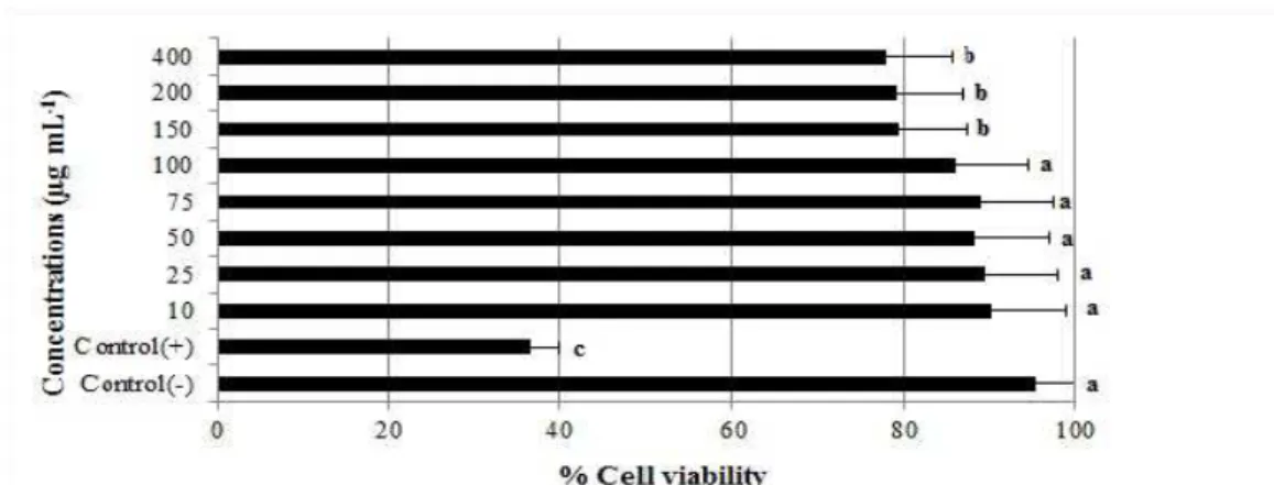
Here, the cytotoxicity was determined using a colorimetric method with the MTT assay and an
enzymatic method using LDH assay. The effects
of GYZ (10- 400 µg/mL-1) on cell growth after 24
h are shown in Figure1.MTT assay demonstrated
that GYZ significantly suppressed the
proliferation of PBL cells at high concentrations. Cell numbers clearly reduced compared to the control group.
The cytotoxic effects of different concentrations of GYZ on PBL cells at 24 h are represented in
Figure 2. Evidently, 150, 200 and
400 µg/mL-1concentrations of GYZ increased the
release of LDH enzyme.
Figure 1 - Effect of GYZ on cell viability under different concentrations on lymphocytes cells using the MTT assay.
Figure 2 - The effects of GYZ on lactate dehydrogenase (LDH) levels in human peripheral blood lymphocytes cells. The abbreviations are as in Figure 1.
The serum total antioxidant capacity (TAC) and the total oxidant status (TOS) were evaluated by using an automated colorimetric measurement method. As shown from the results presented in Table 1, two concentrations of GYZ (25 and 50
µg/mL-1) resulted a significant increase of TAC
levels on human lymphocytes cells compared with the controls. On the other hand, GYZ did not change the TOS levels in cultured lymphocytes at all the concentrations.
The effects of GYZ on the number MN in human lymphocytes cultures are shown in Figure 3. Compared to negative-control group, Mitomycin
C (10-7 M) caused considerable increases of MN
frequencies. GYZ at all applied concentrations
(10-400 µg/mL-1) did not indicate significant
differences in the number of MN compared with the control groups.
The observed CA rate of control and experimental groups are shown in Figure 4. Compared to
negative-control group, Mitomycin C (10-7 M)
caused considerable increases of CA frequencies.
Results indicated that different concentrations of GYZ did not change the rate of CAs when compared with the control values.
Table 1 - The levels of total antioxidant capacity (TAC) and total oxidant status (TOS) in human peripheral blood lymphocytes cultures treated with GYZ for 24 h.
Parameters Concentrations
(µg/mL-1)
TAC (Trolox Equiv. / mmol L-1)
TOS (H2O2
Equiv. / µmol -1)
Control (-) 6.24±0.71b 11.58±1,38a
Control(+) 12.46±1.49d 39.14±4.59c
10 6.23±0.74b 11.61±1.39a
25 9.12±0.91c 11.68±1.40a
50 9.32±0.93c 11.72±1.41a
75 6.44±0.76b 11.69±1.38a
100 6.38±0.68b 11.73±1.42a
150 6.21±0.66b 11.84±1.39a
200 5.56±0.63a 11.89±1.43a
400 5.23±0.57a 12.13±1.45b Ascorbic acid (10 µm) and hydrogen peroxide (25 µm) were also used as the positive controls in TAC and TOS analysis, respectively. Different superscript letters in the same column indicate significant differences (P < 0.05).
Figure 4 - The rate of chromosomal aberrations (CAs) in cultured human peripheral blood lymphocytes exposed to different concentrations of GYZ.The abbreviations are as in Figure 1.
DISCUSSION
In the current study, PBLs were treated with different concentrations of GYZ. MTT and LDH assays were used to examine the cytotoxicity
effect of GYZ in vitro. MTT assay is a
colorimetric method for determining the number of viable cells in proliferation. According to results of MTT analysis, the lower concentrations
of the GYZ (10-100 µg/mL-1) had no effect in any
of the parameters assessed (Fig.1). The highest
doses (150, 200 and 400 µg/mL-1) of GYZ were
cytotoxic for the PBL cells. Lactate
dehydrogenase (LDH) is a stable cytosolic enzyme released upon membrane damage in necrotic cells. LDH released into the medium provides an index of cell death and membrane permeability to LDH and an increase in LDH activity in the medium occurs as a result of cell membrane fragmentation and enzyme leakage (Yokogawa et al. 2004). According to results of LDH analysis, the highest doses (150, 200 and 400 µg/mL) of GYZ resulted increases in the LDH levels compared with the controls (Fig. 2). These results were in agreement with the previous studies, which have reported that GYZ and azulene derivates could exhibit cytotoxic effects on cell lines such as human gingival fibroblast (HGF), pulp cell (HPC), periodontal ligament fibroblast (HPLF) and human tumor cell lines, including submandibular gland carcinoma (HSG), oral squamous cell carcinoma (HSC-2, HSC-3), promyelocytic leukemia (HL-60) (Wakabayashi et al. 2003; Fiori et al. 2011).
The antioxidant/oxidant effects of GYZ have not been investigated on PBL cells. For assessing the
antioxidant/oxidant effects of GYZ, total
antioxidant capacity (TAC) and total oxidant status (TOS) assays were performed in this study. The level of TAC increased after treatment by GYZ (25 and 50 µg/mL) compared to control group (Table 1). Similar to these findings, previous studies showed that GYZ inhibited the
rat hepatic microsomal membrane peroxidation in
vitro (Kourounakis et al. 1997a; Dovolou et al.
2011). In vitro and in vivo studies on the effect of
GYZ on rat hepatic cytochrome P450 (CYP) showed that GYZ inhibited CYP1A2 activity (Kourounakis et al. 1997b). Rekka et al. (2002) reported that GYZ considerably inhibited the lipoxygenase activity in rat hepatic microsomal membrane lipids, applying the 2-thiobarbituric acid test. Pratsinis and Haroutounian (2002)
demonstrated that 3-vinylguaiazulene (a
guaiazulene derivate) had antioxidant properties
assayed by 2,2-diphenly-1-picrylhydrazly
(DPPH). GYZ applications at all the tested concentrations did not show any significant increases of TOS levels in both the cultures.
Many studies have demonstrated the in vitro or in
vivo genotoxicity of several sesquiterpenes but not
GYZ in different cell types. This study evaluated the genetic effect of GYZ on human lymphocytes
culture using by micronuclei (MN) and
chromosomal aberration (CA) assays. The findings showed that GYZ was neither cytotoxic
nor genotoxic in vitro. Similar to these findings,
Al-Zubairi et al. (2010) reported that zerumbon (a
ginger) did not induce genotoxicity in the cultured human peripheral blood lymphocytes. Di Sotto et al. (2010) reported that beta-caryophyllene exhibited antigenotoxic activity in human PBL cells against the toxicity of ethyl methanesulfonate (EMS) and colcemid (COL). On the other hand,
Gil da Costa et al. (2012) found that ptaquiloside
(a norsesquiterpene) was genotoxic in the cultured human lymphocytes by using sister chromatid exchange (SCE) test. Besides, gossypol was also
genotoxic in human lymphocyte cells by using
SCE (Best and McKenzie 1988).These divergent
results suggested the relevance of the chemical structure in the biological effect of sesquiterpenes and indicated the importance of using various test models to reach a valid conclusion.
Consequently, GYZ could be used as a suggested natural antioxidant for therapeutic, pharmaceutical and many health care products and solutions.
ACKNOWLEDGMENTS
The authors are grateful to all volunteers for the blood samples.
REFERENCES
Afoulous S, Ferhout H, Raoelison EG, Valentin A, Moukarzel B, Couderc F, et al. Chemical composition and anticancer, antiinflammatory, antioxidant and antimalarial activities of leaves essential oil of Cedrelopsis grevei. Food Chem Toxicol. 2013; 56: 352-362.
Al-Zubairi AS, Abdul AB, Syam MM. Evaluation of the genotoxicity of zerumbone in cultured human peripheral blood lymphocytes. Toxicol In Vitro. 2010; 24: 707-712.
Andersen FA. Thirty-seventh report of the cosmetic ingredient review expert panel on the safety assessment of cosmetic ingredients. Int J Toxicol. 1999; 18: 59.
Barlak Y, Değer O, Colak M, Karataylı SC, Bozdayı AM, Yücesan F. Effect of Turkish propolis extracts on proteome of prostate cancer cell line. Proteome Sci. 2011; 9: 74.
Best RG, McKenzie WH. Variable Sister-chromatid exchange response in human lymphocytes exposed ın vitro to gossypol acetic acid. Mutat Res. 1988; 206: 227-233.
Bubols GB, Vianna Dda R, Medina-Remon A, von Poser G, Lamuela-Raventos RM, Eifler-Lima VL, et al. The antioxidant activity of coumarins and flavonoids. Mini Rev Med Chem. 2013; 13: 318-334.
Cirić A, Karioti A, Koukoulitsa C, Soković M, Skaltsa H. Sesquiterpene lactones from Centaurea zuccariniana and their antimicrobial activity. Chem Biodivers. 2012; 9: 2843-2853.
Çelik K, Toğar B, Turkez H, Taşpınar N. In vitro
cytotoxic, genotoxic and oxidative effects of acyclic sesquiterpene farnesene. Turk J Biol. 2014; 38: 253-259.
Davıs ME, Croteau R. Cyclization enzymes in the biosynthesis of monoterpenes, sesquiterpenes, and diterpenes topics in current. Top Curr Chem. 2000; 209: 54-92.
Di Sotto A, Mazzanti G, Carbone F, Hrelia P, Maffei F. Inhibition by beta-caryophyllene of ethyl methanesulfonate-induced clastogenicity in cultured human lymphocytes. Mutat Res. 2010; 699: 23-28. Dovolou E, Clemente M, Amiridis GS, Messinis IE,
Kallitsaris A, Gutierrez-Adan A, et al. Effects of guaiazulene on in vitro bovine embryo production and on mRNA transcripts related to embryo quality.
Reprod Domest Anim. 2011; 46: 862-869.
Erel O. A novel automated direct measurement method for total antioxidant capacity using a new generation, more stable ABTS radical cation. Clin Biochem.
2004; 37: 277-285.
Evans HJ, O’Riordan ML. Human peripheral blood lymphocytes for the analysis of chromosome aberrations in mutagen tests. Mutat Res. 1975; 31: 135-148.
Fiori J, Teti G, Gotti R, Mazzotti G, Falcon M. Cytotoxic activity of guaiazulene on gingival fibroblasts andthe influence of light exposure on guaiazulene-induced cell death. Toxicol In Vitro.
2011; 25: 64-72.
Fiori J, Gotti R, Albini A, Cavrini V. Study on the photostability of guaiazulene by high-performance liquid chromatography/mass spectrometry and gas chromatography/mass spectrometry. Rapid Commun Mass Spectrom. 2008; 22: 2698-2706.
Geyikoğlu F, Turkez H. Protective effect of sodium selenite against the genotoxicity of aflatoxin B-1 in human whole blood cultures. Braz Arch Biol Technol. 2006; 49: 393-8.
Gil da Costa RM, Oliveira PA, Bastos MM, Lopes CC,
Lopes C. Ptaquiloside-induced early-stage urothelial lesions show increased cell proliferation and intact β -catenin and e-cadherin expression. Environ Toxicol.
2014; 29: 763-769
Goze İ, Alim A, Sihoglu A, Sökmen M, Sevgi K, Tepe B. Screening of the antioxidant activity of essential oil and various extracts of Origanum rotundifolium
Boiss. from Turkey. J Med Plant Res. 2009; 3: 246-54.
Higa T, Sakemi S. Guaiazulene derivatives and their methods of use. US Patent No. US 4755529 A. 1988; 5-7.
Hussain SM, Hess KL, Gearhart JM, Geiss KT, Schlager JJ. In vitro toxicity of nanoparticles in BRL 3A rat liver cells. Toxicol In Vitro. 2005; 19: 975-983.
IPCS. (International Program on Chemical Safety), Environmental Health Criteria 46. Guidelines for the Study of Genetic Effects in Human Populations. WHO, Geneva. 45-1985; 54 pp.
Jahangir T, Khan TH, Prasad L, Sultana S. Alleviation of free radical mediated oxidative and genotoxic effects of cadmium by farnesol in Swiss albino mice. Redox Rep. 2005; 10:303-310.
Jin L, Zhang Y, Yan L, Guo Y, Niu L. Phenolic compounds and antioxidant activity of bulb extracts of six Lilium species native to China. Molecules.
2012; 17: 9361-9378.
Kourounakis AP, Rekka EA, Kourounakis PN.
Antioxidant activity of guaiazulene and protection against paracetamol hepatotoxicity in rats. J Pharm Pharmacol. 1997a; 49: 938-942.
Kourounakis AP, Rekka EA, Kourounakis PN. Effect of guaiazulene on some cytochrome P450 activities. Implication in the metabolic activation and hepatotoxicity of paracetamol. Arch Pharm (Weinheim). 1997b; 330: 7-11.
Kundu A, Saha S, Walia S, Shakil NA, Kumar J, Annapurna K. Cadinene sesquiterpenes from
Eupatorium adenophorum and their antifungal activity. J Environ Sci Health B. 2013; 48: 516-522. Lewerenz V, Hanelt S, Nastevska C, El-Bahay C,
Rouhrdanz E, Kahl R. Antioxidants protect primary rat hepatocyte cultures against acetaminophen-induced DNA strand breaks but not against acetaminophen-induced cytotoxicity. Toxicol. 2003; 191: 179-118.
López-Alarcón C, Denicola A. Evaluating the antioxidant capacity of natural products: a review on chemical and cellular-based assays. Anal Chim Acta. 2013; 763: 1-10.
Mitjavila MT, Moreno JJ. The effects of polyphenols on oxidative stress and the arachidonic acid cascade: Implications for the prevention/treatment of high prevalence diseases. Biochem Pharmacol. 2012; 84: 1113-1122.
Naveena BM, Sen AR, Kingsly RP, Singh DB, Kondaiah N. Antioxidant activity of pomegranate rind powder extract in cooked chicken patties. Int J Food Sci & Technology. 2008; 43:1807-1812.
Pratsinis H, Haroutounian SA. Synthesis and antioxidant activity of 3-substituted guaiazulene derivatives.Nat Prod Lett. 2002; 16: 201-205. Rekka E, Chrysselis M, Siskou I, Kourounakis A.
Synthesis of new azulene derivatives and study of their effect on lipid peroxidation and lipoxygenase activity. Chem Pharm Bull (Tokyo). 2002; 50: 904-907.
Shi Q, Fu J, Ge D, He Y, Ran J, Liu Z, et al. Huperzine A ameliorates cognitive deficits and oxidative stress in the hippocampus of rats exposed to acute hypobaric hypoxia. Neurochem Res. 2012; 37: 2042-2052.
Tanaka M, Yamaki F, Saitoh M, Nakazawa T, Tanaka H, Noguchi K, et al. Relaxant action of azulene-carboxamidine derivative N1, N1-dimethyl-N2-(2-pyridilmethyl)-5-isopropyl-3,8-dimethylazulene-1- carboxamidine (HNS-32) in pig coronary artery.
Pharm Pharmacol Commun. 2000; 6: 397-404. Turkez H, Geyikoglu F, Aslan A, Karagöz Y, Turkez
O, Anar M. Antimutagenic effects of lichen Pseudovernia furfuracea (L.) Zoph. extracts against the mutagenicity of aflatoxin B1 in vitro.Toxicol Ind Health. 2010; 26: 625-631.
Wakabayashi H, Nishishiro M, Arikawa S, Hashimoto K, Kikuchi H, Nishikawa H, et al. Cytotoxic activity of azulenes against human oral tumor cell lines.
Anticancer Res. 2003; 23: 4747-4756.
Wang T, Jiang A, Zhang J, Jing F. Apoptosis induction by E2F decoy DNA of the prostate cancer cell line.
Braz Arch Biol Technol. 2010; 53: 327-334.
Yanagisawa T, Kosakai K, Tomiyama T, Yasunami M,
Takase K. Studies on anti-ulcer agents. II. Synthesis and antiulcer activities of 6-isopropylazulene-1- sodium sulfonate derivatives. Chem Pharm Bull (Tokyo). 1990; 38: 3355-3358.
Yokogawa K, Watanabe M, Takeshita H, Nomura M, Mano Y, Miyamoto K. Serum aminotransferase activity as a predictor of clearance of drugs metabolized by CYP isoforms in rats with acute hepatic failure induced by carbon tetrachloride. Int J Pharm. 2004; 269: 479-489.
Yu WY, Kan WJ, Yu PX, Li MM, Song JS, Zhao F. Anti-inflammatory effect and mechanism of artemisinin and dihydroartemisinin. Zhongguo Zhong Yao Za Zhi. 2012; 37:2618-2621.