! !
! ! ! !
! ! !
! ! ! !
! ! ! ! ! ! ! !
LUCIANE DIAS DE OLIVEIRA
ANTIMICROBIANOS E EXTRATOS NATURAIS PARA
São José dos Campos 2015
LUCIANE
DIAS DE OLIVEIRA
ANTIMICROBIANOS E EXTRATOS NATURAIS PARA
USO EXTERNO NA ODONTOLOGIA
Tese apresentada ao curso de Odontologia do Instituto de Ciência e
Tecnologia, UNESP – Univ Estadual Paulista, Campus de São José
dos Campos, como parte dos requisitos para obtenção do título de
LIVRE DOCENTE nas disciplinas Farmacologia e Terapêutica Clínica,
BANCA EXAMINADORA
Prof. Tit. Antonio Olavo Cardoso Jorge Instituto de Ciência e Tecnologia Universidade Estadual Paulista – UNESP Campus de São José dos Campos
Profa. Adj. Rosilene Fernandes da Rocha Instituto de Ciência e Tecnologia Universidade Estadual Paulista – UNESP Campus de São José dos Campos
Prof. Tit. João Ernesto de Carvalho Faculdade de Ciências Farmacêticas Universidade Estadual de Campinas – UNICAMP
Campus de Campinas
Profa. Tit. Maria Cristina Volpato Faculdade de Odontologia de Piracicaba Universidade Estadual de Campinas – UNICAMP Campus de Piracicaba
Prof. Associado Caio Cezar Randi Ferraz Faculdade de Odontologia de Piracicaba Universidade Estadual de Campinas – UNICAMP
“
A minha alma descansa somente em DEUS;
Dele vem a minha salva
çã
o.
Somente Ele
é
a rocha que me salva;
Ele
é
minha torre segura!
”
DEDICATÓRIA
Dedico minha Livre-Docência a DEUS, por me dar saúde, força,
segurança, proteção e amor! Por me dar o dom da vida acadêmica.
Obrigada Senhor por cuidar de todos os detalhes e por todas as bênçãos
que tem derramado em minha vida. Glória a Ti, em nome de Jesus!
Ao meu marido Cláudio, por estar todos os dias ao meu lado me
apoiando e me incentivando. Por ser este marido tão presente e especial
que Deus colocou em minha vida. Obrigada amor, por dividir comigo todos
os desafios e todas as vitórias de nossas vidas! Que Deus continue
abençoando nosso amor e união pra todo o sempre. Te Amo!
Ao meu filho Guilherme, que é meu maior presente de Deus, por todo
amor, carinho e travessuras, por completar minha vida! Filho, com você eu
aprendo todos os dias a dádiva de ser mãe e agradeço a Deus por ter me
enviado exatamente você, que preenche nossa casa e nossos corações com
seu jeito “mandão e carinhoso de ser”. Te Amo!
Aos meus pais Alberto e Dirce, por todo amor e dedicação. Por não
medirem esforços pela minha formação. Cada vitória minha é uma vitória pra
vocês. Obrigada por serem tão presentes na minha vida, por serem pais
maravilhosos e avós melhores ainda! Amo vocês!!
Ao meu eterno filhote “Johnny” (meu beagle), que sempre estava ao
meu lado me dando carinho incondicional enquanto escrevia minha dissertação,
AGRADECIMENTOS
Ao Instituto de Ciência e Tecnologia da Universidade Estadual Paulista “Júlio de Mesquita Filho”, representado pelo diretor Prof. Tit. Estevão Tomomitsu Kimpara e pela vice-diretora Profa Adj. Rebeca di Nicoló, por toda minha formação (graduação, mestrado, doutorado e pós-doutorado) e pela oportunidade de seguir minha profissão e meu dom e por permitir meu crescimento acadêmico científico. Obrigada Unesp São José!
Ao Prof. Tit. Antonio Olavo Cardoso Jorge, por ser meu eterno mestre. Obrigada professor por todos os ensinamentos, incentivos, apoios e oportunidades que o senhor me deu (e me dá ainda!). Obrigada pela orientação no Doutorado e supervisão no Pós-Doutorado. Obrigada pelo convívio diário, pelas conversas no café, pela amizade que temos. Eu te admiro muito!
À Profa Adj. Rosilene Fernandes da Rocha por me apresentar a Farmacologia!! Obrigada pela grande oportunidade que a senhora me deu. Obrigada por todos os ensinamentos, por toda a paciência que teve comigo e obrigada principalmente pela amizade que cultivamos. Tenho um carinho enorme pela senhora! Sinto falta das nossas conversas, dos nossos cafezinhos. A senhora é muito importante pra mim!
À Profa Marcia Carneiro Valera, por todo incentivo, apoio e parcerias. Obrigada Marcia por me ajudar desde o início da minha linha de pesquisa durante o mestrado. Eu te admiro muito como docente, pesquisadora e como amiga. Sei que posso contar sempre com você e com o Emmanuel. Agradeço a Deus por ter colocado vocês em meu caminho, pois são muito especiais pra mim!!
À amiga Profa Alessandra, pela amizade de “longa data”, pelo convívio Vocês são muito competentes e muito especiais pra mim!
À amiga Profa Juliana, pelo convívio desde a graduação. Obrigada pela amizade, incentivo e carinho!
À amiga Profa Samira, por toda amizade, parceria, e pelo convívio diário. Tenho um grande carinho por você Samy!
À amiga Profa Graziella, pela grande amizade que cultivamos, principalmente nos últimos anos. Obrigada pela parceria na Farmacologia e nas pesquisas. Tenho um carinho muito especial por você.
À Profa Ana Sueli, com quem divido a disciplina Terapêutica. Obrigada pelos ensinamentos, pelo carinho e pela amizade.
Ao Programa de Pós Graduação em Biopatologia Bucal, representado pela coordenadora Profa Juliana Campos Junqueira, por me receber como aluna e como docente e por me dar oportunidade de crescer cientificamente.
Ao Departamento de Biociências e Diagnóstico Bucal, na pessoa do chefe Prof. José Benedito Oliveira Amorim, por me acolher como docente e pesquisadora. Aos atuais professores do Departamento: Yasmin Rodarte Carvalho, Ana Lia Anbinder, Luiz Eduardo Blumer Rosa, Marianne Spalding, Estela Kaminagakura Tango, Janete Dias Almeida, Ana Sueli Rodrigues Cavalcante, Antonio Olavo Cardoso Jorge, Juliana Campos Junqueira, Samira Esteves Afonso Camargo, Horácio Faig Leite, Simone Helena Ferreira Gonçalves, Luana Marotta Reis de Vasconcellos, Lucio Murilo dos Santos, Miguel Angel Castillo Salgado, Walter Domingos Niccoli Filho e Monica Fernandes Gomes, pela boa convivência que temos, por tornarem agradável nosso ambiente de trabalho e pelo apoio que sempre tive.
A todos os técnicos do Departamento pelo convívio salutar.
Aos meus alunos da Pós-Graduação: Felipe, Jonatas, Adeline, Daiane, Nadia, Karen, Carlos Eduardo. Vocês me ensinam todos os dias a arte de orientar! Obrigada por toda dedicação, por cuidarem do nosso laboratório, pelas pesquisas desenvolvidas e principalmente pela amizade que temos!
Aos alunos da graduação, por me ensinarem a ser docente! Vocês dão sentido a minha carreira, pois quero sempre aprender mais pra ensinar o melhor a vocês. Tenho um agradecimento especial a turma de formandos 2015 do curso integral T58, que me homenageou como patrona e “nome de Turma”. Este reconhecimento não tem preço! Obrigada de todo coração!
A todos os funcionários do ICT – UNESP que fazem parte direta ou indiretamente do meu crescimento. Obrigada pela ajuda e colaboração.
À Fundação de Amparo à Pesquisa do Estado de São Paulo – FAPESP, que, por meio de suas diversas linhas de fomento (bolsas de IC, mestrado, doutorado, pós-doutorado, auxílio à Pesquisa), financiou e permitiu a realização desta coletânea de trabalhos aqui apresentados.
Aos meus irmão Ricardo e Andrea, pelo amor que nos une. Vocês são muito especiais na minha vida. Obrigada por todo carinho e incentivo. Amo vocês. Agradeço meus cunhados Fabiane e Wetse por toda amizade. Agradeço meu sobrinho Ricardinho, que torce por mim e me incentiva sempre. Agradeço meus sobrinhos Ravi e Pedro, e minhas afilhadas princesas Gabriela e Luna. Amo vocês!!
Aos meus sogros D. Graça e S. Antonio, obrigada pelo amor, carinho, apoio, incentivo. Vocês são bênçãos em minha vida. Amo vocês!
A minha avó Maria, que com 97 anos acompanha todas as conquistas da minha vida. Obrigada minha vozinha linda! Te amo!
SUMÁRIO
RESUMO ... 10
ABSTRACT ... 12
1 INTRODUÇÃO ... 14
2 PROPOSIÇÃO ... 18
3 ARTIGOS ... 21
3.1 In vitro effects of calcium hydroxide and polymyxin B on endotoxins in root canals... 21
3.2 In vitro effects of endodontic irrigants on endotoxins in root canals... 31
3.3 Action of propolis and medications against Escherichia coli and endotoxin in root canals... 40
3.4 Efficacy of endodontic treatment for endotoxin reduction in primarily infected root canals and evaluation of cytotoxic effects... 46
3.5 In vitro antimicrobial activity of auxiliary chemical substances and natural extracts on Candida albicans and Enterococcus faecalis in root canals... 52
3.6 Cytotoxicity of brazilian plant extracts against oral microorganisms of interest to dentistry... 59
3.7 Control of microorganisms of oral health interest with Arctium lappa L. (burdock) extract non-cytotoxic to cell culture of macrophages (RAW 264.7)... 68
3.9 Persea americana glycolic extract: in vitro study of
antimicrobial activity against Candida albicans biofilm and
cytotoxicity evaluation... 83
3.10 Intracanal medications neutralize the effects of
lipoteichoic acid to induce the production of proinflammatory
cytokines and nitric oxide in macrophages RAW 264.7: in
vitro study... 89
4 CONSIDERAÇÕES FINAIS ... 105
Oliveira LD. Antimicrobianos e extratos naturais para uso externo na Odontologia. [tese]. São José dos Campos (SP): Instituto de Ciência e Tecnologia, UNESP - Univ Estadual Paulista; 2015.
RESUMO
O objetivo desta tese foi reunir os resultados de estudos que avaliaram os efeitos in vitro e in vivo de irrigantes endodônticos e medicações intracanais, incluindo própolis e extratos de plantas, sobre micro-organimos, endotoxinas (LPS) e ácido lipoteicóico (LTA) em canais radiculares, bem como a citotoxicidade e atividade antimicrobiana de extratos de plantas contra micro-organismos de interesse para Odontologia. No estudo dos efeitos do hidróxido de cálcio e polimixina B sobre endotoxinas em canais radiculares, observou-se que estas medicações intracanais detoxificaram endotoxinas alterando sua capacidade de estimular a produção de anticorpos por linfócitos B. No próximo estudo, foram avaliados os efeitos in vitro de irrigantes endodônticos (NaOCl 2,5% e 5%; clorexidina 2%, hidróxido de cálcio 0,14% e polimixina B) sobre endotoxinas em canais radiculares. Somente hidróxido de cálcio e polimixina B conseguiram neutralizar endotoxinas quando utilizados como irrigantes, sendo que NaOCl e clorexidina não foram efetivos. Quanto à análise da ação de extrato de própolis como irrigante dos canais radiculares sobre Escherichia coli e endotoxinas, verificou-se que o extrato de própolis promoveu eliminação completa de
E. coli e redução significativa de endotoxinas, porém, somente com a
óleo de Ricinus comunis sobre Candida albicans e Enterococcus faecalis em canais radiculares e verificou-se potencial antimicrobiano do extrato de Z. officinale para uso intracanal. Em outro estudo, foi analisada a atividade antimicrobiana e anti-endotoxina do extrato de Z. officinale e verificou-se que este extrato é um forte agente antimicrobiano para C.
albicans, E. faecalis e E. coli, porém não neutraliza endotoxinas. Diante
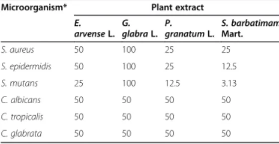
da grande diversidade de plantas e seu uso restrito na Odontologia, foi analisada a citotoxicidade e atividade de extratos de plantas (Equisetum
arvense, Glycyrrhiza glabra, Punica granatum, Stryphnodendron barbatimam e Arctium lappa) sobre micro-organismos de interesse
odontológico (Staphylococcus aureus, Staphylococcus epidermidis,
Streptococcus mutans, Candida albicans, Candida tropicalis, and Candida glabrata), em culturas planctônicas e biofilmes. Todos os
extratos foram efetivos sobre os micro-organismos avaliados, em diferentes concentrações, sendo que os extratos de G. glabra e A. lappa foram os menos citotóxicos para macrófagos e o extrato de E. arvense apresentou citotoxicidade superior a 50%. O extrato de P. americana foi avaliado sobre biofilme de C. albicans e apresentou potencial antifúngico, embora tenha sido citotóxico para macrófagos em altas concentrações. Como conclusão geral, os estudos demostraram que: a) para neutralização de endotoxinas (LPS) e ácido lipoteicóico (LTA) o hidróxido de cálcio é o agente antimicrobiano mais eficaz, possibilitando a neutralização de seus efeitos citotóxicos; b) os extratos de gengibre (Z.
officinale), barbatimão (S. barbatiman), bardana (A. bappa), romã (Punica granatum), alcaçuz (G. glabra), cavalinha (E. arvense) e abacateiro (P. americana) são importantes agentes antimicrobianos sobre bactérias e
leveduras de interesse odontológico, sendo os extratos de bardana e alcaçuz os menos citotóxicos para macrófagos. Com isso, os extratos de plantas apresentam potencial antimicrobiano que deve ser mais explorado e seu uso clínico odontológico mais difundido.
Oliveira LD. Antimicrobials and natural extracts for external use in
Dentistry. [thesis]. São José dos Campos (SP): Institute of Science and Technology, UNESP - Univ Estadual Paulista; 2015.
ABSTRACT
This thesis aimed to collect results from studies that evaluated in vitro and in vivo effects of endodontic irrigants and intracanal medication, such as propolis and plant extracts on microorganisms, endotoxins (LPS) and lipoteichoic acid (LTA) in root canals, as well as cytotoxicity and antimicrobial activity of plant extracts against microorganisms of interest
for Dentistry.! In the study of the effects of calcium hydroxide and
analyzed antimicrobial and anti-endotoxin activity of Z. officinale extract and it was found that this extract is a strong antimicrobial agent for C.
albicans, E. faecalis and E. coli, but it is not able to neutralize endotoxins.
In the presence of the great diversity of plants and their restricted use in Dentistry, it was analyzed plant extracts (Equisetum arvense, Glycyrrhiza
glabra, Punica granatum, Stryphnodendron barbatimam and Arctium lappa) cytotoxicity and antimicrobial activity against microorganisms of
Dental interest (Staphylococcus aureus, Staphylococcus epidermidis,
Streptococcus mutans, Candida albicans, Candida tropicalis and Candida glabrata) in planktonic cultures and biofilm. All extracts were effective
against the microorganisms evaluated, at different concentrations, being
G. glabra and A. lappa extract the least cytotoxic for macrophages while E. arvense extract showed cytotoxicity higher than 50%. P. americana
extract was evaluated on C. albicans biofilm and showed antifungal potential, although it was cytotoxic to macrophages in high concentrations. As a general conclusion, the studies demonstrated that: a) in order to neutralize endotoxins (LPS) and lipoteichoic acid (LTA), calcium hydroxide is the most effective antimicrobial agent, allowing neutralization of their cytotoxic effects; b) ginger (Z. officinale), barbatimam (S. barbatiman), burdock (A. lappa), pomegranate (Punica granatum), licorice (G. glabra), horsetail (E. arvense) and avocado (P. american) are important antimicrobial agents against bacteria and yeast of Dental interest, being burdock and licorice extracts the least cytotoxic to macrophages. On the basis of the evidences available, it can be concluded that the plant extracts showed antimicrobial potential that should be further explored and more widespread in Dental clinic use.
1 INTRODUÇÃO
As principais alterações patológicas que acometem a
polpa, os tecidos periapicais e a mucosa bucal é de etiologia microbiana,
sendo que diferentes micro-organismos e seus produtos apresentam
significante papel na indução e manutenção destas lesões. Na área
endodôntica, estudos demonstraram importante relação entre infecção
polimicrobiana dos canais radiculares, especialmente causada por
bactérias anaeróbias Gram-negativas, e sinais e sintomas clínicos, como
dor espontânea, dor à palpação, edema e exsudato purulento (Gomes et
al., 2007; Siqueira Jr et al., 2008; Siqueira & Roças, 2013; Ahmed et al.,
2013). Estas bactérias possuem na membrana externa da parede celular
endotoxinas que são complexos lipopolissacarídicos (LPS), responsáveis
por amplificar a resposta inflamatória, imunológica e citotóxica, sendo de
grande importância métodos de remoção ou neutralização de LPS nas
infecções bucais. Endotoxinas são detectadas em 100% das coletas
iniciais de canais radiculares com polpa necrosada, com níveis
significativamente maiores em dentes com sintomatologia (Martinho et al.,
2008; Martinho et al., 2011; Oliveira et al., 2012; Cardoso et al., 2015;
Herrera et al., 2015). Com isso, o tratamento destas infecções deve não
somente destruir micro-organismos como também inativar suas
endotoxinas e demais produtos tóxicos.
Desde 1975, quando Schein & Shilder relataram pela
primeira vez a participação de LPS nas infecções endodônticas, busca-se
Oliveira et al., 2007, Vianna et al., 2007; Martinho et al., 2008; Gomes et
al., 2009; Maekawa et al., 2011; Oliveira et al., 2012; Marinho et al.,
2015). Por outro lado, estudos in vitro e in vivo demonstraram que o
hidróxido de cálcio e polimixina B como medicação intracanal ou irrigantes
dos canais radiculares detoxificaram endotoxinas, alterando a propriedade
de LPS de induzir produção de anticorpos por linfócitos B e citocinas
pró-inflamatórias por macrófagos (Oliveira et al., 2005; 2007; 2012). Outras
pesquisas demonstraram significativa redução dos níveis de LPS nos
canais radiculares quando hidróxido de cálcio foi usado como medicação
(Maekawa et al., 2011; Xavier et al., 2013; Sousa et al., 2014; Adl et al.,
2015), porém, as substâncias químicas auxiliares mais utilizadas durante
o preparo dos canais não são efetivas sobre LPS, sendo necessário
ampliar os estudos de substâncias alternativas, como extratos naturais,
que apresentem efetiva ação antimicrobiana e que possam contribuir para
ampliação da terapia endodôntica. Própolis e extratos de plantas tem sido
estudados como agentes antimicrobianos e anti-inflamatórios e têm
demonstrado resultados promissores, incluindo desinfecção dos canais
radiculares (Valera et al., 2010; Zare-Jahromi et al., 2012; Tyagi et al.,
2013; Bhandari et al., 2014; Valera et al., 2015).
Apesar da população mundial utilizar desde a antiguidade
extratos de plantas para o tratamento de diversas enfermidades, diante da
grande diversidade de plantas, são poucas as pesquisas que comprovam
sua eficácia. Na Odontologia, o uso clínico de extratos naturais é muito
restrito, de modo que estudos científicos específicos precisam ser
realizados para promover sua inclusão em enxaguatórios bucais,
dentifrícios, irrigantes e medicações intracanais, pomadas, entre outros,
visando direcionar suas indicações terapêuticas. Em 2015, Farooqui et al.
relataram que as combinações sinérgicas de agentes antimicrobianos
com diferentes mecanismos de ação têm possibilitado estratégias de
sucesso no combate a infecções envolvendo bactérias multirresistentes.
Juglans regia (nogueira) e oxacilina reverteu a resistência a oxicilina de
Staphylococcus aureus. Anand et al. (2015) analisaram a atividade
antimicrobiana de extratos de Anacardium occidentale (caju) e Mangifera
indica (manga) sobre micro-organismos de interesse odontológico
(Enterococcus faecalis, S. aureus, Streptococcus mutans, Escherichia coli
e Candida albicans) e verificaram que os extratos de manga e caju
promoveram significativa redução dos biofilmes microbianos e foram
menos citotóxicos para cultura de fibroblastos gengivais em comparação
com enxaguatórios bucais (clorexidina 0,2% e iodopovidona 0,2%).
Outros estudos demonstraram efetiva ação antimicrobiana de extratos de
plantas, como Pineapple, Emex spinosa , Olea europaea, Mastic gum,
Inula viscosa, Camellia sinensis, Punica granatum, Psidium guajava,
Arctium lappa, Acacia nilotica e Schinus terebinthifolius Raddi, Persea
americana, Anacardium occidentale, sobre micro-organismos
cariogênicos, periodontopatógenos e sobre leveduras do gênero Candida
(Anand et al., 2015; Shekar et al., 2015; Jesus et al., 2015; Praveen et al.,
2014, Donia et al., 2014; Oliveira et al., 2014; Vieira et al. 2014;
Karygianni et al., 2014; Araghizadeh et al., 2013; Oliveira et al., 2013).
Em 2015, Shekar et al. revisaram dez extratos de plantas
no cuidado de saúde bucal abordando o cenário atual e necessidades
futuras e concluíram que a efetiva ação antimicrobiana da combinação de
extratos de plantas sobre patógenos cariogênicos e
periodontopatogênicos pode levar ao desenvolvimento de métodos
inovadores capazes de inibir simultaneamente as duas doenças bucais
mais comuns, além de retardar o desenvolvimento de resistência
microbiana. Apesar do uso clínico ainda ser restrito na Odontologia, os
estudos com extratos de plantas estão crescendo muito. Só no ano de
Os extratos de plantas apresentam diferentes finalidades
terapêuticas e diante da grande diversidade os estudos envolvendo
substâncias naturais representam importante campo de pesquisa que
pode trazer grandes benefícios à terapia odontológica, sendo de grande
interesse estudar seus efeitos sobre micro-organismos e seus produtos,
incluindo os envolvidos nas infecções endodônticas e periodontais, cárie e
candidose. Estes estudos são importantes para comprovar os diferentes
efeitos benéficos dos extratos, os quais podem ser inseridos em
formulações de uso odontológico, tais como medicações e irrigantes
intracanais, enxaguatórios bucais, dentifrícios, gel, pomadas, entre outros.
Assim, com a necessidade atual de ampliar as estratégias
antimicrobianas, o uso de extratos de plantas na Odontologia, para
prevenção e tratamento de infecções bucais, é uma terapia alternativa
2 PROPOSIÇÃO
A proposta desta tese foi demonstrar os resultados de
estudos realizados in vitro e in vivo dos efeitos de agentes
antimicrobianos e extratos naturais, para uso externo na Odontologia,
sobre diferentes micro-organismos de interesse odontológico e sobre
seus produtos (LPS e ácido lipoteicóico - LTA). Sua estrutura foi dividida
em capítulos que correspondem aos artigos publicados ou enviados para
publicação, seguindo a sequência abaixo:
a) Avaliação in vitro dos efeitos do hidróxido de cálcio e
polimixina B sobre endotoxinas em canais radiculares.
(artigo relacionado ao Processo FAPESP 00/09427-7,
publicado no periódico Journal of Dentistry, 2005
Feb;33(2):107-14. doi:10.1016/j.jdent.2004.08.008,
QUALIS A1, Impacto 2,749, citações no ISI=18, Scopus=
18, Google Scholar= 31);
b) Efeitos in vitro de irrigantes endodônticos sobre
endotoxinas em canais radiculares. (artigo relacionado ao
Processo FAPESP 02/06467-3, publicado no periódico
Oral Surgery Oral Medicine Oral Pathology Oral Radiology
and Endodontics, 2007 jul 104(1):135-42.
doi:10.1016/j.tripleo.2006.11.037, QUALIS A2, Impacto
Processo FAPESP 05/57668-7, publicado no periódico
Oral Surgery Oral Medicine Oral Pathology Oral Radiology
and Endodontics, 2010 oct 110(4):e70-4. doi:
10.1016/j.tripleo.2010.01.029., QUALIS A2, Impacto
1,261, citações no ISI=11, Scopus= 18, Google Scholar=
27);
d) Eficácia do tratamento endodôntico para redução de
endotoxinas em canais radiculares com infecção primária
e avaliação dos efeitos citotóxicos (artigo relacionado aos
Processos FAPESP 05/57668-7 e 08/53863-8, publicado
no periódico Journal of Endodontics, 2012
Aug;38(8):1053-7. doi: 10.1016/j.joen.2012.04.015.
QUALIS A1, Impacto 2,788, citações no ISI=7, Scopus=
13, Google Scholar= 20);
e) Atividade antimicrobiana in vitro de substâncias químicas
auxiliares e extratos naturais sobre Candida albicans e
Enterococcus faecalis em canais radiculares. (artigo
publicado no periódico Journal of Applied Oral Science,
2013 mar-apr 21(2):118-23. doi: 10.1590/
1678-7757201302135. QUALIS A2, Impacto 0,923, citações no
ISI=3, Scopus= 3, Google Scholar= 11);
f) Citotoxicidade de extratos de plantas brasileiras contra
micro-organismos orais de interesse para Odontologia.
(artigo publicado no periódico BMC Complementary &
Alternative Medicine, 2013 Aug;15(13):208.
g) Controle de micro-organismos de interesse odontológico
com extrato de Arctium lappa L. (bardana) não citotóxico
para cultura de macrófagos (RAW 264.7). (artigo
relacionado ao Processo FAPESP 07/55867-8, publicado
no periódico Archives of Oral Biology, 2014
Aug;59(8):808-14. doi: 10.1016/ j.archoralbio.2014.05.013
QUALIS A2, Impacto 1,735, citações no ISI=1, Scopus= 1,
Google Scholar= 3);
h) Acão in vitro da atividade antimicrobiana e anti-endotoxina
do extrato de Zingiber officinale como substância química
auxiliar e como medicação associada ao hidróxido de
cálcio e clorexidina. (artigo publicado no periódico Acta
Odontologica Scandinavica, 2015; 73(7):556-61. doi:
10.3109/00016357.2014.949846. QUALIS B2, Impacto
1,03);
i) Avaliação in vitro da atividade antimicrobiana do extrato
glicólico de Persea americana sobre biofilme de Candida
albicans e avaliação de sua citotoxicidade. (artigo
relacionado ao processo FAPESP 15/08776-3, aceito para
publicação no Scientific World Journal, 2015 oct 15:1-5.
doi: 10.1155/2015/531972, QUALIS B2, Impacto 1,73);
j) Avaliação in vitro da ação de medicações intracanais em
neutralizar os efeitos do ácido lipoteicóico (LTA) de induzir
a produção de citocinas pró-inflamatórias e óxido nítrico
3 ARTIGOS
3.1 In vitro effects of calcium hydroxide and polymyxin B on
endotoxins in root canals*
ABSTRACT
Objectives. To evaluate the effects of intracanal medicaments on
endotoxins in root canals. Methods. Seventy-five freshly extracted
maxillary incisors were used in this study. The crowns of teeth were
sectioned near the CEJ in order to standardize the root length to 14 mm.
The root canals were instrumented to an apical size #50 file and irrigated
with 1% sodium hypochlorite solution and sterilized with 60 Co gamma
irradiation. Standardized suspension containing Escherichia coli endotoxin
was inoculated into the 60 root canals. The specimens were randomly
assigned to 5 groups (n=15), according to the intracanal medicament
used: (G1) calcium hydroxide; (G2) polymyxin B; (G3) combination
neomycin–polymyxin B-hydro- cortisone; (G4) positive control (no
intracanal medicament); (G5) negative control (no endotoxin and no
intracanal medicament). After 7 days, the detoxification of endotoxin was
evaluated by Limulus lysate assay and antibody production in
B-lymphocytes culture. Results. Groups 1, 2 and 5 presented the best
results by Limulus lysate and were significantly different to groups 3 and 4
(p<0.05). Stimulation of antibodies production in cell culture by groups 1
and 6 was smaller and statistically different than groups 2, 3, 4 and 5
(p<0.05). Groups 2 and 5 induced a small increase in the antibodies
_____________________
production in relation to the groups 1 and 6. Groups 3 and 4 induced a
significant increase of antibodies production (p<0.05). Conclusions. The
calcium hydroxide and polymyxin B intracanal medicaments detoxified
endotoxin in root canals and altered the properties of LPS to stimulate the
antibody production by B-lymphocytes. The combination neomycin–
In vitro effects of calcium hydroxide and polymyxin
B on endotoxins in root canals
L.D. Oliveiraa,*, M.V.P. Lea˜oa, C.A.T. Carvalhob, C.H.R. Camargob,
M.C. Valerab, A.O.C. Jorgea, C.S. Unterkirchera
a
Department of Oral Biosciences and Diagnosis, School of Dentistry, Paulista State University-UNESP, Av. Eng. Francisco Jose´ Longo 777, Sa˜o Jose´ dos Campos 12245-000, SP Brazil
b
Department of Restorative Dentistry, School of Dentistry, Paulista State University-UNESP, Av. Eng. Francisco Jose´ Longo 777, Sa˜o Jose´ dos Campos 12245-000, SP Brazil
Received 6 March 2004; received in revised form 10 August 2004; accepted 13 August 2004
KEYWORDS
Endotoxin; Root canal; Calcium hydroxide; Polymyxin B
Summary Objectives. To evaluate the effects of intracanal medicaments on endotoxins in root canals.
Methods. Seventy-five freshly extracted maxillary incisors were used in this study. The crowns of teeth were sectioned near the CEJ in order to standardize the root length to 14 mm. The root canals were instrumented to an apical size #50 file and irrigated with 1% sodium hypochlorite solution and sterilized with 60 Co gamma irradiation. Standardized suspension containing Escherichia coli endotoxin was inoculated into the 60 root canals. The specimens were randomly assigned to 5 groups (nZ15), according to the intracanal medicament used: (G1) calcium
hydroxide; (G2) polymyxin B; (G3) combination neomycin–polymyxin B-hydro-cortisone; (G4) positive control (no intracanal medicament); (G5) negative control (no endotoxin and no intracanal medicament). After 7 days, the detoxification of endotoxin was evaluated by Limulus lysate assay and antibody production in B-lymphocytes culture.
Results. Groups 1, 2 and 5 presented the best results by Limulus lysate and were significantly different to groups 3 and 4 (p!0.05). Stimulation of antibodies production in cell culture by groups 1 and 6 was smaller and statistically different than groups 2, 3, 4 and 5 (p!0.05). Groups 2 and 5 induced a small increase in the antibodies production in relation to the groups 1 and 6. Groups 3 and 4 induced a significant increase of antibodies production (p!0.05).
Conclusions. The calcium hydroxide and polymyxin B intracanal medicaments detoxified endotoxin in root canals and altered the properties of LPS to stimulate the antibody production by B-lymphocytes. The combination neomycin–polymyxin B-hydrocortisone did not detoxified endotoxin.
q2004 Elsevier Ltd. All rights reserved.
Journal of Dentistry (2005)33, 107–114
www.intl.elsevierhealth.com/journals/jden
0300-5712/$ - see front matterq2004 Elsevier Ltd. All rights reserved. doi:10.1016/j.jdent.2004.08.008
* Corresponding author. Address: Disciplina Microbiologia, Faculdade de Odontologia de Sa˜o Jose´ dos Campos-UNESP, Av. Eng. Francisco Jose´ Longo 777, Sa˜o Jose´ dos Campos, Caixa Postal 314, CEP 12245-000, SP Brazil. Tel.:C55 12 3939 2235.
Introduction
The fundamental role of bacteria in the aetiology of endodontic lesions has been intensively studied since 1965.1Analyses of the microflora in infected root canals with necrotic pulps have revealed that Gram-negative anaerobic bacteria are the predo-minant microorganisms.2,3Tani-Ishii et al.4 charac-terized the root canal microflora present during the active phase of lesion development in mice and demonstrated that the microflora becomes increas-ingly Gram-negative and anaerobic with the emer-gence of Peptostreptococcus, Bacteroides,
Prevotella and Neisseria during the period of rapid lesion expansion.
Gram-negative bacteria contain endotoxin in their outer cell membrane.5 This endotoxin or lipopolysaccharides (LPS) is released during cell duplication or after cell death. Endotoxins are responsible for several important biological effects as: polymorphonuclear leucocytes (PMNs), release of collagenase, macrophages activation and release of bioactive inflammatory mediators such as the ciclooxygenase pathway products, platelet activat-ing factor (PAF), tumour necrosis factor (TNF), interleukins (IL-1, IL-6 and IL-8), superoxide (O2
K
), nitric oxide, interferona,bandg,6,7,8complement system activation, B-lymphocytes polyclonal acti-vation, cytotoxicity, hemodinamics modifications, fever induction and osteoclasts attraction.9,10 These factors are important in the development and maintenance of inflammatory reaction and bone resorption in the periapical region.11
Many investigators reported the important role of endotoxin in the pathogenesis of periapical lesions.8,12–14Dahle´n et al.,15Nelson-Filho et al.16 and Silva et al.17studied the effect of endotoxin on the periapical tissues of monkeys or dogs. These authors inserted LPS into root canals of animals and evaluated the radiographic and histological appear-ance of the periapical region. Inflammatory reac-tions and bone resorption occurred in the periapical tissues of all the experiment teeth. Khabbaz et al.18,19demonstrated that endotoxins are pre-sent in carious lesions of symptomatic and asymp-tomatic teeth and in the pulpal tissue of the carious teeth, indicating that LPS may play an important role in the pathogenesis of human pulpal diseases. The major goal of endodontic treatment is the elimination of bacteria from the root canal system by mechanical and chemical means and a wide variety of antimicrobial agents have been used for this purpose.6 However, it is possible that even though bacteria are removed from the root canal, endotoxins capable of maintaining or to induce
the apical periodontitis can remain,8 interfering in the success of endodontic treatment. Therefore, the treatment of infected root canals should not only be concerned with bacterial death, but also the inactivation of endotoxin.16,17
Calcium hydroxide has been recommended for use as intracanal medicament based on its high alkalinity, tissue dissolving property and antimicro-bial effects on Gram-positive and Gram-negative bacteria, commonly found in infected root canals.20 Safavi and Nichols6,7 and Barthel et al.8 demon-strated that calcium hydroxide alters some biologi-cal properties of bacterial LPS as the ability to stimulate prostaglandin E2and TNF-aproduction by monocytes.
Polymyxin B (PmB), a cyclic cationic polypeptide antibiotic, prevents the lethal effect of endotoxin in chick embryos, mice, dogs, goats, foal, horse.21–24 In these animal models, polymyxin B inhibits LPS-induced intravascular coagulation, the generalized Shwartzman reaction, LPS-induced macrophage production of interferon-g, TNF-aand interleukin-1 and endotoxin-mediated shock.25Polymyxin B pre-sents important results in endotoxin systemic detoxifying, however, their effects on LPS in root canal system have received little attention.
Despite of the presence of endotoxins in teeth with vital pulp or with necrotic pulp and apical periodontitis has already been showed since 1975 by Schein and Schilder,3little research has been performed to evaluate the effectiveness of intra-canal medicaments on bacterial LPS using teeth as system model. The purpose of this study was to evaluate in vitro the effects of intracanal medica-ments: calcium hydroxide, polymyxin B and the combination neomycin–polymyxin B-hydrocortisone on endotoxins in root canals.
Materials and methods
Specimen preparation
standardize the root length to approximately 14 mm. Working length was determined by sub-tracting 1 mm from this measurement.
Canals were enlarged to a size #50 K-file (Maillefer, Michigan, USA), which served as the master apical file. Step-back cleaning and shaping was performed with recapitulation followed by irrigation with 1% sodium hypochlorite solution and the cervical third was enlarged with size #3 and #4 Gates-Glidden burs. During instrumentation, the root canals were irrigated with 5 ml of 1% sodium hypochlorite for each file used.
The canals were dried with sterile paper points (Dentsply Ind. Com. LTDA, RJ, Brazil) and apical region was sealed with light-cured resin composites (3M Dental Products, St Paul, USA). The outer surfaces of the specimens were covered with two layers of epoxy adhesive (Araldite—Brascola, SP, Brazil), except the cervical opening.
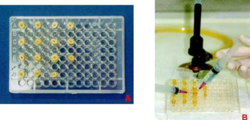
Specimen sterilization
All the specimens were sterilized by autoclaving (20 min at 1218C) and randomly assigned to five cell culture plates (96-wells, Costar, NY, USA), with 15 teeth each (Fig. 1A). The specimens and all the materials used in the experiment were irradiated with 60 Co gamma-rays for degradation of pre-existing LPS.30
Contamination with endotoxin
Escherichia coli055:B5 endotoxin (Sigma, St Louis, USA) was used for the experiments. Under sterile laminar flow, 10ml of a standard solution containing endotoxin were inoculated into the root canals of
60 specimens using a micropipette. Teeth were sealed with pyrogen-free cotton ball and the plates containing the specimens were closed, sealed and incubated for 24 h at 378C in a humidified atmosphere.
Experimental groups
After 24 h, the cervical sealing was removed and the canals lumen was filled with intracanal medicaments.
The 75 specimens were divided into five groups
(nZ15), according to the intracanal medicament
used, as follows:
Group 1: calcium hydroxide (Calen, S.S. White, Rio de Janeiro, Brazil), inserted with ML syringe (SS White, Rio de Janeiro, Brazil);
Group 2: polymyxin B solution (10,000 UI/ml) (Ophtalmos Fo´rmulas Oficiais Ltda, Sa˜o Paulo, Brazil), inserted with non-pyrogenic 1.0 ml plas-tic syringes and 13 mm needles (Injex, Sa˜o Paulo, Brazil);
Group 3: the combination neomycin–polymyxin B-hydrocortisone (OTOSPORIN, Glaxo Wellcome, SP, Brazil), inserted with non-pyrogenic 1.0 ml plastic syringes and 13 mm needles (Injex, Sa˜o Paulo, Brazil);
Group 4 (control group): did not receive any medicament (positive control);
Group 5 (control group): did not receive endo-toxin and any medicament (negative control).
All the specimens were sealed with a pyrogen-free cotton ball. The plates containing the speci-mens were closed, sealed and incubated at 378C in
Figure 1 Model system: (A) Distribution of teeth (nZ15) in cell culture plates (96 wells); (B) Irrigation with
pyrogen-free water to remove all intracanal medicament.
a humidified atmosphere for 7 days. After this period, the root canals were irrigated with 3 ml of pyrogen-free water (Fig. 1B) to remove all intraca-nal medicaments and dried with pyrogen-free paper points. Each specimen was filled with pyrogen-free water and 130ml of root canal content was collected with a non-pyrogenic 1.0 ml plastic syringe (Injex, Sa˜o Paulo, Brazil) to verify the detoxification of endotoxin.
Detoxification of endotoxin—Limulus amebocyte lysate assay
The detoxification of endotoxin was evaluated by Limulus amebocyte lysate assay (Sigma, USA). Limulus is prepared from a lysate of the circulating amebocytes of the horseshoe crab,Limulus poly-phemus. When exposed to minute quantities of endotoxins, the lysate increases in opacity as well as viscosity, forming a hard gel. An aliquot of canal content (100ml) was put into a pyrogen-free tube containing 100ml of Limulus lysate, according to the manufacture’s instructions. The tube was gently mixed and incubated at 378C for 1 h. These proce-dures were repeated for all the specimens. After 1 h incubation, the tubes were gently removed and slowly inverted 1808while observing for evidence of gelation. The formation of a hard gel, which permits complete inversion of the tube without disruption of gel, was considered as positive test. All other results: soft gels, turbidity, increase in viscosity, clear liquid were considered negative test.
Detoxification of endotoxin—antibody production in lymphocyte B culture
Three BALB/c mice with approximately 6 months of age were sacrificed and the spleen of each animal was removed and macerated in a pyrogen-free Falcon tube containing RPMI 1640 medium (Sigma Chemical Company, St Louis, USA), to obtain B-lymphocytes. The tube was centrifuged at 534.8!g for 10 min and the pellet was resuspended in 30 ml of RPMI 1640 medium supplemented with 10% bovine fetal serum. In order to prevent contami-nation of the culture, sterile conditions were maintained by sterile laminar flow.
For cell viability, the trypane blue exclusion assay (0.4%) (Sigma, St Louis, USA) and a buffer to promote the erythrocytes rupture were used. The viable cells count was 4.36!106cells/ml. Approxi-mately 1!106viable cells (250ml) were transferred for each well of non-pyrogenic cell culture plates (24-well, Costar, NY, USA). These plates were incubated for 24 h at 378C in a humidified
atmosphere of 5% carbon dioxide (CO2). B-lympho-cytes of each well of cell cultures plates were stimulate with the remaining 30ml of each root canal content. Fifteen wells contained only pure cell culture consisted the cell culture control group (G6). The plates were incubated at 378C in a humidified atmosphere of 5% CO2and 95% air for 4 days. The enzyme-linked immunosorbent assay (ELISA) was used to determine of total IgM levels in the culture supernatant.
ELISA was performed in three microplates (96-well, Hemobag, Sa˜o Paulo, Brazil). All the plates were coated with mouse anti-immunoglobulin M (Sigma, St Louis, USA) in the concentration of 0.5 mg/ml. The plates were incubated for 2 h at 378C and maintained in the refrigerator until used. In the day of the experiment, the plates were blocked with 0.5% gelatin (G) in phosphate buffered saline (PBS) and 0.2% BSA (45 min at 378C). Culture supernatants (100ml) were added to each well and incubated for 2 h at 378C, followed by washing with 0.1% Tween-20 in PBS (PBS-T). All the tests were performed in dupli-cate. Anti-mouse IgM peroxidase conjugate, diluted 1000 times with PBS-GT (0.5% gelatin and 0.1% Tween 20 in PBS), was added to the plates and incubated for 1 h at 378C. After washing with PBS-T, peroxidase enzyme activity was detected by addition of the substrate, 50ml of o -phenyle-nediamine (1 mg/ml) in a solution of 0.1 M citrate and 0.03% H2O2 for 10 min. Finally, the reaction was stopped with 2.5 N H2SO4. The optical density (OD) of the wells was read at an absorbance of 490 nm on a Bio-Rad Microplate reader Model 3550 (Bio-Rad Laboratories, Hercules, CA). For each specimen, the mean values of OD were obtained and the results were statistically analysed.
Statistical analysis
The results obtained with Limulus lysate assay were statistically analysed using non-parametric Mann– Whitney test, attributing scores 1 to the specimens that presented positive results and score 0 to the specimens that presented negative results. The ELISA results were statistically analysed using ANOVA and Tukey’s test. In all cases,pvalues!
0.05 were considered as statistically significant.
Results
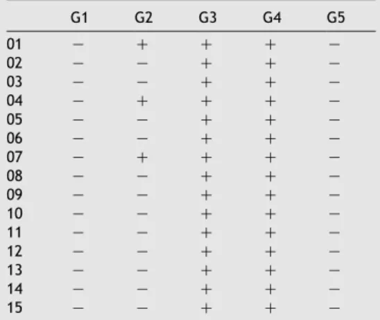
The results of Limulus lysate assay are presented in
specimens of groups 3 and 4 presented gel formation. Fig. 2A and B shows the formation or not of hard gel by Limulus amebocytes lysate. There were no statistically significant differences among groups 1, 2 and 5 (pO0.05) and between groups 3 and 4 (pO0.05). However, the groups 1, 2 and 5
were significantly different from groups 3 and 4 (p!
0.05).
The values of OD obtained by ELISA for all the experimental groups are presented in Table 2. Stimulation of antibodies (IgM) production in B-lymphocytes culture by groups 1 and 6 was smaller and statistically different than groups 2, 3, 4 and 5 (p!0.05). Groups 2 and 5 induced a small increase in the antibodies (IgM) production in relation to the groups 1 and 6. Groups 3 and 4 induced a significant increase of IgM production, statistically different to the other groups (p!0.05).
Discussion
Kakehashi et al.1firstly showed the essential role of bacteria and their products in the development of periapical lesions. Elimination of microorganisms from root canals system by antimicrobial agents has been a major goal of endodontic treatment.26 However, treatment of root canals in teeth with pulp necrosis should not only be concerned with bacterial death, but also the inactivation of endotoxin.16,17Endotoxin has an important role in the inflammatory and immunologic responses of pulpal and periapical tissues, including bone resorption.8,11,12
The presence of endotoxin in teeth with vital or necrotic pulps has already been proven by many researchers,3,18,27however, few studies have eval-uated the effects of intracanal medicaments on LPS using teeth as a model system. Endotoxins as well as bacteria are capable to diffuse through dentinal tubules which are present not only in the canal lumen but also in the entire root canal system.28,29 In the present research, we used single-root human teeth extracted for several reasons, independently of diagnosis, because the endotoxin possibly pre-sent in these teeth was degraded by 608C gamma irradiation.30Selection of teeth was made on the basis of relative dimensions and similarity in morphology and the apical 3 mm of each root were removed to minimize variations such as cracks, apical resorptions and ramifications.
The results of this study showed that calcium hydroxide medication intracanal for 7 days has a detoxifying effect on endotoxin in root canal system. These results are similar to other studies using different methodology that demonstrated calcium hydroxide could alter the biological proper-ties of LPS.6–8,16
Safavi and Nichools6related that calcium hydrox-ide treatment of LPS release elevated quantities of
Table 1 Results obtained by Limulus lysate assay.
G1 G2 G3 G4 G5
01 K C C C K
02 K K C C K
03 K K C C K
04 K C C C K
05 K K C C K
06 K K C C K
07 K C C C K
08 K K C C K
09 K K C C K
10 K K C C K
11 K K C C K
12 K K C C K
13 K K C C K
14 K K C C K
15 K K C C K
G1, calcium hydroxide; G2, polymyxin B; G3, combination neomycin–polymyxin B-hydrocortisone; G4, positive control; G5, negative control.
Figure 2 Results obtained by Limulus lysate assay: (A) gel formation; (B) No gel formation.
hydroxy fatty acids by hydrolysis of ester bonds in the lipid moiety of bacterial LPS. This result suggests that detoxification of LPS by calcium hydroxide may be one of the mechanisms by which this agent exerts its beneficial effects in clinical endodontics. According to Safavi and Nichools7 the prostaglandin E2 production was inhibited in monocytes culture stimulated with calcium hydroxide-treated LPS. These experiments suggested that the biological properties of LPS require the presence of ester-linked hydroxy fatty acid and these linkages are destroyed by treatment with calcium hydroxide. In 1997, Barthel et al.8also evaluated whether the toxic potential of anE. coli
LPS could be reduced or eliminated by calcium hydroxide. It was concluded that calcium hydroxide is able to eliminate the ability of anE. coliLPS to stimulate TNF-a production in peripheral blood monocytes. In this study, we verified that calcium hydroxide is able to alter the ability of endotoxin to stimulate antibodies production in B-lymphocytes culture. The absence of hard gel formation in the Limulus amebocyte lysate assay showed also that calcium hydroxide is able to detoxify LPS, probably by hydrolysis of the link ester-hydroxy fatty acids.
Polymyxin B was also effective in LPS detoxifing in root canal system. Only three teeth treated with this medicament induced the gel formation in the Limulus lysate assay and B-lymphocytes activation was significantly smaller than groups 3 and 4 (p!
0.05). These results are in accordance with other authors22,24 that used polymyxin B treatment in
Gram-negative microorganisms sepsis and endo-toxic shock.
The polymyxin B neutralizing effect in LPS activity was observed for the first time by Neter31 that incubated LPS with polymyxin B and verified that the erythrocytes agglutination, normally induced by LPS, was inhibited. Since then, other authors have demonstrated that polymyxin B can inhibit the endotoxic lethality in chicken, rats, rabbits, dogs and horses.22–24Rifkind and Palmer21 and Rifkind22 showed that polymyxin B prevents endotoxic lethality in chicken embryos and rats, and neutralize the Schwartzman’s reaction in rabbits. They postulated a mechanism of action in which polymyxin B, a cationic antibiotic, covalently couples with the electronegative charge of the A lipid in endotoxin molecule. Morrison and Jacobs32 confirmed this theory in vitro. Polymyxin B has the ability to bind with high affinity to the A lipid portion, altering the three-dimensional confor-mation of the LPS molecule. This conforconfor-mational alteration possibly enables the binding of the complex endotoxin–polymyxin B to monocytes CD14 receptor, inhibiting the liberation of inflam-matory mediators such as the TNF.23In the present study, polymyxin B enabled the B-lymphocytes activation by LPS and also inhibited the gel formation in Limulus amebocyte lysate assay, probably due to the conformational alteration of the endotoxin–polymyxin B complex.
Endotoxins may also evoke pain due to Hage-man factor activation33 or by neurotoxic
Table 2 Optical density (OD) values obtained in groups.
G1 G2 G3 G4 G5 G6
01 0.690 1.033 1.206 1.292 0.763 0.725
02 0.825 0.924 1.230 1.284 0.722 0.491
03 0.909 0.994 1.270 1.260 0.822 0.504
04 0.680 0.996 1.254 1.287 0.994 0.489
05 0.841 0.866 1.200 1.268 0.779 0.614
06 0.735 0.878 1.121 1.308 0.819 0.656
07 0.730 0.933 1.115 1.350 0.849 0.562
08 0.873 0.875 1.205 1.196 0.806 0.675
09 0.856 0.695 1.079 1.314 0.963 0.678
10 0.739 0.865 1.152 1.287 0.952 0.491
11 0.556 0.875 1.288 1.242 0.990 0.764
12 0.569 0.671 1.191 1.265 1.004 0.766
13 0.735 0.753 1.181 1.273 0.966 0.698
14 0.740 0.831 1.185 1.197 0.945 0.782
15 0.399 0.846 1.127 1.213 1.007 0.764
Means 0.725 0.869 1.187 1.269 0.892 0.643
SD 0.136 0.103 0.059 0.045 0.100 0.111
G1, calcium hydroxide; G2, polymyxin B; G3, combination neomycin–polymyxin B-hydrocortisone; G4, positive control; G5, negative control; G6, cell culture control group.
properties. Schein and Schilder,3 Horiba et al.27 and Khabbaz et al.19 showed that symptomatic teeth present greater amount of endotoxin than asymptomatic teeth. Many dentists use the combination neomycin–polymyxin B-hydrocorti-sone to treat teeth with vital pulp due to its satisfactory anti-inflammatory action and capacity of preserving the periapical tissues.34 In the present study, this medicament showed gel formation in all the tested specimens and high values of OD, inducing significant increase in antibodies (IgM) production. This value was very similar to those obtained in G4 (no medication). These results disagree with those obtained by other authors35,36 that used this medicament when treating of supurative medium otitis caused by Gram-positive and negative microorganisms. However, none of these studies evaluated the neutralizing activity of the combination neomy-cin–polymyxin B-hydrocortisone on LPS. Lopes and Siqueira37 studied the antimicrobial activity of this medicament on salivary microorganisms. After aerobiosis and anaerobiosis incubation, it was observed that this medication produced inhibitory effects only in oxygen presence. There-fore, it was effective against aerobic and facultative microorganisms but not against strictly anaerobic bacteria. In the present study, we investigated the detoxifying capacity of the combination neomycin–polymyxin B-hydrocorti-sone on the LPS and did not observe any activity. This absence of activity could be due to the solution pH, as we observe that polymyxin B presented a pH value of approximately 6.4 while the combination neomycin–polymyxin B-hydrocor-tisone presented a pH value of 5.4. Another possibility is the interference of one of the components on the polymyxin B. These and other alterations could have affected the endo-toxins detoxifying action. In this way, the combination neomycin–polymyxin B-hydrocorti-sone should have limited use in the endodontics.
Conclusions
Under the conditions of this study, it can be concluded that the calcium hydroxide and poly-myxin B intracanal medicaments detoxified endo-toxin in root canals and altered the properties of LPS to stimulate the antibodies production by B-lymphocytes. The combination neomycin–poly-myxin B-hydrocortisone did not detoxify endotoxin.
Acknowledgements
The authors gratefully acknowledge the financial support provided by FAPESP (Fundac¸a˜o de Amparo a` Pesquisa do Estado de Sa˜o Paulo).
References
1. Kakehashi S, Stanley HR, Fitzgerald RJ. The effects of surgical exposures of dental pulps in germ-free and conventional laboratory rats.Oral Surgery, Oral Medicine, Oral Pathology1965;20:340–9.
2. Wittgow Jr. WC, Sabiston Jr. CB. Microorganisms from pulpal chambers of intact teeth with necrotic pulps.Journal of Endodontics1975;1(5):168–71.
3. Schein B, Schilder H. Endotoxin content in endodontically involved teeth.Journal of Endodontics1975;1(1):19–21. 4. Tani-Ishii N, Wang CY, Tanner A, Stashenko P. Changes in
root canal microbiota during the development of rat periapical lesions. Oral Microbiology and Immunology 1994;9(2):129–35.
5. Petsch D, Anspach FB. Endotoxin removal from protein solutions.Journal of Biotechnology2000;76(2/3):97–119. 6. Safavi KE, Nichols FC. Effects of calcium hydroxide on
bacterial lipopolysaccharide.Journal of Endodontics1993; 19(2):76–8.
7. Safavi KE, Nichols FC. Alteration of biological properties of bacterial lipopolysaccharide by calcium hydroxide treat-ment.Journal of Endodontics1994;20(3):127–9. 8. Barthel CR, Levin LG, Reisner HM, Trope M. TNF-arelease in
monocytes after exposure to calcium hydroxide treated Escherichia coliLPS.International Endodontic Journal1997; 30(3):155–9.
9. Morse DR. Endodontic microbiology in the 1970s. Inter-national Endodontic Journal1981;14(2):69–79.
10. Farber PA, Seltzer S. Endodontic microbiology. I. Etiology. Journal of Endodontics1988;14(7):363–71.
11. Wang C, Stashenko P. The role of interleukin-1ain the pathogenesis of periapical bone destruction in a rat model system.Oral Microbiology and Immunology1993;8(1):50–6. 12. Dwyer GT, Torabinejad M. Radiographic and histologic evaluation of the effect of endotoxin on the periapical tissues of the cat.Journal of Endodontics1981;7(1):31–5. 13. Pitts DL, Williams BL, Morton Jr. TH. Investigation of the role
of endotoxin in periapical inflammation.Journal of Endo-dontics1982;8(1):10–18.
14. Yamasaki M, Nakane A, Kumazawa M, Hashioka K, Horiba N, Nakamura H. Endotoxin and Gram-negative bacteria in the rat periapical lesions.Journal of Endodontics1992;18(10): 501–4.
15. Dahle´n G, Magnusson BC, Mo¨ller A. Histological and histochemical study of the influence of lipopolysaccharide extracted fromFusobacterium nucleatumon the periapical tissues in the monkeyMacaca fascicularis.Archives of Oral Biology1981;26:591–8.
16. Nelson-Filho P, Leonardo MR, Silva LAB, Assed S. Radio-graphic evaluation of the effect of endotoxin (LPS) plus calcium hydroxide on apical and periapical tissues of dogs. Journal of Endodontics2002;28:694–6.
17. Silva LAB, Nelson-Filho P, Leonardo MR, Rossi MA, Pansani CA. Effect of calcium hydroxide on bacterial endotoxin in vivo.Journal of Endodontics2002;38:94–8.
18. Khabbaz MG, Anastasiadis PL, Sykaras SN. Determination of endotoxins in caries: association with pulpal pain. Inter-national Endodontic Journal2000;33(2):132–7.
19. Khabbaz MG, Anastasiadis PL, Sykaras SN. Determination of endotoxins in the vital pulpl of human carious teeth: association with pulpal pain.Oral Surgery, Oral Medicine, Oral Pathology2001;91(5):587–93.
20. Barbosa CAM, Gonc¸alves RB, Siqueira Jr. JF, Uzeda M. Evaluation of the bacterial activities of calcium hydroxide, chlorhexidine, and camphorated paramonochlorophenol as intracanal medicament. A clinical and laboratory study. Journal of Endodontics1997;23(5):297–300.
21. Rifkind D, Palmer JD. Neutralization of endotoxin toxicity in chick embryos by antibiotics.Journal of Bacteriology1966; 92(4):815–9.
22. Rifkind D. Prevention by polymyxin B of endotoxin lethality in mice.Journal of Bacteriology1967;93(4):1463–4. 23. Bucklin SE, Lake P, Lo¨gdberg L, Morrison DC. Therapeutic
efficacy of a polymyxin B-dextran 70 conjugate in exper-imental model of endotoxemia.Antimicrobial Agents and Chemotherapy1995;39(7):1462–6.
24. Parviainen AK, Barton MH, Norton NN. Evaluation of polymyxin B in an ex vivo model of endotoxemia in horses. American Journal of Veterinary Research2001;62(1):72–6. 25. Evans ME, Feola DJ, Rapp RP. Polymyxin B sulfate and
colistin: old antibiotics for emerging multiresistant Gram-negative bacteria.The Annals of Pharmacotherapy1999; 33(9):960–7.
26. Bystro¨m A, Sundqvist G. Bacteriologic evaluation of the effect of 0.5 percent sodium hypochlorite in endodontic therapy.Oral Surgery, Oral Medicine, Oral Pathology1983; 55:307–12.
27. Horiba N, Maekawa Y, Abe Y, Ito M, Matsumoto T, Nakamura H. Correlations between endotoxin and clinical symptoms or radiolucent areas in infected root canals.Oral Surgery, Oral Medicine, Oral Pathology1991;71(4):492–5.
28. Nissan R, Segal H, Pashley D, Stevens R, Trowbrigde H. Ability of bacterial endotoxin to diffuse through human dentin.Journal of Endodontics1995;21(2):62–4. 29. Oliveira LD, Carvalho CAT, Koga-Ito CY, Valera MC,
Jorge AOC. Avaliac¸a˜o in vitro da capacidade de difusa˜o da endotoxina pelos tu´bulos dentina´rios.Pesquisa Odontolo´gica Brasileira2003;17(special issue):173.
30. Csako G, Elin RJ, Hochstein D, Tsai CM. Physical and biological properties of US standard endotoxin EC after exposure to ionizing radiation.Infection and Immunity1983; 41(1):190–6.
31. Neter E. Bacterial hemagglutination and hemolysis. Bac-teriological Reviews1956;20:166–88.
32. Morrison DC, Jacobs DM. Binding of polymyxin B to the lipid A portion of bacterial lipopolysaccharides.Immunochemistry 1976;13(10):813–8.
33. Seltzer S, Farber PA. Microbiologic factors in endodontology. Oral Surgery, Oral Medicine, Oral Pathology 1994;78: 634–45.
34. Holland R, et al. Emprego da associac¸a˜o corticostero ´ide-antibio´tico durante o tratamento endodoˆntico. Revista Paulista de Odontologia1980;1(2):4–7.
35. Tong MCF, Woo JKS, Hasselt CAV. A double-blind compara-tive study of ofloxacin otic drops versus neomycin–polymyxin B-hydrocortisone otic drops in the medical treatment of chronic suppurative otitis media.The Journal of Laryngology and Otology1996;4:309–14.
36. Miro´ NMD. Controlled multicentes study on chronic suppura-tive otitis media treated with topical applications of ciprofloxacin 0,2% solution in single-dose containers or combination of polymyxin B, neomycin, and hydrocortisone suspension.Otolaryngology Head and Neck Surgery2000; 123(5):617–23.
37. Lopes HP, Siqueira Junior JF.Endodontia: biologia e te´cnica. Rio de Janeiro, Brazil: MEDSI; 1990.
3.2 In vitro effects of endodontic irrigants on endotoxins in root
canals*
ABSTRACT
Objective. The objective of this study was to evaluate the effects of
endodontic irrigants on endotoxins in root canals. Study design.
Ninety-eight single-root human teeth were used. Escherichia coli endotoxin was
inoculated into 84 root canals. All root canals were enlarged and assigned
to 7 groups (n = 14), according to solution used. Group 1 (G1): 2.5%
NaOCl; G2: 5.25% NaOCl; G3: 2% chlorhexidine; G4: 0.14% calcium
hydroxide; G5: polymyxin B; G6: positive control, saline solution; G7:
negative control (no endotoxin). Two samplings of root canal were
accomplished: immediate and after 7 days. Detoxification of endotoxin
was evaluated by Limulus assay and antibody production in B-lymphocyte
culture. Results were analyzed by Kruskal-Wallis/Dunn and
ANOVA/Tukey. Results. At the immediate and second samplings, groups
G4, G5, and G7 presented the best results, significantly different from
groups G1, G2, G3, and G6 (P < 0.05). Conclusions. Calcium hydroxide
and polymyxin B detoxified endotoxin in root canals and altered properties
of LPS to stimulate the antibody production by B-lymphocytes. Sodium
hypochlorite and chlorhexidine did not detoxify endotoxin.
_____________________
In vitro effects of endodontic irrigants on endotoxins in root canals
Luciane Dias de Oliveira, MsC, PhD,aAntonio Olavo Cardoso Jorge, MsC, PhD,b Cláudio Antonio Talge Carvalho, MsC, PhD,cCristiane Yumi Koga-Ito, MsC, PhD,dand Marcia Carneiro Valera, MsC, PhD,eSao Paulo, Brazil
UNESP PAULISTA STATE UNIVERSITY
Objective.The objective of this study was to evaluate the effects of endodontic irrigants on endotoxins in root canals.
Study design.Ninety-eight single-root human teeth were used.Escherichia coliendotoxin was inoculated into 84 root canals. All root canals were enlarged and assigned to 7 groups (n!14), according to solution used. Group 1 (G1): 2.5% NaOCl; G2: 5.25% NaOCl; G3: 2% chlorhexidine; G4: 0.14% calcium hydroxide; G5: polymyxin B; G6: positive control, saline solution; G7: negative control (no endotoxin). Two samplings of root canal were accomplished: immediate and after 7 days. Detoxification of endotoxin was evaluated by Limulus assay and antibody production in B-lymphocyte culture. Results were analyzed by Kruskal-Wallis/Dunn and ANOVA/Tukey.
Results.At the immediate and second samplings, groups G4, G5, and G7 presented the best results, significantly different from groups G1, G2, G3, and G6 (P".05).
Conclusions.Calcium hydroxide and polymyxin B detoxified endotoxin in root canals and altered properties of LPS to stimulate the antibody production by B-lymphocytes. Sodium hypochlorite and chlorhexidine did not detoxify endotoxin.(Oral Surg Oral Med Oral Pathol Oral Radiol Endod 2007;104:135-42)
Bacteria and their by-products have a fundamental role in the initiation and perpetuation of pulpal and perira-dicular disease. Analyses of the microflora in infected root canals with necrotic pulps have revealed that Gram-negative anaerobic bacteria are the predominant microorganisms,1,2mainly during the active phase of rapid lesion expansion.3
Gram-negative bacteria contain endotoxin in their outer cell membrane.4This endotoxin or
lipopolysac-charide (LPS) is released during cell duplication or after cell death. Endotoxins are responsible for several important biological effects, such as polymorphonu-clear leucocytes (PMN), release of collagenase, macro-phage activation, and release of bioactive inflammatory mediators such as the cyclooxygenase pathway prod-ucts; platelet-activating factor (PAF); tumor necrosis factor (TNF); interleukins (IL-1, IL-6, and IL-8);
su-peroxide (O2
–); nitric oxide; interferon
#,$, and%5-7; complement system activation; B-lymphocyte poly-clonal activation; cytotoxicity; hemodinamics modifi-cations; fever induction; and osteoclast attraction.8
These factors are important in the development and maintenance of inflammatory reaction and bone resorp-tion in the periapical region.9
The inoculation of LPS into root canals of many animals (monkeys, dogs, cats) induced inflammatory reactions and bone resorption in the periradicular tissues of all the experiment teeth,10-15
demonstrat-ing the important role of endotoxin in the pathogen-esis of periapical lesions. Khabbaz et al.16,17showed the presence of endotoxins in carious lesions of symptomatic and asymptomatic teeth and in the pul-pal tissue of the carious teeth, suggesting that LPS may play an important role in the pathogenesis of human pulpal diseases.
The major goal of endodontic treatment is the elim-ination of bacteria from the root canal system by me-chanical and chemical means and a wide variety of antimicrobial agents have been used for this purpose.5
However, it is possible that even though bacteria are removed from the root canal, endotoxins capable of maintaining or inducing apical periodontitis can re-main,7interfering in the success of endodontic treat-ment. Therefore, the treatment of infected root canals should not only be concerned with bacterial death, but also the inactivation of endotoxin.11,12
During instrumentation of root canal, frequent irri-gation is recommended with the main purpose of re-moving debris from the canal, killing microorganisms,
The authors gratefully acknowledge the financial support provided by FAPESP (Fundação de Amparo à Pesquisa do Estado de São Paulo, to L.D.O.).
Received from the School of Dentistry, Paulista State University-UNESP.
aDepartment of Oral Biosciences and Diagnosis.
bChairman of Microbiology and Immunology, Department of Oral
Biosciences and Diagnosis.
cDepartment of Restorative Dentistry. dDepartment of Oral Biosciences and Diagnosis. eDepartment of Restorative Dentistry.
Received for publication Feb 28, 2006; returned for revision Sep 13, 2006; accepted for publication Nov 22, 2006.