Re nal e ffe cts o f uro guanylin
and guanylin
in vivo
1Department of Internal Medicine, Division of Infectious Diseases,
VA Medical Center, and 2Department of Physiology and Biophysics, University of Kentucky, Lexington, KY, USA
3Division of Gastroenterology and Division of Molecular and Cellular Physiology,
University of Cincinnati, Cincinnati, O H, USA
4Department of Pharmacology,Truman VA Medical Center, University of Missouri,
Columbia, MO , USA S.L. Carrithers1, M.J. Hill1,
B.R. Johnson1, S.M. O ’Hara1, B.A. Jackson2, C.E. O tt2, J. Lorenz3, E.A. Mann3, R.A. Giannella3, L.R. Forte4 and R.N. Greenberg1
Abstract
Uroguanylin and guanylin are newly discovered endogenous heat-stable peptides that bind to and activate a membrane bound guanylyl cyclase signaling receptor (termed guanylyl cyclase C; GC-C). These peptides are not only found in blood but are secreted into the lumen of the intestine and effect a net secretion of electrolytes (Na+, K+, Cl-, HCO
3-) and fluid into the intestine via a cyclic guanosine-3',5'-monophosphate (cGMP) mechanism. GC-C is also the receptor for
Escherichia coli heat-stable enterotoxin (STa) and activation by STa results in a diarrheal illness. Employing mouse renal in vivo models, we have demonstrated that uroguanylin, guanylin, and STa elicit natriuretic, kaliuretic, and diuretic effects. These biological responses are time- and dose-dependent. Maximum natriuretic and kaliuretic effects are observed within 30-40 min following infusion with phar-macological doses of the peptides in a sealed-urethra mouse model. Our mouse renal clearance model confirms these results and shows significant natriuresis following a constant infusion of uroguanylin for 30 min, while the glomerular filtration rate, plasma creatinine, urine osmolality, heart rate, and blood pressure remain constant. These data suggest the peptides act through tubular transport mechanisms. Consistent with a tubular mechanism, messenger RNA-differential display PCR of kidney RNA extracted from vehicle- and uroguanylin-treated mice show the message for the Na+/K+ ATPase g-subunit is down-regulated. Interestingly, GC-C knockout mice (Gucy2c -/-) also exhibit significant uroguanylin-induced natriuresis and kaliuresis in vivo, suggesting the presence of an alternate receptor signaling mech-anism in the kidney. Thus, uroguanylin and guanylin seem to serve as intestinal and renal natriuretic peptide-hormones influencing salt and water transport in the kidney through GC-C dependent and independ-ent pathways. Furthermore, our recindepend-ent clinical probe study has re-vealed a 70-fold increase in levels of urinary uroguanylin in patients with congestive heart failure. In conclusion, our studies support the concept that uroguanylin and guanylin are endogenous effector pep-tides involved in regulating body salt and water homeostasis. Co rre spo nde nce
R.N. Greenberg
Department of Internal Medicine Division of Infectious Diseases University of Kentucky VA Medical Center
800 Rose Street, UKMC-MN668-A Lexington, KY 40536-0084 USA
Fax: + 1-606-323-1020 E-mail: rngree01@ pop.uky.edu Presented at the Meeting “NO Brazil, Basic and Clinical Aspects of Nitric O xide”, Foz do Iguaçu, PR, Brazil, March 10-13, 1999.
Received May 28, 1999 Accepted July 8, 1999
Ke y wo rds
·Guanylyl cyclase C ·Heat-stable enterotoxin ·Kidney
·Natriuresis
Intro ductio n
A considerable body of evidence sup-ports a role for the guanylyl cyclase C (GC-C)-cyclic guanosine-3',5'-monophosphate (cGMP) pathway in the control of fluid and electrolyte transport in mammals (1). Early studies on the mechanism of action of the
Escherichia coli heat-stable enterotoxin
(STa), which are secreted by enteric bacteria and cause travelers diarrhea, demonstrated its signal transduction to be initiated by GC-C (1). However, the STa/GGC-C-GC-C ligand recep-tor axis has been shown to utilize an endog-enous pathway utilizing cell-specific secre-tion of STa-like mimic peptides involving the intestine and other organs (2). Uroguany-lin and guanyUroguany-lin, two newly identified en-dogenous peptides of 13-15 amino acids, were shown to bind to and activate GC-C in the intestinal mucosa and cause significant elevation of intracellular cGMP resulting in increased Cl
secretion from the gut (3,4). Our objective was to assess the role of uroguanylin and guanylin, which we have recently shown to increase cGMP accumula-tion in the intestinal and renal epithelium (5,6). Uroguanylin and guanylin are initially expressed as inactive propeptides. Subse-quent activation of the active peptide forms is accomplished by cleavage and release of the COOH-terminal amino acid moieties (2). Biologically inactive (prohormone form) and active moieties of uroguanylin and guanylin circulate in the plasma of mammals, suggest-ing an endocrine mechanism for their action in addition to the autocrine and paracrine fashion in which they have been shown to act in the intestine (2,7).
Most of the research to date has been involved in the characterization of the uro-guanylin and uro-guanylin signaling pathway in the intestine. However, due to the role that these peptides play in electrolyte and water homeostasis, the kidney should be investi-gated. The overall goal of the studies out-lined in this report was to test the
fundamen-tal hypothesis that uroguanylin and guanylin act as natriuretic and diuretic peptide hor-mones which act on the kidney to regulate sodium homeostasis during normal salt in-take and disorders involved in imbalances of fluid and electrolytes. Our studies will focus on the biological actions of uroguanylin and guanylin on renal function employing an in vivo sealed or catheterized urethra mouse model. These experiments are followed by a study measuring the urinary uroguanylin lev-els in patients with congestive heart failure, a disorder characterized by sodium retention and fluid overload with resulting pulmonary and peripheral congestion.
Expe rim e ntal pro ce dure s
Suckling mo use inte stinal fluid assay
Uroguanylin, guanylin, and STa were tested for their ability to induce intestinal fluid accumulation in newborn mice as de-scribed previously (5). ICR-Harlan Sprague-Dawley (HSD) suckling mice 2-4 days old were dosed peros with 0.1 ml of test solu-tion. The injections contained vehicle (HEPES) alone or the peptides with and without the protease inhibitors chymostatin (0.1 mmol) and aprotinin (0.67 units). After administration of vehicle or peptide ago-nists, the mice were kept at room tempera-ture for 3 h. The mice were then killed, intestinal and body weights measured, and a ratio of the intestinal weight to remaining body weight was calculated. A ratio of 0.0875 represents one mouse unit of activity. The secretion activity (one unit) of STa was 3.75 ng/mouse (5); uroguanylin was 41.5 ng/ mouse, and guanylin was142.9 ng/mouse.
The se ale d-m o use re nal functio n assay
methoxyflurane in a desiccator until the mouse was unconscious. At this time, the abdominal region was shaved and a 25-gauge needle was inserted through the skin to the visible bladder. Urine was aspirated and the bladder emptied. The urethra was cannu-lated by PE-5 tubing or sealed shut with Krazy glue and the mouse was placed in a restrainer to limit excessive movement. Fifty microliters of test solution (0.2% Evans blue dye (indicator for intravenous injec-tion) with or without peptide) was injected into the tail vein. After the indicated time, the animal was again exposed to methoxy-flurane, the bladder contents emptied, and the urine volume recorded. The mouse was then sacrificed by open chest pneumothorax. Urine sodium, potassium, and osmolality were measured. Sodium and potassium lev-els were determined by flame photometry (Instrumentation Laboratory Autocal Flame Photometer model 643, Lexington, MA, USA) and osmolality was measured by an osmometer (Wescor 5100B Vapor Pressure Osmometer, Logan, UT, USA). Osmolality was not reported because the values did not differ significantly. Prior to the experiment, the mice were allowed to drink ad libitum. Their diet had consisted of 18% crude pro-tein, 9% crude fat, 4% crude fiber, and 11% crude moisture (Prolab Rat, Mouse, Hamster 2000, Harlan, Indianapolis, IN, USA).
Cyclic GMP accumulatio n assay in T84 ce lls
The detection and quantification of uro-guanylin extracted from urine samples was performed using a de novo T84 cell stimula-tion bioassay by previously described meth-ods (8). T84 cells were grown as monolayers in 24-well plates as described previously (9). The bioactivity, which is represented as pmol cGMP produced min-1 ml-1, was a direct
measure of the amount of GC-C stimulating peptide present in a particular sample. Uro-guanylin bioactivity was assessed at acidic pH (5.5) (8). Briefly, the sample was layered
onto the T84 cell monolayers for 40 min at 37oC. The media was removed, and the cells
were washed twice with serum-free media. Cells were lysed and peptide-induced cGMP was measured using a specific RIA (a-cGMP antibody was kindly provided by Donald C. Robertson, Ph.D., University of Idaho, Mos-cow, ID, USA), and protein was measured (BioRad, Hercules, CA, USA) using bovine serum albumin as standard. The effects ob-served on cGMP accumulation in this assay under acidic conditions reflect primarily, if not completely, the bioactivity of uroguany-lin in the urine. The effects of other known urine peptides were compared with those of synthetic uroguanylin and guanylin. Under acidic conditions employed in this bioassay, guanylin, the other known endogenous GC-C activating peptide, is approximately 150 times less potent than uroguanylin. Sensitiv-ity for this bioassay approaches 1 pmol cGMP well-1 mg protein-1, which is similar to the
sensitivity found previously (4,8). Peptides of the atrial natriuretic peptide (ANP) family (10-6 M; ANP, brain natriuretic peptide
(BNP), C-type natriuretic peptide (CNP), urodilatin) do not stimulate increases in in-tracellular cGMP in this assay, nor does sodium nitroprusside (10-3
M) or urinary cGMP (since it is removed during the pre-parative Sep-Pak step) (3,4,10).
Me sse nge r RNA diffe re ntial display PCR
Differential display (11) was performed on 100 ng of normalized DNase-treated RNA in a 20-µl reverse transcription reaction con-taining 50 mM Tris-HCl, pH 8.3, 75 mM KCl, 3 mM MgCl2, 10 mM DTT, 20 µM each
dNTP, 2.5 µM anchored primer, and 15 U/µl MMLV reverse transcriptase (Life Technolo-gies, Gaithersburg, MD, USA). Anchored primers were a series of three 21-mers, each with an EcoRI restriction site, T11 stretch and
a single 3' base, either G, A, or C. The reaction was incubated 60 min at 37oC and
then for 5 min at 95o
C, followed by a 4o
hold. Utilizing a GeneAmp PCR kit (Perkin-Elmer, Norwalk, CT, USA), 20-µl PCR re-actions, with 2 µl of the reverse transcriptase reaction as template, were prepared in dupli-cate containing 10 mM Tris-HCl, pH 8.3, 50 mM KCl, 1.5 mM MgCl2, 0.001% gelatin,
20 µM each dNTP, 1 µM anchored primer, 1 µM arbitrary decamer-primer (GEP Services, Oklahoma City, OK, USA), 12.5 µCi (1 Ci = 37 GBq) 35
S-dATP (DuPont/NEN, Wilming-ton, DE, USA), and 0.5 U of AmpliTaq per reaction. Ten different arbitrary primers were employed in PCR. Following an initial 60-s denaturation step at 95o
C, thermal cycling consisted of: 94o
C, 30 s; 40o
C, 120 s; 72o
C, 30 s; 40 cycles. A final extension of cDNA at 72o
C for 5 min completed PCR cycling, which was followed by a 4o
C hold. Ampli-fied samples were subjected to electrophore-sis under denaturing conditions using acry-lamide (6%)/urea (40%) DNA sequencing gels prepared with 1x TBE buffer (89 mM Tris base, 89 mM boric acid, 2 mM EDTA, pH 8.0). Five microliters of formamide load-ing dye (95% formamide, 20 mM EDTA, 0.05% bromophenol blue, 0.05% xylene cyanol, pH 8.0) was added to each 20 µl
sample, then heat-denatured at 90o
C for 5 min before loading 8 µl per well with dupli-cate reactions. The gels were run at 85 W for 4 h, dried onto Whatman 3-mm paper, and exposed to X-ray film for 2-4 days at -70oC.
The cDNA bands that were visually selected for sub-cloning showed a difference in the display pattern between the Ugn-treated and vehicle (saline)-treated mice. Luminescent paint dots were used to align the autoradio-gram and gel, and only the single most in-tense center band of a multiple band series was excised from the dried gel. This excised band with attached Whatman paper was re-hydrated in 100 µl of water and eluted at room temperature for 10 min followed by 100o
C for 15 min. After cooling, the paper and gel pieces were removed and DNA was precipitated, washed in ethanol, and resus-pended in H2O. The band was amplified by
PCR again, and an aliquot was analyzed on a 1.5% agarose gel to confirm amplification of the cDNA band of interest. The product was ligated into the pNoT7 shuttle vector (5 Prime-3 Prime, Inc., Boulder, CO, USA) for sequencing, which was performed by the University of Kentucky Sequencing Core Facility.
Re sults
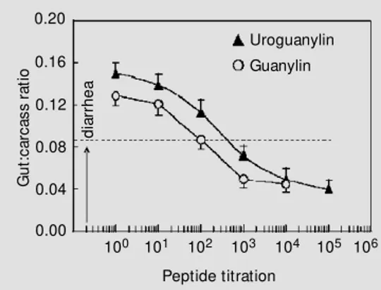
Uroguanylin and guanylin are endoge-nous heat-stable peptides of 13-15 amino acids containing 2 disulfide bonds that are mimicked by STa in primary and secondary structures (Figure 1). Figure 1 shows the primary structures of STa, uroguanylin, and guanylin. Both uroguanylin and guanylin are intestinal peptides that activate guanylyl cy-clase molecules on the luminal surface of the intestinal epithelium. We assessed the po-tency of uroguanylin and guanylin with the suckling mouse intestinal fluid assay. Figure 2 demonstrates that uroguanylin is more po-tent in causing intestinal secretion of fluid than guanylin, but neither peptide is as po-tent in the diarrheal response as STa:
UROGUANYLIN
Figure 1 - Primary structures of uroguanylin, guanylin, and the Escherichia coli heat-stable
enterotoxin (STa). * Not show n are additional amino acid residues on the amino-terminus of STa that are “ N-S-S” for STa-h (isolated originally from human) and “ H-T” for STa-p (isolated originally from sw ine).
Opossum
Human
Rat
GUANYLIN
Opossum
Human
Mouse/Rat
E. coli STa-h*
guanylin, 142.9 ng/mouse < uroguanylin, 41.5 ng/mouse < STa, 3.75 ng/mouse (5). This assay was employed with these pep-tides because we needed a quantitative as-sessment of both uroguanylin and guanylin before in vivo and in vitro renal function assays could be performed.
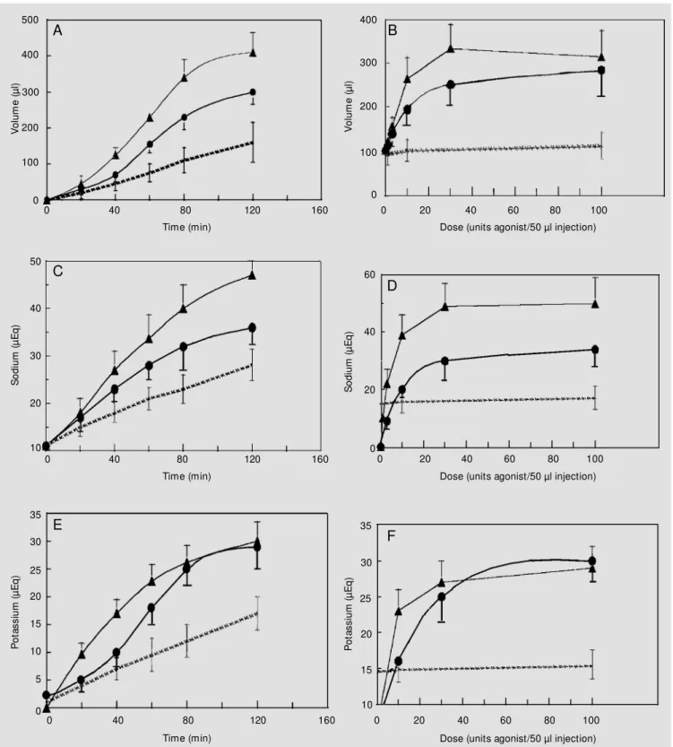
To address the question whether urogua-nylin and guaurogua-nylin affect renal function in vivo, experiments were designed to measure the urine output, sodium and potassium ex-cretion, and urine osmolality after intrave-nous injections of the agonist-peptides. We also asked the question if these effects medi-ated by uroguanylin and guanylin occurred in a time-dependent and dose-response man-ner. The sealed-mouse renal function assay was employed to test these questions. Figure 3 (left panels) demonstrates that both uro-guanylin and uro-guanylin caused an increase in urine volume, and total urinary sodium and potassium excretion in a time-dependent fash-ion when compared to control (vehicle)-in-jected mice. The urine osmolalities did not change (data not shown). Eighty minutes post-injection showed the highest signifi-cant increase in urine volume for both the uroguanylin- and guanylin-stimulated re-sponses to exogenous peptide treatment. Also, uroguanylin was more active in the in vivo renal function assay that guanylin (even when the guanylin injection solution con-tained the protective protease inhibitor cock-tail of chymostatin and aprotinin) (5). This pattern was shown to be consistent in the natriuretic and kaliuretic effects mediated by uroguanylin and guanylin. A dose-response for diuresis, natriuresis, and kaliuresis was also observed for uroguanylin and guanylin (Figure 3, right panels). As previously dem-onstrated in the time course for the renal effects of uroguanylin and guanylin, uronylin elicited a greater natriuresis than gua-nylin. Maximal diuresis, natriuresis, and ka-liuresis were observed at 80 min post-injec-tion with approximately 10-30 units of pep-tide.
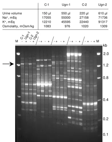
To examine the molecular events induced by uroguanylin during the sealed-urethra mouse renal function assay, we employed differential display PCR of RNA from treated and control mice (Figure 4). Control mice were treated with an intravenous injection of vehicle only, whereas the experimental mice were treated with bioactive uroguanylin (10 units of peptide (as quantitated by the suck-ling mouse assay) for 80 min). An increase in urine volume, and total urinary sodium and potassium was observed for two mice. The kidneys were removed for RNA imme-diately following the assay endpoint. Re-verse transcription followed by PCR and electrophoresis demonstrated that multiple amplified products are up- or down-regu-lated. The arrow shown in Figure 4 is the PCR-amplified product that we chose to ex-pand upon. This down-regulated DNA was cut out of the gel, subcloned, and sequenced. The protein translated from this gene was the Na+
/K+
ATPase g-subunit. We suggest that uroguanylin treatment iv induces a down-regulation of the gene that constitutes one of the major protein sodium pumps in the neph-ron.
To determine if the effects of uroguany-lin in the kidney are mediated by its receptor, GC-C, we employed the sealed-mouse renal function assay on GC-C deficient mice (12). These transgenic mice were developed to test for the impact of GC-C mediated events in the mammal. Using an optimum time and concentration of uroguanylin in the assay,
Figure 2 - Assessment of po-tency for uroguanylin and guany-lin em ploying t he suckguany-ling mouse intestinal fluid assay. U-roguanylin (triangles) and guany-lin (circles) w ere serially diluted before a 0.1-ml solution in HEPES w as injected into suckling mice (N = 4-6 mice per dose). Diar-rhea is assessed as the gut/car-cass ratio over 0.0875 (dashed line). Results are reported as the mean ± SEM .
G
u
t:
c
a
rc
a
s
s
r
a
ti
o
0.20
100 101 102 103 104 105 106
Peptide titration 0.16
0.12
0.08
0.04
0.00
Uroguanylin Guanylin
d
ia
rr
h
e
V
o
lu
m
e
(
µ
l)
500
400
300
200
100
0
V
o
lu
m
e
(
µ
l)
400
300
200
100
0
0 40 80 120 160
Time (min)
0 20 40 60
Dose (units agonist/50 µl injection)
80 100
S
o
d
iu
m
(
µ
E
q
)
50
40
20
10 30
S
o
d
iu
m
(
µ
E
q
)
60
40
20
0
0 40 80 120 160
Time (min)
0 20 40 60
Dose (units agonist/50 µl injection)
80 100
P
o
ta
s
s
iu
m
(
µ
E
q
)
35
30
20
15 25
10
5
0
P
o
ta
s
s
iu
m
(
µ
E
q
)
35
30
25
20
10 15
0 40 80 120 160
Time (min)
0 20 40 60
Dose (units agonist/50 µl injection)
80 100
Figure 3 - Uroguanylin and guanylin elicit a time-dependent (using 10 units peptide) and dose-dependent (using 80 min) increase in urine volume, and
total urinary sodium and potassium in the sealed-mouse renal function assay. Panels A, B, Increase in diuresis w ith 10 units of uroguanylin and
guanylin; Panels C, D, increase in natriuresis; Panels E, F, increase in kaliuresis. Uroguanylin (triangles), guanylin (circles), control (dotted lines). Data
points are represented as the mean ± SEM of six or more mice.
A B
D C
GC-C null mice responded to exogenous uroguanylin in a similar fashion to normal wildtype mice (Table 1). The osmolalities for each animal are similar. These data sug-gest that in the kidney, uroguanylin, and possibly guanylin, are mediated by a GC-C independent mechanism.
D iscussio n
Uroguanylin and guanylin are two newly identified intestinal peptides that cause Cl
-secretion into the lumen of the gut via cGMP mechanisms. We tested the hypothesis that due to their secretagogue nature both pep-tides should influence renal function. We tested our hypothesis employing an in vivo
model; the sealed-mouse renal function as-say. Through comparing the effects of uri-nary fluid volume and increased sodium and potassium levels in the urine of vehicle sham-injected mice to agonist-treated mice, one can obtain information about the renal func-tion of a peptide-agonist. We demonstrated that both uroguanylin and guanylin induce diuresis, natriuresis, and kaliuresis in vivo in a time- and dose-dependent fashion. Uro-guanylin is more active in causing a natri-uretic effect in our model. However, guany-lin appears to have as much of an effect on kaliuresis as uroguanylin, suggesting that guanylin could act through a different mech-anism of action than uroguanylin.
Differential display RT-PCR of the RNA extracted from the kidneys of the sealed-mouse assay show that the g-subunit of Na+
/ K+
ATPase is down-regulated by uroguany-lin treatment. It is not known if down-regula-tion of the g-subunit influences reabsorption of sodium in the nephron. However, inhibi-tion of the channel protein does result in a loss of reabsorbed sodium by the kidney. Thus, further studies are required in order to determine a direct role for uroguanylin and the Na+
/K+
ATPase.
Lastly, uroguanylin must exert its effects through a GC-C independent fashion in the
Figure 4 - M essenger RNA differential display profile of mouse kidney treated w ith and w ithout uroguanylin. A sealed-mouse renal function assay (80 min) w as performed in duplicate w ith injections containing vehicle w ith and w ithout 10 units of uroguanylin. The table (above the autoradiogram) show s the raw data (total volume, sodium, and potassium) from this particular experiment employing the sealed-mouse renal function assay. C-1, C-2 are control-vehicle-treated mice and Ugn-1, Ugn-2 are mice treated w ith uroguanylin. The slash mark separates the series of samples, w hich are in the order of C-1, 1, C-2, Ugn-2. “ -” and “ +” indicate w hich mice w ere treated w ithout and w ith uroguanylin. Differential display w as performed according to the M ethods section. M , Size markers in kilobases (kb). The arrow indicates the excised band in lane 1 that w as amplified, sequenced, and show n
to be a message for the mouse Na+/K+ ATPase g-subunit.
Table 1 - GC-C null mice respond to exogenous uroguanylin.
Thirty units of uroguanylin (Ugn) w as administered iv to mice through the tail vein.
After 60 min, the urine from the animal w as removed and analyzed for sodium,
potassium, and osmolality. Gucy2c (-/-) mice represent the GC-C knockout mice and
the w ildtype (+/+) strain is homogenous for the GC-C gene and contains GC-C (12).
Strain Treatment Urine volume Sodium Potassium Osmolality N
(µl) (µEq/ml) (µEq/ml) (mOsm/kg)
Gucy2c -/- Control 133 10083 19406 924 3
+ Ugn 210 38278 38664 991 4
Wildtype +/+ Control 110 21155 11643 848 4
+ Ugn 236 46926 27656 816 4
ICR/HSD Control 138 27750 18152 855 12
+ Ugn 202 32829 26053 927 12
C-1 Ugn-1 C-2 Ugn-2
Urine volume 150 µl 550 µl 220 µl 610 µl
Na+, mEq 17055 55000 27158 71736
K+, mEq 12210 45595 22440 91317
Osmolality, mOsm/kg 1083 976 1020 1309
M - + - + - + - + - + - + - + - + - + - + M
2.0
1.2
0.8
0.4
0.2
0.1 kb
C-1 Ug n-1
C-2Ug n-2
kidneys of the GC-C null mice. The receptor for uroguanylin could be another isoform of GC-C. It would follow that another receptor for the uroguanylin/guanylin family of re-ceptors exists due to the fact that there are multiple receptors for the atriopeptins (guanylyl cyclase A, guanylyl cyclase B, and the natriuretic peptide receptor C). Recently, another GC-C isoform has been cloned from opossum kidney distinct from GC-C (13). However, it has yet to be defined that this particular GC-C isoform is present in non-marsupial mammals. Secondly, it has yet to be determined that this isoform of GC-C exists in the intestine of opossum, since in-testinal GC-C has not been cloned from this animal.
In conclusion, uroguanylin and guanylin are intestinal (and possibly renal) peptides that may play a role in fluid and electrolyte balance. Exogenous uroguanylin and
guany-lin increase sodium and potassium excretion in a time- and dose-dependent fashion. Their mechanism(s) of action in the kidney may be both GC-C dependent and independent due to the fact that the GC-C null mice respond to uroguanylin with a significant diuresis, natri-uresis, and kaliuresis. Uroguanylin causes a series of molecular changes in the renal RNA expression pattern that may or may not con-tribute to alterations in sodium excretion. Thus, in order to attribute a more direct role for the physiological function of uroguany-lin and guanyuroguany-lin peptides on renal function, mouse renal clearance studies need to be performed. Our laboratory has successfully set up these models and thus, we will be able to investigate various questions as to whether these peptide-induced effects on the kidney proceed through hemodynamic and/or tubu-lar transport mechanisms.
Re fe re nce s
1. Field M , Graf Jr LH, Laird WJ & Smith PL
(1978). Heat-stable enterotoxin of
Esche-richia coli: In vitro effects on guanylate cyclase activity, cyclic GM P concentration,
and ion transport in small intestine.
Pro-ceedings of the National Academy of Sci-ences, USA, 75: 2800-2804.
2. Forte LR & Hamra FK (1996). Guanylin and uroguanylin: intestinal peptide hor-mones that regulate epithelial transport. New s in Physiological Sciences, 11: 17-24.
3. Currie M G, Fok KF, Kato J, M oore RJ, Hamra FK, Duffin KL & Smith CE (1992). Guanylin: an endogenous activator of
in-testinal guanylate cyclase. Proceedings of
the National Academy of Sciences, USA, 89: 947-951.
4. Kita T, Smith CE, Fok KF, Duffin KL, M oore WM , Karabotsos PJ, Kachur JF, Hamra FK, Pidhorodeckyj NV, Forte LR & Currie M G (1994). Characterization of human u-roguanylin: A member of the guanylin
peptide family. American Journal of
Phys-iology, 266: F342-F348.
5. Greenberg RN, Hill M , Crytzer J, Krause WJ, Eber SL, Hamra FK & Forte LR (1997).
Comparison of effects of uroguanylin,
guanylin, and Escherichia coli heat-stable
enterotoxin STa in mouse intestine and kidney: evidence that uroguanylin is an
intestinal natriuretic hormone. Journal of
Investigative M edicine, 45: 276-283. 6. Fonteles M C, Greenberg RN, Crytzer J,
M onteiro HSA, Currie M G & Forte LR (1998). Natriuretic and kaliuretic activities of guanylin and uroguanylin in the isolated
perfused rat kidney. American Journal of
Physiology, 44: F101-F111.
7. Fan X, Wang Y, London RM , Eber SL, Krause WJ, Freeman RH & Forte LR (1997). Signaling pathw ays for guanylin and uroguanylin in the digestive, renal, central nervous, reproductive, and
lym-phoid systems. Endocrinology, 138:
4636-4648.
8. Hamra FK, Eber SL, Chin DT, Currie M G & Forte LR (1997). Regulation of intestinal uroguanylin/guanylin receptor-mediated
responses by mucosal acidity.
Proceed-ings of the National Academy of Sciences,
USA, 94: 2705-2710.
9. Carrithers SL, Barber M T, Bisw as S, Parkinson SJ, Park PK, Goldstein SD &
Waldman SA (1996). Guanylyl cyclase C is a selective marker for colorectal tumors in
human extraintestinal tissues.
Proceed-ings of the National Academy of Sciences,
USA, 93: 14827-14832.
10. Kuhn M , Kulalsiz H, Aderm ann K, Rechkemmer G & Forssmann WG (1994). Radioimmunoassay for circulating human
guanylin. FEBS Letters, 341: 218-222.
11. Liang P, Averboukh L & Pardee AB (1993). Distribution and cloning of eukaryotic mRNAs by means of differential display:
refinements and optimization. Nucleic
Ac-ids Research, 21: 3269-3275.
12. M ann EA, Jump M L, Wu J, Yee E & Giannella RA (1997). M ice lacking the guanylyl cyclase C receptor are resistant
to STa-induced intestinal secretion.
Bio-chemical and Biophysical Research Com-munications, 239: 463-466.
13. London RM , Eber SL, Visw esw ariah SS, Krause WJ & Forte LR (1999). Structure and activity of OK-GC: a kidney receptor guanylate cyclase activated by guanylin
peptides. American Journal of