and CCA1 Transcriptional Activity Is Essential for Clock
Temperature Compensation in Arabidopsis
Sergi Portole´s, Paloma Ma´s*
Consortium CSIC-IRTA-UAB, Centre for Research in Agricultural Genomics (CRAG), Department of Plant Molecular Genetics, Barcelona, Spain
Abstract
Circadian rhythms are daily biological oscillations driven by an endogenous mechanism known as circadian clock. The protein kinase CK2 is one of the few clock components that is evolutionary conserved among different taxonomic groups. CK2 regulates the stability and nuclear localization of essential clock proteins in mammals, fungi, and insects. Two CK2 regulatory subunits, CKB3 and CKB4, have been also linked with theArabidopsis thalianacircadian system. However, the biological relevance and the precise mechanisms of CK2 function within the plant clockwork are not known. By using ChIP and Double–ChIP experiments together within vivoluminescence assays at different temperatures, we were able to identify a temperature-dependent function for CK2 modulating circadian period length. Our study uncovers a previously unpredicted mechanism for CK2 antagonizing the key clock regulator CIRCADIAN CLOCK-ASSOCIATED 1 (CCA1). CK2 activity does not alter protein accumulation or subcellular localization but interferes with CCA1 binding affinity to the promoters of the oscillator genes. High temperatures enhance the CCA1 binding activity, which is precisely counterbalanced by the CK2 opposing function. Altering this balance by over-expression, mutation, or pharmacological inhibition affects the temperature compensation profile, providing a mechanism by which plants regulate circadian period at changing temperatures. Therefore, our study establishes a new model demonstrating that two opposing and temperature-dependent activities (CCA1-CK2) are essential for clock temperature compensation in Arabidopsis.
Citation:Portole´s S, Ma´s P (2010) The Functional Interplay between Protein Kinase CK2 and CCA1 Transcriptional Activity Is Essential for Clock Temperature Compensation in Arabidopsis. PLoS Genet 6(11): e1001201. doi:10.1371/journal.pgen.1001201
Editor:Gregory P. Copenhaver, The University of North Carolina at Chapel Hill, United States of America ReceivedJune 18, 2010;AcceptedOctober 7, 2010;PublishedNovember 4, 2010
Copyright:ß2010 Portole´s, Ma´s. This is an open-access article distributed under the terms of the Creative Commons Attribution License, which permits unrestricted use, distribution, and reproduction in any medium, provided the original author and source are credited.
Funding:This work was supported by grants to PM from the Spanish Ministry of Science and Education (MEC) (BIO2007-66068), from the EMBO YIP program, and from EUROHORCS (European Heads Of Research Councils) and European Science Foundation (ESF) through the EURYI Award. SP is a recipient of the FPI pre-doctoral fellowship (MEC). The funders had no role in study design, data collection and analysis, decision to publish, or preparation of the manuscript.
Competing Interests:The authors have declared that no competing interests exist.
* E-mail: [email protected]
Introduction
Circadian rhythms are daily biological oscillations driven by an endogenous mechanism known as circadian clock. The phase of the rhythms is synchronized by environmental cues, mostly changes in light and temperature, that occur during the 24-hour day/night cycle. Synchronization ensures adequate timing and allows the rhythmic activities to occur at the most appropriate phase relationships with the environment [1–5]. In many organisms, the reciprocal regulation among key clock genes and proteins sustains molecular oscillations that are translated into metabolic and behavioral rhythms [6–8]. Additional mechanisms involving chromatin remodeling [9,10] and post-translational regulation of clock components [11,12] also contribute to circadian rhythmicity. Despite the conservation of clock mechanisms, the actual molecular components responsible for circadian function are not conserved among phylogenetic kingdoms. A remarkable exception is the protein kinase CK2 (formerly casein kinase 2) with an important function within the plant, fungi, insect and mammalian circadian systems [13].
CK2 is an evolutionarily conserved serine/threonine protein kinase involved in the regulation of key cellular events including tumorigenesis, cell viability and proliferation [14]. CK2 achieves its function by regulating more than 300 putative substrates [15]. The CK2 holoenzyme consists of two catalytic a-subunits and two
Despite all these advances, little is known about the actual mechanism of CK2 function within the Arabidopsis circadian clock. In contrast, a recent study on theNeurospora crassacircadian system has importantly advanced our knowledge of CK2 role controlling a defining property of circadian function [30]. Indeed, to be effective as a timing mechanism, the circadian system must be relatively independent of temperature changes in order to avoid running faster at higher temperatures and slower at lower temperatures [31]. This property was proposed to rely on a compensating mechanism, which would allow the clock to buffer its period length against changes in temperature [32]. The recent study by Mehra et al., assigns a role for CK2 in the mechanism underlying temperature compensation in Neurospora. The mechanism seems to involve CK2-mediated phosphorylation at specific sites of the period-controlling clock protein FREQUENCY (FRQ). The CK2-mediated phosphorylation of FRQ targets the protein for degradation preferentially at high temperatures, thereby contribut-ing to temperature compensation in Neurospora [30].
Experimental data and computer modeling studies have suggested different mechanisms contributing to temperature compensation. One of the mechanisms include the counterbalance of opposing biochemical functions that have similar temperature coefficients [33]. Consistently, the contrasting effects of inter- and intra-molecular interactions of PER were proposed as a basic mechanism underlying temperature compensation in Drosophila [34]. The key role of Drosophila PER was further reinforced by natural variation studies showing thatperpolymorphisms might help to fine-tune the circadian clock to different thermal environments [35]. A more recent study has also shown that the interaction between the Drosophila circadian photoreceptor CRYPTO-CHROME (CRY) and the protein complex composed of PER and TIM are also critical for temperature compensation in Drosophila [36]. In other model organisms, the mechanisms by which the circadian clock compensates its period length over a range of temperatures are less well known. In the cyanobacterium
Synechococcus elongatus, the ATPase activity of the essential clock
component KaiC was found to be temperature compensated. The authors suggested that temperature compensation in Synechococ-cus could be driven by this biochemical reaction [37]. In plants, the temperature compensation of leaf movement rhythms was uncov-ered by a quantitative genetic approach [38]. The identified genes included the flowering time regulatorFLOWERING LOCUS C[39] and the flowering and clock-related geneGIGANTEA(GI) [40]. GI was proposed to play a role extending the temperature range over which rhythms can be maintained [40]. By using mutant plants and analyzing the expression of the central clock genesCCA1[41], and
LHY[42] andTIMING OF CAB EXPRESSION 1(TOC1orPRR1) [43,44], the authors also concluded that a balance between GI and the core component LHY was important at high temperatures, while CCA1 would preferentially function at low temperatures [40]. Although quantitative genetic studies have been a successful approach for the identification of possible elements contributing to temperature compensation, the mechanisms responsible for these clock responses remain to be discovered.
Here, we provide evidence for a role of CK2 modulating temperature responses in Arabidopsis. This function is achieved by antagonizing the clock component CCA1. CK2 does not alter protein accumulation or subcellular localization, as CK2 does in other circadian systems, but affects the transcriptional activity of CCA1. Over-expression, mutation or pharmacological inhibition reveal that both CK2 regulatory function and CCA1 binding are more effective at high temperatures, which provide a mechanism by which plants buffer clock period length against temperature changes.
Results
Genetic interaction of CCA1 and CKB4 in the control of flowering time, hypocotyl elongation, and circadian gene expression
The circadian clock is responsible for the integration of temporal and photic information that regulates hypocotyl elongation [45,46] and flowering time in Arabidopsis [47–49]. Previous studies have shown similar circadian phenotypes ofcca1
mutant and CKB4 over-expressing (CKB4-MYC-ox) plants [29]. We therefore explored the inverse correlation between phenotypes and expression by conducting a genetic study in which CCA1 over-expressing (CCA1-YFP-ox) and cca1-1/lhyRNAi mutant plants [50] were transformed with the CKB4-MYC-ox construct [29]. We used homozygous, single insertion lines (Figure S1) to examine hypocotyl elongation and flowering time as an indication of clock function on these developmental outputs. Analysis of CCA1-YFP-ox/CKB4-MYC-ox plants revealed that over-expres-sion of CKB4 reduced the delayed flowering phenotype of CCA1-YFP-ox plants such that flowering in CCA1- CCA1-YFP-ox/CKB4-MYC-ox plants occurred at almost the same time than in Wild-Type (WT) plants under both Long-Days (LgD,16 h light:8 h dark) (Figure 1A) or Short-Days (ShD, 8 h light:16 h dark) (Figure 1B). In contrast, the early flowering phenotype ofcca1-1/ lhyRNAi mutant plants was not significantly affected by over-expression of CKB4 (Figure S2), which indicates a non-additive interaction and suggests that the effect of CKB4-MYC-ox might require the functional expression of CCA1 and LHY. When hypocotyl elongation was examined under light:dark (LD) cycles, we found that over-expression of CKB4 significantly reduced the long hypocotyl phenotype of CCA1-YFP-ox plants under both LgD (p-value,0.0001) or ShD (p-value,0.0001)(Figure 1C). A similar trend was observed at different fluence rates of constant white light (Figure 1D). Together, these results indicate that the severity of the flowering and hypocotyl phenotypes of CCA1-YFP-ox plants is considerably reduced by over-expression of CKB4. Author Summary
Most organisms are able to rhythmically coordinate their physiology and metabolism in consonance with the day-night cycle. The cellular mechanism responsible for generating the biological rhythms is known as circadian clock. In contrast to many other biochemical reactions, the pace of the clock does not change with temperature (i.e. the clock does not run faster at high temperatures). This is a property known as clock temperature compensation, and despite its importance for circadian function, we know few details about the mechanisms responsible for the temperature-dependent regulation of circadian period length. Plants, as sessile organisms, are particularly sensible to maintaining the clock running at the proper pace, regardless the external temperature. In our study, we have identified that the activity of two key plant clock components (CK2 and CCA1) is regulated by temperature. Notably, these temperature-dependent activities are an-tagonistic, and they counterbalance each other at increasing temperatures so that the clock does not run faster at these high temperatures. When we alter the expression and activity of CK2 and CCA1, we observe that the clock is not longer able to maintain the proper pace, thus demonstrating that the balance between these two activities is essential for clock temperature compensation in plants.
As CCA1 regulates the expression of the evening-expressed clock gene TOC1 [51,52], we next explored whether over-expression of CKB4 altered the repressive function of CCA1. Luminescence from WT, CCA1-YFP-ox, CKB4-MYC-ox and CCA1-YFP-ox/CKB4-MYC-ox plants expressing the TOC1
promoter fused to the luciferase (TOC1:LUC) was examined under 12 h light/12 h dark (LD) conditions. As expected, TOC1:LUC
expression in CCA1-YFP-ox plants exhibited a very reduced amplitude compared to WT plants (Figure S3). In contrast, higher amplitude and slightly advanced phase ofTOC1promoter activity was observed in CKB4-MYC-ox plants (Figure S3). Noticeably, the double over-expressing CKB4 and CCA1 plants displayed circadian waveforms very similar to those observed in WT plants (Figure S3). Consistent with the studies of hypocotyl length and flowering time, these results suggest that theTOC1:LUCrepression by CCA1-YFP-ox is reduced by over-expression of CKB4. These results are also in agreement with the notion that CKB4 interferes with CCA1 function within the circadian clock.
Molecular interaction of CCA1 and CKB4 in the nucleus Previous studies have shown the interaction of CCA1 with CKB1, CKB2 and CKB3, three members of the CK2 regulatory subunit family with high sequence homology to CKB4 [26]. To examine the possible physical association of CCA1 with CKB4, we performedin vivoco-immunoprecipitation assays using the CCA1-YFP-ox/CKB4-MYC-ox plants. Immunoprecipitation of CCA1 with the anti-GFP antibody (a-GFP) and subsequent detection with the anti-MYC antibody (a-MYC) revealed a band with a relative molecular mass of about 40 kDa, coincident with the expected size of CKB4-MYC (Figure 2A,a-MYC) indicating that CCA1 and CKB4 can be found in the same protein complex in Arabidopsis plants. The absence of signal in single CCA1-YFP-ox extracts (Figure 2A, a-MYC) and single CKB4-MYC-ox (not shown) revealed the specificity of the interaction. Detection using the GFP antibody showed a protein band with a molecular mass of 110 kDa that coincided with the predicted size of the CCA1:YFP
fusion protein and confirmed the immunoprecipitation of CCA1 (Figure 2A,a-GFP). The interaction was also examined in plants expressing CCA1 under its own promoter (CCA1pro:CCA1-MYC/cca1-1) [53]. The results confirmed that CCA1 and CK2 regulatory subunits are present in the same protein complex (Figure S4) as revealed after immunoprecipitation with an antibody to the human regulatory subunit CK2B (a-CKB) followed by detection with the a-MYC antibody. The a-CKB antibody efficiently recognizes the Arabidopsis CK2 regulatory subunits (Figure S4). In agreement with previous studies showing that CK2 phosphorylates CCA1 [26], our immunoprecipitation assays with anti-GFP and subsequent detection with an antibody that specifically recognizes phosphorylated serine residues (a-PSer) revealed that the pattern of CCA1 phosphorylation was increased in CCA1-YFP-ox/CKB4-MYC-ox plants as compared with single CCA1-YFP-ox plants (Figure 2A, a-PSer). The data was reinforced by two-dimensional protein gel analysis followed by immunoblotting with the GFP antibody (Figure 2A, lower panels). The results showed that over-expression of CKB4 enriched CCA1 spots with a lower isoelectric point, which most likely correspond to CCA1 phosphorylated isoforms. Together, these results are consistent with previous published studies and suggest that CK2 phosphorylates CCA1 most likely by direct interaction with the CK2 regulatory subunits.
We next examined thein vivosubcellular localization of CCA1 and CKB4 interaction by exploiting Bimolecular Fluorescent Complementation (BiFC) assays [54]. We analyzed plants over-expressing both CKB4 fused to the C-terminal fragment of the Yellow Fluorescent Protein (YFP) and CCA1 fused to the YFP N-terminal fragment. Confocal microscopy analysis revealed fluo-rescent signals accumulating in the nucleus (Figure 2B, CCA1-nYFP-ox, CKB4-cYFP-ox). As fluorescence would be only visualized if the two fragments of YFP are in such a close proximity that the fluorophore is reconstituted, our results reflect thein vivo interaction of CCA1 and CKB4 in the nucleus. The subcellular localization of CCA1 and CKB4 interaction is similar Figure 1. Genetic interaction of CCA1 and CKB4.Flowering time of plants grown under LgD (16 h light:8 h dark) (A) or ShD (8 h light:16 h dark) (B). The number of leaves at flowering and the number of days to flowering are presented. Data is shown as means6SEM of three independent experiments. (C) Hypocotyl lengths of seedlings grown under LgD or ShD. Data is shown as means6SEM of 15–20 seedlings. (D) Hypocotyl lengths of seedlings under different intensities of white light (LL). Data is shown as means6SEM of 15–20 seedlings. All the experiments were performed at least twice with similar results to those shown here.
to the localization observed in plants expressing the proteins fused to the full-length YFP. In both cases, the localization was mostly nuclear, in speckles and homogeneous distribution throughout the nucleoplasm for CKB4 (Figure 2B, CKB4-YFP) and mostly in nuclear speckles for CCA1 (Figure 2B, CCA1-YFP). Similar fluorescent signals were observed in CCA1-ox plants over-expressing CKB4 fused to full-length YFP or in CKB4-MYC-ox plants over-expressing CCA1 fused to full-length YFP (Figure S4). In contrast, no evident fluorescence could be detected when BiFC experiments were performed with plants over-expressing both TOC1 (TOC1-nYFP-ox) and CKB4-cYFP-ox (Figure S4).
CK2 activity antagonizes CCA1 regulatory function without affecting CCA1 protein accumulation
As phosphorylation targets many clock proteins for degradation [55], we compared by Western-blot analysis, CCA1 protein abundance in CCA1-YFP-ox and CCA1-YFP-ox/CKB4-MYC-ox plants over a diurnal cycle. Our results showed a very similar pattern of CCA1 accumulation in both genotypes at every time point examined (Figure 3A–3C). Furthermore, analysis of CKB4 and CCA1 protein abundance in different CCA1-YFP-ox lines transformed with the CKB4-MYC-ox construct showed a lack of correlation between increasing concentrations of CKB4 and decreasing amounts of CCA1 (Figure 3D). These results strongly suggest that over-expression of CKB4 does not modulate CCA1 protein accumulation. Our previous studies have shown that CKB4-MYC-ox plants exhibit increased CK2 activity that correlates with the circadian phenotypes observed in CKB4-MYC-ox plants [28]. Next we explored whether decreasing CK2 activity could alter the CCA1 protein accumulation or CCA1 repressive function. As a long-term and constant depletion of CK2 activity is lethal for plants [56], the CCA1 protein abundance was analyzed in the presence or in the absence of specific CK2 inhibitors such as DRB (5,6-dichloro-1-beta-D-ribofuranosylbenzimidazole) or DMAT (2-dimethylamino-4,5,6,7-tetrabromo-1H-benzimid-azole) [57]. In a complementary approach, CCA1 protein
abundance was examined in plants expressing a dominant negative CK2 alpha subunit (CKA3-) under the control of the Dexameth-asone (Dex) inducible promoter [56]. Our studies showed that treatment with the inhibitors (Figure 3E) or the induction of CKA3-by Dex (Figure 3F) did not significantly alter CCA1 protein accumulation as compared with non-treated plants. In clear contrast, TOC1:LUC expression was markedly affected by the decreased pattern of CK2 activity. Indeed, treatment with DMAT or with DRB progressively reduced the amplitude and lengthened the period ofTOC1:LUCexpression in a dose-dependent manner (Figure 3G and Figure S5). Similar effects were observed after treatment of CKA3- plants with Dex (Figure 3H) whileTOC1:LUC
expression was not affected when WT plants were treated with Dex (not shown). This is noteworthy, as decreasing CK2 activity leads to the opposite phenotypes of those observed in CKB4-MYC-ox plants. Therefore, CK2 activity is important in controlling the circadian waveform ofTOC1:LUCexpression. As CKB4-MYC-ox reduces the severity of CCA1 repressive function on TOC1
expression, we reasoned that decreasing CK2 activity should have the opposite effects. To explore this hypothesis, we examined
TOC1:LUCexpression in CCA1-YFP-ox/CKA3- plants before and after treatment with Dex. Our studies showed a very reduced amplitude of TOC1:LUC expression in CCA1-YFP-ox/CKA3-plants treated with Dex (Figure 3H). Therefore, and consistently with our hypothesis, the repressive function of CCA1 appears to be enhanced by decreasing CK2 activity. Together, these results show that CK2 and CCA1 have opposing functions in the regulation of
TOC1expression and suggest a possible role for CK2 antagonizing CCA1 regulatory activity.
CK2 antagonizes CCA1 binding to the promoters of its target genes
As CK2 activity reduces the severity of CCA1-YFP-ox phenotypes but this effect is not due to increased degradation of CCA1, we reasoned that CK2 might modulate CCA1 transcrip-tional regulatory function. To explore this possibility, we examined Figure 2. Molecular interaction of CCA1 and CKB4.(A) Western-blot analysis of protein extracts immunoprecipitated with anti-GFP antibody (a-GFP) and subsequent detection of CKB4-MYC (a-MYC), CCA1-YFP (a-GFP) or phosphorylated CCA1 isoforms (CCA1-P)(a-PSer). Lower panels show the immunoblot analysis of two-dimensional gels detecting CCA1-YFP witha-GFP. In all cases, plants were grown under LD conditions and samples were collected at ZT3. All the experiments were performed at least twice with similar results to those shown here. (B) Confocal microscopy analysis of seedlings over-expressing CCA1 fused to the N-terminal fragment of YFP and CKB4 fused to the C-terminal fragment (top panel). Images of CCA1 (middle panel) and CKB4 (lower panel) fused to full-length YFP are also shown. Scale bar 0.2mm.
doi:10.1371/journal.pgen.1001201.g002
by chromatin immunoprecipitation (ChIP) assays, the in vivo
binding of CCA1 to the promoters of its target genes. CCA1 was proposed to be part of a morning loop regulating the expression of both morning- and evening-expressed genes [58]. However, direct
in vivobinding to oscillator genes was only demonstrated forTOC1
[52]. Our ChIP assays with CCA1-YFP-ox plants confirmed the binding of CCA1 to the TOC1 promoter (Figure S6) and also revealed the physical association of CCA1 to the promoters of the oscillator genes PRR7, PRR9 and LUX (LUX ARRHYTHMO) (Figure S6). We next examined the effects of over-expressing CKB4 by comparing the binding of CCA1 in single CCA1-YFP-ox and double CCA1-YFP and CKB4-MYC over-expressing plants. Our results showed that over-expression of CKB4 considerably decreased the binding of CCA1 to the promoters of the morning-expressed genesPRR7andPRR9(Figure 4A) as well as the evening-expressed genes TOC1 and LUX (Figure 4B).
Conversely, decreasing CK2 activity by treatment with DRB (Figure 4C and 4D) or by inducing the dominant negative CK2 mutant (Figure 4E and 4F) had the opposite effect, with an evident increment of CCA1 binding to these promoters. Q-PCR analysis revealed the significance of the binding changes (Figure S6) with p-values,0.001 in all cases. Furthermore, DRB treatment of CCA1-YFP-ox/CKB4-MYC-ox plants significantly decreased the effects of CKB4 over-expression as compared with non-treated plants (Figure S6), suggesting that phosphorylation is indeed important for CKB4 regulation of CCA1 activity. The link between CCA1 phosphorylation and function was also reinforced in studies in which WT andcca1-11mutant plants [59] were treated with DRB. Our results showed that treatment of WT plants expressing the promoter of the clock-controlled geneCAB2(CHLOROPHYLL A/ B BINDING PROTEIN 2, or LIGHT HARVESTING COMPLEX B1, LHCB1*1) fused to luciferase [60] considerably lengthened Figure 3. CK2 regulatesTOC1:LUCexpression without altering CCA1 protein accumulation.Immunodetection of CCA1 protein witha-GFP in CCA1-YFP-ox (A) or CCA1-YFP-ox/CKB4-MYC-ox plants (B). Seedlings were entrained under LD cycles and samples were collected at the indicated ZT. Quantification of CCA1-YFP protein accumulation is shown in (C). Means6SD of two independent experiments are represented relative to the maximum value and normalized to the RUBISCO protein. (D) Western-blot analysis of CKB4-MYC (a-MYC) or CCA1-YFP (a-GFP) in CCA1-YFP-ox, CKB4-MYC-ox and different CCA1-YFP-ox/CKB4-CKB4-MYC-ox lines. Western-blot analysis of CCA1-YFP protein accumulation in CCA1-YFP-ox plants in the presence (+) or absence of 150mM DRB (E) or in CCA1-YFP-ox/CKA3- plants with (+) or without 1mM Dex (F). Samples were collected at ZT3 and ZT15. (G)TOC1:LUCluminescence in WT plants treated with DRB (0, 50, 75, 150mM) or (H) in CKA3- and CCA1-YFP-ox/CKA3- plants in the absence or in the presence of 1mM Dex added at CT2 (arrow). Seedlings were entrained under LD cycles and transferred to LL prior luminescence recording. Data are represented as means6SEM of luminescence signals from at least 12 independent plants. All the experiments were performed at least twice with similar results to those shown here.
circadian period (Figure S7). However, the effects of DRB were considerably reduced in cca1-11 mutant plants as compared to
cca1-11untreated plants (Figure S7). A similar trend was observed forTOC1:LUCexpression incca1-1/lhy-11mutant plants [61], in which the up-regulation ofTOC1by the absence of the CCA1 and LHY repressors was not importantly affected by DRB treatment (Figure S7). The alteration ofTOC1:LUCexpression in cca1-1/lhy-11mutant plants was more severe than the circadian phenotypes previously described [61]. This might be due to different growth conditions, different intensities of light different regimes of entrainment and/or the different reporters. Together, our results indicate that the circadian function of CK2 is mostly mediated by CCA1/LHY, which reinforces the link between these components. Our results also indicate that CK2 activity antagonizes CCA1 regulation of circadian gene expression by interfering with the binding of CCA1 to the promoters of the oscillator genes. A phosphorylation-dependent inactivation of transcriptional activity was previously reported to be important in other circadian systems [62].
Proper regulation of CK2 activity is important for temperature compensation in Arabidopsis
Our results show that CK2 activity regulatesTOC1expression. AsTOC1is also modulated by temperature [40], we next explored
whether CK2 regulatory functions were affected by temperature. To that end, we compared the waveforms of TOC1:LUC
expression in WT, CKB4-MYC-ox and CCA1-YFP-ox/CKB4-MYC-ox plants synchronized for 7 days under LD cycles at 22uC and then transferred to 12uC, 22uC or 27uC. Our results showed that over-expression of CKB4 resulted in higher amplitude and slight advanced phase ofTOC1promoter activity at 27uC (Figure S8), a phenotype slightly more severe but following the same trend than that observed at 22uC (Figure S8). Analysis of TOC1:LUC
expression at 12uC revealed that CKB4-MYC-ox plants exhibited a lower amplitude and a slightly advanced phase compared to WT (Figure S8). The differential changes in gene expression by temperature could be fully appreciated in Figure S9 showing point-by-point comparisons ofTOC1:LUCexpression in WT and CKB4-ox at 27uC and 12uC. These results indicate that CK2 function regulatingTOC1expression is differentially modulated by temperature. This notion was in agreement with our pharmaco-logical studies using the CK2 inhibitor DRB. Treatment with DRB at 27uC resulted in a delayed phase and lower amplitude of
TOC1:LUCexpression whereas at 12uC, only subtle phenotypes of
TOC1:LUCphase or amplitude were observed (Figure S10). Thus, decreasing CK2 activity has stronger effects at high temperatures. If CK2 regulation ofTOC1expression is differentially modulated by temperature, then the CK2 activity counteracting CCA1 Figure 4. CK2 antagonizes CCA1 binding to the promoters of its target genes.ChIP analysis of CCA1 binding to the promoters of the morning-expressed genesPRR7andPRR9(A, C and E) or the evening-expressed genesTOC1andLUX(B, D and F). Analysis was performed in CCA1-YFP-ox and CCA1-CCA1-YFP-ox/CKB4-MYC-ox plants (A and B) or in CCA1-CCA1-YFP-ox plants collected after treatment for 48 h with 150mM DRB (+DRB) (C and D). As a control, samples were similarly processed in the absence of DRB (-DRB). (E and F) ChIP analysis of CCA1-YFP-ox/CKA3- plants collected after induction with 1mM Dex for 48 h (+Dex). As a control, samples were similarly processed but in the absence of Dex (-Dex). Input DNA was used as a control. Plants were synchronized under LD cycles and samples were collected at ZT3. In all cases, the experiments were performed at least three times with similar results to those shown here.
doi:10.1371/journal.pgen.1001201.g004
repressive function should become also most apparent at the higher range of temperatures. Indeed, our analysis with the double CCA1-YFP-ox/CKB4-MYC-ox plants revealed very similar waveforms ofTOC1:LUCexpression in WT and CCA1-YFP-ox/ CKB4-MYC-ox plants at both 27uC and 22uC (Figure S8) suggesting that over-expression of CKB4 efficiently reverted the repressive function of CCA1 at these temperatures. However, at 12uC, the amplitude ofTOC1:LUCexpression in CCA1-YFP-ox/ CKB4-MYC-ox plants was lower than in WT plants (Figure S8) suggesting that CK2 activity interfering with CCA1 repressive function is less effective or that additional mechanisms are engaged to regulate TOC1 expression at low temperatures. We also observed an earlier declining phase, particularly evident at 27uC. The mechanism behind this phenotype might rely on the direct/ indirect regulation ofTOC1declining phase by CK2/CCA1 and/ or other clock components.
In view of our findings and based on previous studies assigning a major role for CK2 in temperature compensation within the Neurospora circadian system [30], we next examined the possible connection between temperature compensation and CK2 activity in Arabidopsis. As the direct effects of temperature on TOC1
expression might mask real clock period compensation, we monitored overall clock output rates by using the morning-expressed reporterCAB2:LUCand the evening-expressed reporter
CCR2:LUC(COLD-CIRCADIAN RHYTHM–RNA BINDING2). We determined the free-running period (FRP) in plants entrained at 22uC and then transferred to constant light at 12uC, 17uC, 22uC or 27uC. As expected, our results showed that circadian clock function in WT plants was temperature compensated with similar period length at the different temperatures (Figure 5A, 5B, 5D, 5E, and Figure S11). In CKB4-MYC-ox plants, the FRP was shorter than that of WT plants at all temperatures examined. However, the period shortening forCAB2:LUC(Figure 5A) andCCR2:LUC
(Figure 5D) became more severe at the higher end of the temperature range. The temperature dependency of period shortening was also evident when the inverse of the period (as an indication of the oscillator rate) was plotted against temperature (Figure 5B and 5E). The linear regression analysis showed that the
slope was significantly deviated from zero (p-values,0.0001) in CKB4-MYC-ox plants but not in WT (Figure 5B and 5E). These results were in agreement with our studies using CK2 inhibitors in which the increased FRP could be correlated with increasing amounts of the inhibitor (Figure 5C and 5F). Noticeably, period lengthening was more severe at high temperatures (Figure 5C and 5F and Figure S11). Therefore, manipulation of CK2 activity by over-expression of CKB4 or by pharmacological inhibition modulates the temperature compensation profiles. Increased CK2 activity leads to under-compensated clock function (i.e.the clock runs faster at high temperatures) whereas decreased CK2 activity results in a slightly over-compensation (i.e.the clock runs slower at high temperatures). The temperature dependent function of CK2 is manifested by the different phenotypes of clock outputs at various temperatures such that period compensation is not properly achieved when CK2 activity is mis-regulated.
Temperature modulates both CK2 and CCA1 regulatory activities
In a last part of our study, we were interested in determining the mechanism underlying the temperature-dependent CK2 function in the Arabidopsis circadian clock. Our results indicated that CK2 interferes with the binding of CCA1 to the promoters of its target genes. Therefore, we next explored whether the temperature-dependent phenotypes of CK2 could be mechanistically linked with altered CCA1 activity. We examined by ChIP assays the CCA1 binding activity in CCA1-YFP-ox and CCA1-YFP-ox/ CKB4-MYC-ox plants entrained at 22uC and then transferred to 12uC or 27uC. Q-PCR analysis of CCA1-YFP-ox plants revealed significantly increased amplification at 27uC compared to 12uC (Figure 6A and Figure S12) suggesting that CCA1 binding is regulated by temperature. Previous studies have shown that the temperature dependence of the heat capacity is a thermodynamic property of the majority of sequence-specific DNA-protein interactions [63]. Compared to CCA1-YFP-ox, a much reduced amplification was observed when CCA1-YFP-ox/CKB4-MYC-ox plants were examined (Figure 6A), with reduced differences in CCA1 binding between 12uC and 27uC (Figure S12). These
Figure 5. A link between CK2 and temperature compensation in Arabidopsis.Free-running periods ofCAB2:LUC(A) andCCR2:LUC(D) in WT and CKB4-MYC-ox plants entrained under LD cycles at 22uC and subsequently transferred to LL conditions at 12uC, 17uC, 22uC or 27uC. Data are represented as means6SEM of the period estimated of approximately 12 plants (from two independent experiments). The inverse of the period (oscillator rate) fromCAB2:LUC(B) andCCR2:LUC(E) was used to extrapolate the linear regression of the temperature response. Free-running periods forCAB2:LUC(C) andCCR2:LUC(F) of WT plants entrained under LD cycles at 22uC and subsequently transferred to LL conditions at 17uC, 22uC and 27uC. Plants were treated with 0, 75, 150, 200mM DRB. Data are the means6SEM of the period estimated of approximately 12 plants.
results indicate a temperature-dependent regulation of both CCA1 binding activity and the CK2 antagonistic function. The differences in binding cannot be attributable to changes in CCA1 and CKB4 mRNA (not shown) or protein accumulation (Figure 6B, 6C, and Figure S12) did not vary at different temperatures. In contrast, the CCA1 phosphorylated isoforms were considerably increased at 27uC, as revealed by the immunoprecipitation assays with anti-GFP antibody followed by detection with anti-P-Ser antibody (Figure 6D). To verify that the observed effects were not due to artefactual protein over-expression, we performed ChIP assays with a CCA1prom:CCA1-HA-YFPline, which expresses CCA1 under its own promoter [53]. This line displays a circadian period (25.660.6) slightly longer than WT plants (24.360.1) [53]. Using this line, we confirmed the CCA1 binding to its target loci and verified a regulatory role of temperature on this binding (Figure S12). Our conclusions were also in agreement with studies of TOC1:LUC-expressing double mutant cca1-1/lhy-11 plants [61]. The results showed that the luminescence signals damped high at 27uC (Figure 6E) indicating that TOC1 repression was clearly alleviated by the absence of CCA1 and LHY. However, this function was not so evident at 12uC, withcca1-1/lhy-11plants displaying intermediate lumines-cence signals (Figure 6F). These results also suggest that at 12uC
and in the absence of CCA1 and LHY, additional factors contribute toTOC1 repression. The previously postulated major role for CCA1 at low temperatures [40] might be reflecting the decreased regulatory function of CK2 at the lower range of temperatures. In consonance with this hypothesis, our studies showed that circadian gene expression was clearly affected in CCA1-ox and in cca1-11 mutant plants at high temperatures (Figure S13) and the DRB effects were more severe at 27uC than at 12uC (Figure S13). In agreement with these results, we observed more evident effects of DRB on CCA1 binding at high than at low temperatures (Figure S14). Altogether, our results suggest that the temperature-dependent balance between CCA1 binding activity and CK2 opposing function contributes to proper temperature compensation in Arabidopsis.
CCA1 dephosphorylated isoforms are preferentially bound to the promoters of the oscillator genes
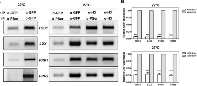
Our results indicate that CK2 interferes with CCA1 function and suggest an inverse correlation between CK2 activity and CCA1 binding. We next performed Double ChIP assays at temperatures of maximal CCA1 binding (22uC and 27uC) in an attempt to estimate the phosphorylated CCA1 fraction that is associated with the promoters. We performed a double round of Figure 6. Effects of temperature and CK2 kinase function on CCA1 binding activity.(A) ChIP analysis of CCA1 binding to the promoters of TOC1,LUX, PRR7andPRR9in plants entrained under LD cycles at 22uC and transferred to 12uC or 27uC. Samples were collected after 35 h under LL. Input DNA was used as a control. Immunodetection of (B) CCA1-YFP and (C) CKB4-MYC protein accumulation in CCA1-YFP-ox/CKB4-MYC-ox plants grown under the same conditions described in (A). Similar protein transference in each lane was verified by staining with Red Ponceau. (D) Western-blot analysis of CCA1-YFP-ox/CKB4-MYC-ox protein extracts immunoprecipitated with anti-GFP antibody (a-GFP) with subsequent detection of phosphorylated CCA1 isoforms (CCA1-P)(a-PSer) at 12uC or 27uC. Luminescence ofTOC1:LUCexpression in WT andcca1-1/lhy-11plants grown under LD cycles at 22uC and subsequently transferred to 27uC (E) or 12uC (F). Plots are means6SEM of 12 individual seedlings. The white and solid boxes correspond to the light and dark periods, respectively. The experiments were performed at least twice with similar results to those shown here. doi:10.1371/journal.pgen.1001201.g006
immunoprecipitation, firstly, with thea-GFP antibody to immu-noprecipitate total CCA1 protein bound to the target promoters and secondly, with thea-PSer antibody to discriminate between phosphorylated and dephosphorylated CCA1 isoforms. By virtue of doubling-up the immunoprecipitation round, we expected to get PCR amplification only if CCA1 phosphorylated isoforms were preferentially bound to the target promoters (Figure S15). Our results showed slightly above background amplification that was observed at both 22uC or 27uC and for all the promoters examined (Figure 7A and 7B). This corresponds to only a minimal fraction of the total amount of CCA1 bound to the promoters, as assayed by double round of immunoprecipitation with the anti-GFP antibody (Figure 7A and 7B). The reliability of these results is supported by the abundant CCA1 phosphorylated isoforms observed at 27uC (Figure 6D) and the efficient detection of immunoprecipitated CCA1 by the P-Ser antibody (Figure 2A and Figure 6D). In addition, the lack of amplification in the Double ChIP assays was not likely due to reduced efficiency of the technique, as Double-ChIP experiments similarly processed with a-Histone3 (a-H3) antibody followed by a-PSer immunoprecip-itation revealed clear amplified bands in all cases (Figure 7A and Figure S15). Together, the results confirmed and extend our findings suggesting that the complex regulatory interplay between CK2 and CCA1 binding activity is modulated by temperature and contributes to proper temperature compensation within the Arabidopsis circadian clock.
Discussion
Our results show that over-expression of CKB4 reduces the severity of CCA1-YFP-ox phenotypes while the lack of additive phenotypes in CKB4-MYC-ox/cca1-1/lhyRNAi plants indicates that CKB4 and CCA1 function in the same signalling pathway. Repression of TOC1 expression in CCA1-YFP-ox plants was alleviated by over-expression of CKB4, which assigns an important role for CKB4 modulating one of the main feedback
loops described in Arabidopsis [58]. Pharmacological treatment with specific CK2 inhibitors or the use of plants expressing a CK2 inducible dominant mutant reflected the involvement of CK2 activity in this regulation, rather than a holoenzyme-independent function of the CK2 regulatory subunit [64] . In human cells, treatment with DRB or DMAT also results in long-period phenotypes [19-21]. Similarly, decreased CK2 activity causes long-period behavioral rhythms in Drosophila [22,23]. These results highlight a remarkable conservation of CK2 circadian function among very different organisms including plants, insects and mammals.
Based on studies with other circadian systems, it was plausible to assume that phosphorylation of CCA1 by CK2 might mediate changes in CCA1 accumulation or subcellular localization [12]. However, our results assigned a role for CK2 in the control of CCA1 binding activity rather than protein accumulation or localization. Our studies also revealed that CCA1 regulates the expression of morning- and evening-expressed genes most likely by direct binding to their promoters. Therefore, by antagonizing this binding, CK2 can precisely modulate CCA1 regulation of circadian gene expression. The binding of CCA1 to the LIGHT-HARVESTING CHLOROPHYLL A/B1*3(LHCB1*3) promoter was shown to be increased by CK2 [25,26]. However, the authors also reported that in CKB3-ox plants, the induction of theLHCB1*1
gene was reduced compared to WT (Sugano et al., 1999). In our studies, we observed that CK2 antagonizes CCA1 binding to the promoters of the oscillator genes. This function consistently fits with the CKB and CCA1 inverse correlation between phenotypes and expression. Evidence that CK2 phosphorylation decreases protein binding to target promoters was also provided for the bZIP transcription factor HY5 [65]. Our binding analyses were consistent with the Double-ChIP results showing that CCA1 dephosphorylated isoforms were preferentially bound to the promoters of CCA1 target genes. An altered ratio of phosphor-ylated/dephosphorylated isoforms by increased or decreased CK2 activity is fully consistent with the circadian phenotypes observed
Figure 7. Analysis of CCA1 isoforms bound to the promoters of the oscillator genes.(A) Double ChIP assays of CCA1 isoform binding to the promoters ofTOC1,LUX, PRR7andPRR9in CCA1-YFP-ox/CKB4-MYC-ox plants entrained under LD cycles at 22uC and transferred to LL conditions at 22uC or 27uC. Samples were taken after 35 h under LL. The first immunoprecipitation was performed witha-GFP antibody followed by a second round witha-PSer or witha-GFP. The combinations ofa-H3/a-H3 antibodies ora-H3/a-Pser antibodies were used as controls. The experiments were performed at least twice with similar results to those shown here. (B) Q-PCR analysis of CCA1 isoform binding by double ChIP assays. Data are shown as means6SD relative to the maximum value of two independent experiments (*** p-value,0.001).
in CKB4-MYC-ox and CCA1-YFP-ox/CKB4-MYC-ox plants. This inhibitory function is not exclusive of the plant circadian system as a phosphorylation-dependent inactivation of transcriptional function was previously reported in other circadian systems [62].
To be effective as a key mechanism of clock progression, the interplay between CK2 and CCA1 should be in turn precisely regulated. Indeed, we found that different temperatures affect CK2 regulatory function. The higher amplitude ofTOC1:LUCexpression in CKB4-MYC-ox plants was mostly observed at 22uC and 27uC while the activating function of CK2 onTOC1expression was not so clearly observed at 12uC. The lower amplitude of TOC1:LUC
expression in CKB4-MYC-ox plants at 12uC is difficult to explain, but denotes that a different mechanism is engaged at low temperatures and emphasizes the complexity of the temperature response within the Arabidopsis circadian network. This notion is reinforced by the results showing that CK2 also phosphorylates LHY [26]. Thus, it is possible that the previously described role of LHY-GI in clock temperature compensation [40] might be also regulated by CK2. Notably, CCA1 binding activity appeared to be also modulated by temperature in CCA1-ox plants. Despite the lower binding at 12uC, CCA1 is still able to repress TOC1
expression at 12uC as evidenced by our luminescence assays. Furthermore, CK2 activity might also have a residual function as it was able to interfere with CCA1 binding. Therefore, there is a parallelism between the temperature regulation of CCA1 binding and CK2 activity. We proposed that the dynamic regulation of these activities by temperature is important for clock function, as altering the functional expression by over-expression, mutation or pharma-cological inhibition deregulates the temperature-dependent modu-lation of gene expression.
Unlike the temperature dependency of most biological and biochemical activities, the circadian clock sustains period length over a range of constantly maintained temperatures [66]. When we examined clock outputs, we found that the temperature compensation profiles were altered in CKB4-MYC-ox plants compared to those observed in WT. A simple explanation would be that CK2 activity might be compromised only when CKB4 abundance is increased above physiological limits. However, decreased CK2 activity by pharmacological inhibitors also led to altered temperature compensation. Additionally, CKB4 protein abundance did not vary between 12uC and 27uC. Together, these findings indicate that CK2 contributes to proper temperature responses in Arabidopsis: increased CK2 activity led to under-compensation whereas decreased activity resulted in a slightly over-compensated clock. This is noteworthy because previous studies in Neurospora have also implicated CK2 in temperature compensation [30]. Therefore, CK2 integrates a molecular clock component with a regulatory function that is conserved among different circadian systems. However, the specific mechanisms of CK2 function differ among organisms. In Neurospora, the stability of FRQ is controlled, at least in part, by a CK2-dependent phosphorylating mechanism, which facilitates the FRQ protein degradation preferentially at high temperatures [30]. In Arabi-dopsis, CK2 does not affect CCA1 protein accumulation but rather its transcriptional activity. Our results suggest that a precise temperature regulation of CK2 and CCA1 activity shapes the temperature compensation profile. Altering this delicate balance by over-expression, mutation or pharmacological inhibition affects the period compensation. In addition to the CK2-CCA1 counterbalancing loop, other loops and/or different mechanism should also account for precise regulation of temperature compensation. In fact, prior studies have proposed that LHY and GI contribute to temperature compensation in Arabidopsis [40]. Our findings focusing on CCA1 binding and CK2 activities
add exciting insights into the mechanisms of temperature compensation in Arabidopsis. It would be also interesting to perform similar studies with the other three CK2 regulatory subunits in order to highlight functional similarities or divergences in the control of temperature compensation. Our findings are also in line with the notion that evolution might enhance fitness under different climate conditions without directly affecting the expres-sion of central oscillator components but rather modulating their activity. Circadian clock adaptation to different environments might thus rely on key regulatory factors that modulate the oscillator activity. Further studies using different Arabidopsis
ecotypes can provide new insights into how this regulation might have shaped plant adaptation to different climate areas around the globe. The parallelism in temperature dependency of CCA1 and CK2 activities and the inhibitory effect of CK2 on CCA1 function is in agreement with a previously described model for temperature compensation [33]. The model proposes that the temperature independence of circadian period by the clock occurs through the balance between two biochemical activities, each of which has a similar temperature dependency [33]. Our studies would be also in consonance with the notion that temperature compensation is not only determined by central clock components but also by other elements that function intransto regulate the core proteins [40]. A recent computer modeling study has proposed that a switch-like mechanism might regulate period sensitivity through the control of two parameters that are a function of processes such as phosphorylation, ubiquitination or complex formation [67]. It would be interesting to identify a role of these regulatory processes as key modules shaping the temperature compensation profiles. Specifically for phosphorylation, studies in Neurospora [30] and mammals [68] indicate that in contrast to CK2, the activities of other kinases and clock-related phosphorylation events are temperature-insensitive. Our study showing that the CK2 activity antagonizes CCA1 function in a temperature-dependent manner highlights only one of the many aspects contributing to temperature compensation. Further analysis focusing on addition-al components and mechanisms would aid in our understanding of the intricate interacting networks responsible for temperature compensation in Arabidopsis and in other organisms.
Materials and Methods
Plant material, luminescence assays, flowering time, and hypocotyl length analysis
Arabidopsis thalianaseedlings were stratified at 4uC in the dark for 3 days on Murashige and Skoog agar medium supplemented with 3% sucrose and then transferred to light:dark conditions (LD, 12 h light:12 h hours dark) with 60mmol m22s21 of cool white
fluorescent light at 22uC. The list with the different plants and constructs used in this study is shown in Table S1 and S2. For CCA1-ox/CKB4-ox studies, lines 9 and 14 were used (see Figure S1 and Figure S2). Forcca1/lhy/CKB4-ox studies, lines 4 and 5 were used (see Figure 3D and Figure S1). For Luminescence analyses were performed as previously described [28]. In experiments with CK2 inhibitors, one-week old plants were transferred to 96-well plates containing MS medium supplemented with the specified concentration of DRB or DMAT. Luminescence was recorded 24 hours after the seedlings were transferred to the plates. For Dexamethasone induction, 1mM was added to each well at Zeitgeber Time 2 (ZT 2). For flowering time analysis, seeds were stratified in the dark at 4uC for 3 days on soil. Seedlings were grown under Short-Day (ShD, 8 h light:16 h dark) or Long-Day (16 h light:8 h dark) conditions with 60mmol m22
counting the number of days and number of leaves at the time of a 1 cm-high flower bolt. For hypocotyl length assays, seeds were stratified in the dark at 4uC for 4 days on MS medium supplemented with 3% Sucrose. Germination was induced by exposing the seeds to white light (60mmol m22
s21
) for 6 h followed by 18 h under darkness. Seeds were placed under ShD or LgD conditions (60mmol m22 s21) or under continuous white
light (LL) with the specified light intensities. Hypocotyl length was measured after seven days by using the Image J software (http:// rsb.info.nih.gov/ij/).
Western-blot and co-immunoprecipitation analysis Western-blot assays were essentially performed as previously described [69]. Briefly, nine day-old seedlings were ground in liquid nitrogen and proteins were extracted in RIPA buffer (50 mM Tris-HCl pH 8.0, 150 mM NaCl, 1% NP-40 0.1% SDS, 0.5% Sodium Deoxycholate, 0.5% Polyvinylpolypyrrolidone (PVPP), 50mm MG132, 10 mM NaF, 1 mM PMSF, 5mg/ml
Leupeptin, 1mg/ml Aprotinin, 5mg/ml Antipain, 1mg/ml Pepstatin, 5mg/ml Chymostatin). Protein concentration was calculated using the Bradford method (Bio-Rad) and 20-60mg of
total protein was loaded per lane. Proteins were transferred to nitrocellulose membranes and stained with Red Ponceau following standard protocols. Anti-MYC (clone 9E10; Sigma) and Anti-GFP (A11122; Invitrogen) antibodies were used to detect CKB4-MYC and CCA1-YFP proteins, respectively. Protein accumulation was quantified using the LAS-4000 imaging system (Fujifilm-GE Healthcare). In experiments with CK2 inhibitors, 48 hours before sampling, plants were transferred to medium supplemented with 150mM of DRB, 150mM DMAT or 1mM Dex. For co-immunoprecipitation assays, nine day-old seedlings were ground in liquid nitrogen and proteins extracted in RIPA buffer. Extracts were incubated for 4 h at 4uC with Protein G–Sepharose beads (Amersham Biosciences) conjugated with Anti-GFP antibody. Immunocomplexes were washed 5 times with RIPA buffer followed by additional washing with PBS (Phosphate buffer saline). Immunoprecipitated proteins were eluted by adding Laemmli buffer followed by 4 min incubation at 95uC. Anti-MYC and Anti-GFP antibodies were used to detect CKB4-Anti-MYC and CCA1-YFP respectively. The anti-phosphoserine antibody (Anti-PSer, 4A3, Calbiochem) was used to detect phosphorylated isoforms of CCA1. The antibody recognizes serine-phosphorylated residues (in a positively charged amino acid context directly neighbouring the phosphoserine). For interaction of proteins expressed at endogenous levels, we used a polyclonal anti-CK2B antibody to the human CK2B regulatory subunit CK2B (CSNK2B; Abnova). Detection of CCA1 was performed with the Anti-MYC antibody.
Two-dimensional gels and immunoblotting
For two-dimensional gels experiments, nine day-old seedlings were ground in liquid nitrogen and proteins extracted in lysis buffer (7 M Urea, 2 M Thiourea, 4% CHAPS, 18 mM Tris-HCl pH 8.0, 50mm MG132, 10 mM NaF, 1 mM PMSF, 5mg/ml Leupeptin, 1mg/ml Aprotinin, 5mg/ml Antipain, 1mg/ml Pepstatin, 5mg/ml Chymostatin). Protein concentration was
determined using the Bradford method (Bio-Rad) and 40mg of total protein was loaded onto immobilized pH gradient (IPG) strips (7 cm, pH 3–10, Amersham Biosciences) for the first dimension separation. Strips were rehydrated for 6 h at room temperature and the isoelectric point focusing was performed at 30 V for 6.5 h, 500 V for 1 h, 1000 V for 1 h and 5000 V for 7 h. Strips were subsequently equilibrated for 15 min with equilibra-tion buffer I (50 mM Tris-HCl pH 8.8, 6 M Urea, 30% Glycerol,
2% SDS, 10 mg/ml DTT) followed by a 15 min wash with equilibration buffer II (50 mM Tris-HCl pH 8.8, 6 M Urea, 30% Glycerol, 2% SDS, 25 mg/ml Iodoacetamide). For the second dimension, strips were loaded onto SDS-PAGE 8% polyacryl-amide gels followed by blotting to nitrocellulose membranes. Anti-GFP antibody was used to detect CCA1-YFP protein.
Bimolecular Fluorescence Complementation (BiFC) and confocal microscopy
Plants over-expressing both the CCA1 protein fused to the N-terminal fragment of the Yellow Fluorescent Protein (YFP) (nucleotides: 1–462) and the CKB4 protein fused to the C-terminal part of the YFP protein (nucleotides: 463–741) were grown on MS-agar medium supplemented with 3% Sucrose under LD cycles. Fluorescence signals of hypocotyl cells were imaged using an Olympus Fluoview FV1000 confocal microscope using a 515 nm argon excitation laser. CKB4-YFP-ox, CCA1-YFP-ox, TOC1-YFP-ox, CCA1-ox/CKB4-YFP-ox, CCA1-YFP-ox/CKB4-MYC-ox, TOC1-nYFP-ox/CKB4-cYFP-ox were similarly imaged.
ChIP and double-ChIP assays
ChIP assays were performed essentially as previously described [52]. Briefly, fourteen day-old seedlings were fixed in fixation buffer (0.4 M Sucrose, 10 mM Tris-HCl pH 8.0, 1 mM EDTA,1 mM PMSF, 1% Formaldehyde, 0.05% Triton X-100) for 10 min, followed by addition of Glycine 0.125 M and vacuum incubation during 10 min. Seedlings were subsequently ground in liquid nitrogen and extracted in extraction buffer I (0.4 M Sucrose, 10 mM Tris-HCl pH 8.0, 5 mM b-mercaptoethanol, 1 mM PMSF, 5mg/ml Leupeptin, 1mg/ml Aprotinin, 5mg/ml
Anti-pain, 1mg/ml Pepstatin, 5mg/ml Chymostatin and 50mm MG132). Nuclei were then purified by centrifugation and washed with extraction buffer II (0.25 M Sucrose, 10 mM Tris-HCl pH 8.0, 10 mM MgCl2, 1% Triton X-100, 5 mMb -mercapto-ethanol, 1 mM PMSF, 5mg/ml Leupeptin, 1mg/ml Aprotinin, 5mg/ml Antipain, 1mg/ml Pepstatin, 5mg/ml Chymostatin and
50mm MG132). Nuclei were resuspended in nuclei lysis buffer
(50 mM Tris-HCl pH 8.0, 10 mM EDTA, 1% SDS, 5mg/ml Leupeptin, 1mg/ml Aprotinin, 5mg/ml Antipain, 1mg/ml Pepstatin, 5mg/ml Chymostatin and 50mm MG132). Chromatin
was sonicated to approximately 500–1000 bp fragments with a sonicator (Branch). After centrifugation, soluble chromatin was diluted in ChIP dilution buffer (15 mM Tris-HCl pH 8.0, 150 mM NaCl, 1% Triton-X-100, 1 mM EDTA, 1 mM PMSF, 5mg/ml Leupeptin, 1mg/ml Aprotinin, 5mg/ml Antipain, 1mg/ ml Pepstatin, 5mg/ml Chymostatin and 50mm MG132) and
software (Version 1.5.0.39, Roche). Melting peak analysis using the LightCycler 480 Basic software module (Roche) and gel electrophoresis confirmed that primer-dimers or other non-specific products were not present. Crossing point (Cp) calculation was used for quantification using the Absolute Quantification analysis by the 2ndDerivative Maximun method (LightCycler 480 Basic software module, Roche). ChIP values for each set of primers were normalized to Input values. Primers were designed using the PrimerExpress 2.0 software (Applied Biosystems) with lengths of 18-25 nucleotides, PCR amplicon lengths of 80 to 180 bp, 40-60% G:C content and melting point of 58-62uC. The list of primers used for promoter (prom) amplification and for Q-PCR analysis is shown in Table S3. In experiments with CK2 inhibitors or with plants expressing the CK2 inducible mutant, 48 hours before sampling, plants were transferred to medium supplemented with 150mM of DRB, 150mM DMAT or 1mM Dex. Double-ChIP
assays [70] were performed following the ChIP procedure for chromatin extraction and immunoprecipitation followed by one wash with low salt buffer (20 mM Tris-HCl pH 8.0, 150 mM NaCl, 1% Triton X-100, 0.1% SDS and 2 mM EDTA) and two washes with TE buffer (10 mM Tris-HCl pH 8.0, 1 mM EDTA). Immunocomplexes were eluted using 10 mM DTT and incubat-ing at 37uC for 30 min. Chromatin was diluted in 40 volumes of Double-ChIP buffer (15 mM Tris-HCl pH 8.0, 150 mM NaCl, 1% Triton-X-100, 1 mM EDTA, 1 mM PMSF, 5mg/ml Leupeptin, 1mg/ml Aprotinin, 5mg/ml Antipain, 1mg/ml Pepstatin, 5mg/ml Chymostatin and 50mm MG132) and incubated overnight at 4uC with Protein G–Sepharose beads (Amersham Biosciences) conjugated with Anti-PSer or Anti-GFP antibody. Immunocomplexes were washed, eluted, purified and amplified as described for the ChIP protocol.
Statistical analysis
Statistical analyses were performed using the GraphPad Prism software (GraphPad Software, Inc). For hypocotyl length and flowering time experiments, two-tailed t-tests with 99% of confidence were performed. For multiple comparisons, two-way ANOVA followed by Bonferroni post-tests were performed; photoperiod and genotype were considered as variables. For ChIP quantifications, two-way ANOVA tests followed by Bonferroni post-tests were used. Gene and genotype or temperature and genotype were considered as variables. GraphPad Prism software was also used to extrapolate the linear regression of the temperature response. Profiles for bioluminescence experiments, the estimated period length was determined by Fast Fourier Transform/Nonlinear Least Squares method (FFT/NLLS) [71] using a window of 96 hr of data excluding the first 24 hr to avoid any transient effects after transferring to constant conditions.
Supporting Information
Figure S1 CKB4 and CCA1 expression in different CCA1-ox/ CKB4-ox andcca1/lhy/CKB4-ox lines. (A) RT-PCR analysis of
CKB4,CCA1andACTIN2(ACT2) expression in WT and double CCA1 and CKB4 over-expressing plants. Lines 14 and 9 were used for subsequent studies. (B) RT-PCR analysis of CKB4and
ACTIN2(ACT2) expression in WT and incca1-1/lhyRNAi /CKB4-ox plants. Lines 5 and 4 were used for subsequent studies. Seedlings were entrained under LD cycles and samples were collected at Zeitgeber Time 2 (ZT2). (C) Immunodetection of CKB4 protein accumulation incca1-1/lhyRNAi/CKB4-ox plants. CKB4 protein was detected using thea-MYC antibody. Seedlings were entrained under LD cycles and samples were collected at ZT2.
Found at: doi:10.1371/journal.pgen.1001201.s001 (0.21 MB PDF)
Figure S2 Analysis of CCA1 and CKB4 genetic interaction. Flowering time of WT, cca1-1/lhyRNAi, cca1-1/lhyRNAi /CKB4-MYC-ox and CKB4-/CKB4-MYC-ox plants grown under (A, B) Long-Day (LgD, 16 h light:8 h dark) or (C, D) Short-Long-Day (ShD, 8 h light:16 h dark) conditions. Flowering time was measured as the number of leaves at flowering or the number of days to flowering (1-cm-high bolt). Data are shown as means 6 SEM of three independent experiments. Similar results were obtained when flowering time was examined incca1-1/lhy-11andcca1-1/lhy-11/ CKB4-MYC-ox plants.
Found at: doi:10.1371/journal.pgen.1001201.s002 (0.12 MB PDF)
Figure S3 Analysis ofTOC1:LUCdiurnal expression in different genetic backgrounds. TOC1:LUC luminescence in seedlings maintained under LD (12 h light:12 h dark) cycles. Plots represent means6SEM of at least 12 individual seedlings. The white and solid boxes correspond to the light and dark periods, respectively. The experiment was performed three times with similar results to those shown here.
Found at: doi:10.1371/journal.pgen.1001201.s003 (0.09 MB PDF)
Figure S4 Analysis of CCA1 and CKB4 molecular interaction. (A) Western-blot analysis of WT, ox and CCA1-prom:CCA1-MYC/cca1-1plants using an antibody to the human CK2B subunit (a-CKB). The antibody efficiently recognizes the CK2 regulatory subunits of Arabidopsis. (B) Western-blot analysis of Co-IP experiments with plants expressing CCA1 under its own promoter (CCA1prom:CCA1-MYC/cca1-1). Protein extracts were immunoprecipitated with the a-CKB antibody followed by detection with the a-MYC antibody. Plants were grown under LD conditions and samples were collected at ZT1.5. Nuclear localization analysis by confocal microscopy at ZT11 of plants expressing (C) CCA1-ox/CKB4-YFP-ox, (D) CCA1-YFP-ox/ CKB4-MYC-ox, (E) TOC1 fused to the N-terminal fragment of YFP (TOC1-nYFP-ox) and CKB4 fused to the C-terminal fragment of YFP (CKB4-cYFP-ox) or (F) TOC1 fused to full-length YFP (TOC1-YFP-ox).
Found at: doi:10.1371/journal.pgen.1001201.s004 (0.40 MB PDF)
Figure S5 Effects of the CK2 inhibitors DRB and DMAT on
TOC1:LUCexpression. (A) Free-running periods estimated from
TOC1:LUC luminescence signals in WT plants under LL conditions treated with increasing concentrations of DRB. Period was estimated from individual seedlings plotted against their relative amplitude errors. (B) TOC1:LUC luminescence in WT plants treated with increasing concentrations of DMAT. Lumi-nescence was recorded under LD (12 h light:12 h dark) cycles. Data are represented as means6 SEM of luminescence signals from at least 12 independent plants.
Found at: doi:10.1371/journal.pgen.1001201.s005 (0.11 MB PDF)
Figure S6 Effects of CK2 activity on thein vivoCCA1 binding to the promoters of the oscillator genes. (A) ChIP analysis of CCA1 binding to the promoters of the morning-expressed genes,PRR7
ox plants; in (C) CCA1-YFP-ox with 150mM of the CK2 inhibitor
DRB; in (D) CKA3-/CCA1-YFP-ox plants induced with 1mM Dexamethasone (Dex) or in (E) CCA1-YFP-ox/CKB4-MYC-ox plants treated with 150mM of the CK2 inhibitor DRB. Seedlings were grown under LD cycles and collected at ZT3. Data are presented as means 6 SEM relative to the input and to the maximum value of at least three independent experiments (*** p-value,0.001).
Found at: doi:10.1371/journal.pgen.1001201.s006 (0.20 MB PDF)
Figure S7 Effects of temperature and CK2 kinase function on circadian gene expression. Bioluminescence ofCAB2:LUC expres-sion in WT (A) andcca1-11mutant (B) plants in the presence or in the absence of 100mM DRB. Luminescence was measured in
plants entrained under LD conditions at 22uC and transferred to LL conditions. Plots are means 6 SEM of 8-12 individual seedlings. (C) Analysis of circadian period length of CAB2::LUC
expression in WT andcca1-11mutant plants in the presence or in the absence of DRB. Estimated period length was determined as described in Supplemental experimental procedures. (D) TO-C1:LUC luminescence in WT and cca1-1/lhy-11 double mutant plants in the presence or in the absence of 100mM DRB. Luminescence was measured in plants entrained under LD conditions at 22uC and transferred to LL conditions. Plots are means6SEM of 8-12 individual seedlings. The experiments were repeated at least twice with similar results to those shown here. Found at: doi:10.1371/journal.pgen.1001201.s007 (0.14 MB PDF)
Figure S8 Analysis of TOC1:LUC diurnal expression under different temperatures. Luminescence analysis of TOC1:LUC
expression in plants entrained under LD cycles at 22uC and subsequently transferred to LD cycles at 27uC (A, D), 22uC (B, E) or 12uC (C, F). Data is shown as means6 SEM of at least 12 individual seedlings. The experiments were performed at least twice with similar results to those shown here.
Found at: doi:10.1371/journal.pgen.1001201.s008 (0.13 MB PDF)
Figure S9 Analysis ofTOC1:LUCdiurnal expression in CKB4-ox plants under different temperatures. Point-by-point lumines-cence analysis ofTOC1:LUCexpression in plants entrained under LD cycles at 22uC and subsequently transferred to LD cycles at 27uC (A) or 12uC (B). Data is shown as means6SEM of at least 12 individual seedlings. The experiments were performed at least twice with similar results to those shown here.
Found at: doi:10.1371/journal.pgen.1001201.s009 (0.13 MB PDF)
Figure S10 Effects of DRB treatment on TOC1:LUC diurnal expression at different temperatures.TOC1:LUCluminescence in WT plants in the presence or in the absence of 100mM DRB at 27uC or 12uC. Luminescence was measured in plants entrained under LD conditions at 22uC and transferred to LD cycles at 27uC (A) or 12uC (B). Plots are means6SEM of 12 individual seedlings. The experiments were repeated at least twice with similar results to those shown here. The white and solid boxes correspond to the light and dark periods, respectively.
Found at: doi:10.1371/journal.pgen.1001201.s010 (0.11 MB PDF)
Figure S11 Effects of temperature and CK2 kinase function on the free-running period of the clock-output CAB2:LUC. Free-running periods estimated from CAB2:LUCluminescence signals in WT, CKB4-MYC-ox and WT plants treated with 100mM DRB at 12uC (A), 22uC (B) and 27uC (C). Period was estimated from individual seedlings plotted against their relative amplitude errors. Data come from two independent experiments with approximately 6-12 plants per phenotype or treatment.
Found at: doi:10.1371/journal.pgen.1001201.s011 (0.12 MB PDF)
Figure S12 Effects of temperature and CK2 kinase function on CCA1 binding to the promoters of the oscillator genes. Q-PCR analysis of CCA1 binding to TOC1 (A), LUX (B), PRR7 (C) or
PRR9(D) promoters. CCA1-YFP-ox and CCA1-YFP-ox/CKB4-MYC-ox plants were grown under LD cycles at 22uC and transferred to continuous light (LL) at 12uC or 27uC. Samples were collected after 35 h under LL conditions. Data are presented as means6SD relative to the input and to the maximum value of two independent experiments. (E) Q-PCR analysis of CCA1 binding toTOC1, LUX, PRR7,PRR9and a clock unrelated gene (At5g55840) in plants expressing CCA1 under its own promoter (CCA1pro:CCA1-HA-YFP). Plants were grown under LD cycles at 22uC and transferred to continuous light (LL) at 12uC or 27uC. Samples were collected after 49.5 h under LL conditions. (F) Western-blot analysis of CCA1 protein accumulation in CCA1-YFP-ox plants at 12uC and 27uC. Similar protein transference in each lane was verified by staining with Red Ponceau. The experiments were performed twice with similar results to those shown here (** p-value,0.01; *** p-value,0.001).
Found at: doi:10.1371/journal.pgen.1001201.s012 (0.17 MB PDF)
Figure S13 A role for CCA1 regulating circadian gene expression at high temperatures. TOC1:LUC luminescence in WT and CCA1-YFP-ox plants at 27uC (A) or 12uC (B).
TOC1:LUC luminescence in WT and CCA1-YFP-ox plants at 27uC (C) or 12uC (D) in the presence or in the absence of 100mM DRB. (E) CAB2:LUC luminescence in WT and cca1-11 mutant plants at 27uC and in the presence or in the absence of 100mM
DRB (F). Luminescence was measured in plants entrained under LD conditions at 22uC and transferred to LL conditions at the indicated temperatures. Plots are means6SEM of 12 individual seedlings. The experiments were repeated at least twice with similar results to those shown here.
Found at: doi:10.1371/journal.pgen.1001201.s013 (0.15 MB PDF)
Figure S14 Effects of temperature and CK2 kinase function on CCA1 binding to the promoters of the oscillator genes. Q-PCR analysis of CCA1 binding to TOC1, LUX, PRR7 and PRR9
promoters in plants over-expressing CCA1 in the absence (-) or in the presence (+) of 150mM of the CK2 inhibitor DRB. Plants were grown under LD cycles at 22uC and transferred to continuous light (LL) at 12uC (A) or 27uC (B). Data are presented as means6 SD relative to the input and to the maximum value. Samples were collected after 50 h under LL conditions.
Found at: doi:10.1371/journal.pgen.1001201.s014 (0.11 MB PDF)
Figure S15 Double ChIP analysis. (A) Schematic representation depicting a summary of the Double-ChIP assay described in Figure 7. A double round of immunoprecipitation (IP) was performed. In the first round, thea-GFP antibody was used to detect total CCA1-YFP protein bound to chromatin. In a second round, the a-PSer antibody was used to specifically detect the phosphorylated isoforms of CCA1 (P). PCR amplification would be obtained only if the phosphorylated CCA1 isoforms are preferentially bound to chromatin. (B) Double-ChIP assays with the combination ofa-H3/anti-PSer ora-H3/anti-H3 antibodies Data are presented as means6SD relative to the maximum value of two independent experiments.
Found at: doi:10.1371/journal.pgen.1001201.s015 (0.13 MB PDF)
Table S1 Plant Material used in this study.
Found at: doi:10.1371/journal.pgen.1001201.s016 (0.07 MB DOC)
Table S2 Constructs used in this study.