Marina Angélica Marciano(a) Carlos Estrela(b)
Rafael Francisco Lia Mondelli(a) Ronald Ordinola-Zapata(a) Marco Antonio Hungaro Duarte(a)
(a)Department of Operative Dentistry, Endodontics and Dental Materials, Bauru School of Dentistry, Universidade de São Paulo - USP, Bauru, SP, Brazil.
(b) Department of Prevention and Oral Rehabilitation, School of Dentistry, Universidade de Goiás - UFG, Goiânia, GO, Brazil.
Corresponding Author:
Marina Angélica Marciano E-mail: [email protected]
Analysis of the color alteration and
radiopacity promoted by bismuth oxide
in calcium silicate cement
Abstract: The aim of the study was to determine if the increase in ra-diopacity provided by bismuth oxide is related to the color alteration of calcium silicate-based cement. Calcium silicate cement (CSC) was mixed with 0%, 15%, 20%, 30% and 50% of bismuth oxide (BO), de-termined by weight. Mineral trioxide aggregate (MTA) was the control group. The radiopacity test was performed according to ISO 6876/2001. The color was evaluated using the CIE system. The assessments were performed after 24 hours, 7 and 30 days of setting time, using a spec-trophotometer to obtain the ∆E, ∆a, ∆b and ∆L values. The statistical analyses were performed using the Kruskal-Wallis/Dunn and ANOVA/ Tukey tests (p < 0.05). The cements in which bismuth oxide was add-ed showadd-ed radiopacity corresponding to the ISO recommendations ( > 3 mm equivalent of Al). The MTA group was statistically similar to the CSC / 30% BO group (p > 0.05). In regard to color, the increase of bismuth oxide resulted in a decrease in the ∆E value of the calcium sili-cate cement. The CSC group presented statistically higher ∆E values than the CSC / 50% BO group (p < 0.05). The comparison between 24 hours and 7 days showed higher ∆E for the MTA group, with statistical differ-ences for the CSC / 15% BO and CSC / 50% BO groups (p < 0.05). After 30 days, CSC showed statistically higher ∆E values than CSC / 30%BO and CSC / 50% BO (p < 0.05). In conclusion, the increase in radiopacity pro-vided by bismuth oxide has no relation to the color alteration of calcium silicate-based cements.
Descriptors: Endodontics; Bismuth; Color.
Introduction
Mineral trioxide aggregate (MTA) has been used for several proce-dures including vital pulp therapies,1 root fractures,2 perforations3 and
apexiications,4 with a high rate of success. It is well known that calcium
silicate is the main component of MTA.5 Previous studies have shown
that calcium silicate and MTA present similar chemical, physical and biological properties,6-8 with the exception of radiopacity.8-10 Calcium
silicate does not have the minimum radiopacity recommended (3 mm equivalent of Al) by ISO 6876/2001 speciications.11 Thus, a radiopaciier
agent should be added in order to distinguish it from anatomical struc-tures, in clinical conditions.9 Several radiopacity agents have been
pro-posed for calcium silicate cement composition such as zirconium oxide, Declaration of Interests: The authors
certify that they have no commercial or associative interest that represents a conflict of interest in connection with the manuscript.
Submitted: Feb 06, 2013
calcium tungstate, iodoform and bismuth oxide.9,12
Bismuth oxide (Bi2O3) is a yellow substance
com-monly added to various endodontic materials, e.g., MTA, AH 26 and Sealer 26, for the purpose of pro-viding radiopacity.13-15 The use of MTA as a pulp
capping material in esthetic areas requires taking into account the changes in color of the dentin. Teeth discoloration is an undesirable consequence of some materials used in endodontic therapy.16-18 Color
al-teration of calcium silicate-based cements has been reported.17,19-21 It has been suggested that the
radi-opaciier bismuth oxide, present in the MTA com-position, is the main cause of dental discoloration.22
Furthermore, it has been previously demonstrated that AH 26 sealer, which also contains bismuth ox-ide, results in tooth discoloration.18 Thus, it is
possi-ble that the presence of bismuth oxide in calcium sil-icate-based cements and in AH 26 sealer may induce discoloration over time. This hypothesis has been discussed in the literature, but there is no quantita-tive evidence in this regard.16 The aim of the present
study was to investigate if there is a relation between the increase in radiopacity provided by bismuth ox-ide and the color alteration of calcium silicate-based cement. The hypotheses tested were that the increase in the amount of bismuth oxide in the calcium sili-cate cement increases the radiopacity of this cement and interferes with the color of the material.
Methodology
A portion of calcium silicate cement (CSC; Iraja-zinho Votorantim, Cimento Rio Branco, Rio de Ja-neiro, Brazil) was mixed with 0%, 15%, 20%, 30% and 50% of bismuth oxide (BO; Merck, New Jersey, USA), proportioned in weight using an electronic analytic scale (Mettler Toledo PG5002-S, São Pau-lo, Brazil). MTA was used as the control group. The cements were divided into 6 groups:
• Group 1: MTA,
• Group 2: 100g of CSC and 0g of BO,
• Group 3: 85g of CSC and 15g of BO,
• Group 4: 80g of CSC and 20g of BO,
• Group 5: 70g of CSC and 30g of BO and
• Group 6: 50g of CSC and 50g of BO.
The manipulation of the cements was performed
using a total of 1g of cement and 0.4 mL of distilled water.
Radiopacity
Three cylindrical samples were fabricated for each concentration by placing the manipulated ce-ments into metallic rings with a 10 mm internal diameter and a 1 mm thickness (ISO 6876/2001).11
Next, the illed rings were kept at 37°C, until the ce-ments set completely. The thickness was conirmed with a digital caliper (Mitutoyo Corp., Tokyo, Japan) and the samples were radiographed on occlusal ilms (D-speed; Kodak Comp., New York, USA) with an aluminum step-wedge, graduated from 2 to 16 mm (in 2-mm increments). A radiographic unit (Gnatus XR 6010; Gnatus, São Paulo, Brazil) was used with the exposures set at 60 kV, 10 mA, 0.3 seconds and a focus-ilm distance of 30 cm. The radiographs were digitized and analyzed using the Digora 1.51 soft-ware (Soredex, Helsinki, Finland). The radiopacity was determined according to Duarte et al.9
Color assessment
Ten cylindrical stainless steel rings with an inner diameter of 10 mm and thickness of 2 mm were illed with the cements. They were stored in an incubator at 37°C, 100% humidity, for 24 hours to set completely. After the completion of the setting time, the cements were demolded and the baseline color of the speci-mens was measured with a spectrophotometer (Vita Easyshade, Vita Zahnfabrik H. Rauter, Bad Säckin-gen, Germany) against a white Telon background to obtain the values of lightness (L*), red-green axis (a*) and yellow-blue axis (b*). Three measurements were performed for each sample. The samples were then stored at 37°C, 100% humidity, for 30 days, and the measurements were repeated. The color changes (∆E) were calculated based on the ∆L* (L2 – L1), ∆a*
(a2 – a1) and∆b* (b2 – b1) values for each specimen,
according to the following equation:23
∆E = [ (L2 – L1)2 + (a2 – a1)2 + (b2 – b1)2 ]1/2
In which L1, a1 and b1 are the initial assessment
color values and L2, a2 and b2 are the inal
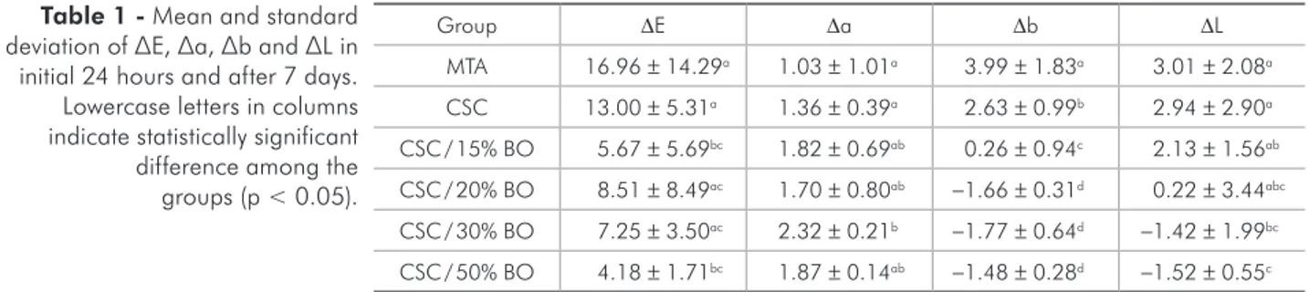
tively. The higher ∆E values were found for MTA at 7 days and for CSC after 30 days. All cements showed
∆E values higher than 4.0. Regarding the ∆a values, all cements showed positive values at 7 and 30 days, indicating a tendency toward the redness axis. The evaluation of ∆b values at 7 days showed positive values for MTA, CSC and CSC / 15% BO, indicating yellowness, whereas the other cements showed nega-tive values, indicating blueness. The analysis of color after 7 days showed higher values of ∆L for MTA and lower values for CSC / 50% BO.
The values of L* represent the value (lightness or darkness); a* is a measure of redness (positive a*) or greenness (negative a*); b* is a measure of yel-lowness (positive b*) or blueness (negative b*); and
∆E represents the color change. A color difference of ∆E < 4.0 has been shown to represent clinically acceptable color matching.24
Statistical analysis
Statistical analysis was performed using the non-parametric Kruskal–Wallis and Dunn tests for color and radiopacity (p < 0.05). Insofar as the values for
∆b showed normality after 7 days, the ANOVA/ Tukey test was used (p < 0.05).
Results
The values for radiopacity and the statistical dif-ferences are represented in Figure 1. All experimen-tal groups in which BO was added presented radi-opacity values higher than a 3 mm equivalent of Al. MTA was signiicantly more radiopaque than CSC, CSC / 15%BO and CSC / 20% BO (p < 0.05). MTA was statistically similar to CSC / 30% BO (p > 0.05). The CSC / 50% BO group was signiicantly more radi-opaque than all the other groups evaluated (p < 0.05). The statistical differences in color of the cements af-ter 7 and 30 days are shown in Table 1 and 2,
respec-Group ∆E ∆a ∆b ∆L
MTA 16.96 ± 14.29a 1.03 ± 1.01a 3.99 ± 1.83a 3.01 ± 2.08a CSC 13.00 ± 5.31a 1.36 ± 0.39a 2.63 ± 0.99b 2.94 ± 2.90a CSC / 15% BO 5.67 ± 5.69bc 1.82 ± 0.69ab 0.26 ± 0.94c 2.13 ± 1.56ab CSC / 20% BO 8.51 ± 8.49ac 1.70 ± 0.80ab −1.66 ± 0.31d 0.22 ± 3.44abc CSC / 30% BO 7.25 ± 3.50ac 2.32 ± 0.21b −1.77 ± 0.64d −1.42 ± 1.99bc CSC / 50% BO 4.18 ± 1.71bc 1.87 ± 0.14ab −1.48 ± 0.28d −1.52 ± 0.55c
Group ∆E ∆a ∆b ∆L
MTA 54.65 ± 33.26a 3.93 ± 1.46a 7.06 ± 2.69a 4.84 ± 3.66ab CSC 66.87 ± 15.80a 3.56 ± 0.51a 7.39 ± 0.81a 7.71 ± 2.61b CSC / 15% BO 41.50 ± 18.54ab 3.59 ± 0.67acd 4.98 ± 1.05ab 6.38 ± 1.88ab CSC / 20% BO 24.30 ± 8.18abc 3.20 ± 0.88acd 2.68 ± 0.34bc 4.38 ± 3.52ab CSC / 30% BO 13.98 ± 6.64bc 3.46 ± 0.25ac 2.13 ± 0.66bc 2.71 ± 2.01a CSC / 50% BO 8.73 ± 0.63c 2.75 ± 0.27bd 1.79 ± 0.11c 2.53 ± 0.50a
Figure 1 - Mean and standard deviation of radiopacity val-ues in millimeters of aluminum equivalent.
Table 1 - Mean and standard deviation of ∆E, ∆a, ∆b and ∆L in initial 24 hours and after 7 days. Lowercase letters in columns indicate statistically significant difference among the groups (p < 0.05).
Discussion
The hypotheses tested in the present study were that an increase in the amount of bismuth oxide in calcium silicate-based cement could increase radi-opacity, and additionally interfere with the color of the cement. In regard to radiopacity, the hypothesis was accepted. Higher amounts of bismuth have sig-niicantly increased the radiopacity of the cement. Radiopacity is an important physical property re-quired for cements.25 A minimal value of radiopacity
is essential to identify the material in the root canal, and allow illing failures to be corrected prior to i-nal restoration.9 The analysis of the results showed
that at least 15% of bismuth oxide must be added to provide the minimum radiopacity required by ISO 6876/2001 speciications (3 mm equivalent of Al).11
Similar results were previously reported by Bueno et al. 26 The results reinforce the need to add a
ra-diopaciier agent to calcium silicate-based cement. 9
The addition of 30% of BO to CSC provided a ra-diopacity statistically similar to that presented by MTA Angelus (p > 0.05). A previous analysis of MTA Angelus showed that the proportion of 20% of bismuth oxide provided a radiopacity correspond-ing to 5.93 mm equivalent of Al.9 This difference in
the results may be associated with the dificulty to obtain homogeneity of CSC and bismuth powders. It is possible that a portion of the cement contained higher or lower amounts of bismuth, and thus inlu-enced the results.
The use of calcium silicate-based cements in vital pulp therapy causes concern regarding the color al-teration of the cement and consequent dentin discol-oration. The methods used to evaluate the color of materials include subjective27 and quantitative
anal-yses.28 It has already been shown that sphere
spec-trophotometers, such as that used in this study, pro-vide a more accurate assessment of the color change than subjective human evaluation.29 This equipment
measures the amount of visible radiant energy re-lected or transmitted by an object, one wavelength at a time for each value, chroma, and hue present in the entire visible spectrum.
The change between the initial and the final color (∆E) specify the color alteration of the mate-rial during a determined period of time. Previous
laboratory and clinical studies have reported dental discoloration after using MTA.17,19,21 Therefore, it is
necessary to study if the agent responsible for the color alteration without the interference of other factors such as blood, dentin or saliva is actually the cement or its metallic components (bismuth). The ∆E observed for all the tested materials was higher than 3.3, which is not considered clinically acceptable.24 Thus, the hypothesis tested was
de-nied. Controversially, the increase of BO decreased the ∆E of the calcium silicate cement. The CSC group presented statistically higher ∆E values than the CSC / 50% BO (p < 0.05). These results could suggest that the main cause of discoloration of the cement was the calcium silicate and not the bismuth oxide. However, it is not possible to state that this change in color is clinically reproduced by the ab-sence of components, such as dentin. It is possible that substances present in the dentin react with the components of CSC, including the bismuth oxide, to induce a chemical reaction of reduction resulting in darkness. Other factors may also be correlated with MTA darkness, such as the light curing per-formed when some restorative materials are used. Vallés et al.30 suggest that an association between
oxygen supply and light curing alter the color of MTA cement.
The radiopaciier bismuth oxide is yellow in col-or; however, contrary to what would be expected, the addition of a high proportion of bismuth oxide to calcium silicate cement did not result in an in-crease in yellowness (∆b). At 7 days of analysis, CSC with 20%, 30% and 50% of BO showed a tendency toward blueness (negative values), whereas CSC, CSC / 15% BO and MTA presented a tendency to-ward yellowness. After 30 days, all cements showed positive values of ∆b, indicating a tendency to pro-duce a yellow color. The indings indicated an in-verse correlation between increase of bismuth oxide and color change in calcium silicate-based cements. However, further evaluations are required to clarify the chemical reactions involved in the color changes of these cements.
Conclusion
radi-opacity provided by bismuth oxide has no relation to the color change in calcium silicate-based ce-ments. Other factors that may be associated with the reaction could cause the result in color change of calcium silicate-based cements.
Acknowledgments
This work was supported by the Coordena-ção de Aperfeiçoamento de Pessoal de Nível Su-perior (CAPES) and Fundação de Amparo à Pes-quisa do Estado de São Paulo (FAPESP), process #2011/13573-3.
References
1. Eskandarizadeh A, Shahpasandzadeh MH, Shahpasandzadeh M, Torabi M, Parirokh M. A comparative study on dental pulp response to calcium hydroxide, white and grey mineral trioxide aggregate as pulp capping agents. J Conserv Dent. 2011 Oct;14(4):351-5.
2. Roig M, Espona J, Mercade M, Duran-Sindreu F. Horizontal root fracture treated with MTA, a case report with a 10-year follow-up. Dent Traumatol. 2011 Dec;27(6):460-3.
3. Mente J, Hage N, Pfefferle T, Koch MJ, Geletneky B, Drey-haupt J, et al. Treatment outcome of mineral trioxide aggre-gate: repair of root perforations. J Endod. 2010 Feb;36(2):208-13.
4. Damle SG, Bhattal H, Loomba A. Apexification of anterior teeth: a comparative evaluation of mineral trioxide aggre-gate and calcium hydroxide paste. J Clin Pediatr Dent. 2012 Spring;36(3):263-8.
5. Song JS, Mante FK, Romanow WJ, Kim S. Chemical analy-sis of powder and set forms of Portland cement, gray Pro-Root MTA, white ProPro-Root MTA, and gray MTA-Angelus. Oral Surg Oral Med Oral Pathol Oral Radiol Endod. 2006 Dec;102(6):809-15.
6. Estrela C, Sydney GB, Bammann LL, Felippe Junior O. Mech-anism of action of calcium and hydroxyl ions of calcium hy-droxide on tissue and bacteria. Braz Dent J. 1995;6(2):85-90. 7. Holland R, Souza V, Murata SS, Nery MJ, Bernabe PF, Oto-boni Filho JA, Dezan Júnior E. Healing process of dog dental pulp after pulpotomy and pulp covering with mineral trioxide aggregate or Portland cement. Braz Dent J. 2001;12(2):109-13. 8. Saidon J, He J, Zhu Q, Safavi K, Spangberg LS. Cell and tissue
reactions to mineral trioxide aggregate and Portland cement. Oral Surg Oral Med Oral Pathol Oral Radiol Endod. 2003 Apr;95(4):483-9.
9. Duarte MAH, El Kadre GDO, Vivan RR, Tanomaru JMG, Tanomaru Filho M, Moraes IG. Radiopacity of portland ce-ment associated with different radiopacifying agents. J Endod. 2009 May;35(5):737-40.
10. Islam I, Chng HK, Yap AU. Comparison of the physical and mechanical properties of MTA and portland cement. J Endod. 2006 Mar;32(3):193-7.
11. International Organization for Standardization. ISO 6876: Dental Root Canal Sealing Materials. Geneva: International Organization for Standardization; 2001. 18 p.
12. Camilleri J, Cutajar A, Mallia B. Hydration characteristics of zirconium oxide replaced Portland cement for use as a root-end filling material. Dent Mat. 2011 Aug;27(8):845-54. 13. Ersev H, Schmalz G, Bayirli G, Schweikl H. Cytotoxic and
mutagenic potencies of various root canal filling materials in eukaryotic and prokaryotic cells in vitro. J Endod. 1999 May;25(5):359-63.
14. Tanomaru-Filho M, Faleiros FBC, Sacaki JN, Duarte MAH, Tanomaru JMG. Evaluation of pH and calcium ion release of root-end filling materials containing calcium hydroxide or mineral trioxide aggregate. J Endod. 2009 Oct;35(10):1418-21.
15. Torabinejad M, Hong CU, McDonald F, Pitt Ford TR. Physical and chemical properties of a new root-end filling material. J Endod. 1995 Jul;21(7):349-53.
16. Krastl G, Allgayer N, Lenherr P, Filippi A, Taneja P, Weiger R. Tooth discoloration induced by endodontic materials: a literature review. Dent Traumatol. 2013 Feb;29(1):2-7 17. Lenherr P, Allgayer N, Weiger R, Filippi A, Attin T, Krastl
G. Tooth discoloration induced by endodontic materials: a laboratory study. Int Endod J. 2012 Oct;45(10):942-9. 18. van der Burgt TP, Mullaney TP, Plasschaert AJ. Tooth
discol-oration induced by endodontic sealers. Oral Surg Oral Med Oral Pathol. 1986 Jan;61(1):84-9.
19. Belobrov I, Parashos P. Treatment of tooth discoloration after the use of white mineral trioxide aggregate. J Endod. 2011 Jul;37(7):1017-20.
20. Bortoluzzi EA, Araujo GS, Tanomaru JMG, Tanomaru-Filho M. Marginal gingiva discoloration by gray MTA: a case re-port. J Endod. 2007 Mar;33(3):325-7.
21. Karabucak B, Li D, Lim J, Iqbal M. Vital pulp therapy with mineral trioxide aggregate. Dent Traumatol. 2005 Aug;21(4):240-3.
22. Steffen R, van Waes H. Understanding mineral trioxide aggre-gate/portland-cement: a review of literature and background factors. Eur Arch Paediatr Dent. 2009 Jun;10(2):93-7. 23. Lenherr P, Allgayer N, Weiger R, Filippi A, Attin T, Krastl
G. Tooth discoloration induced by endodontic materials: a laboratory study. Int Endod J. 2012 Oct;45(10):942-9. 24. Ruyter IE, Nilner K, Moller B. Color stability of dental
25. Tanomaru JMG, Duarte MAH, Gonçalves M, Tanomaru-Filho M. Radiopacity evaluation of root canal sealers con-taining calcium hydroxide and MTA. Braz Oral Res. 2009 Apr-Jun;23(2):119-23.
26. Bueno CE, Zeferino EG, Manhaes Jr LRC, Rocha DG, Cunha RS, De Martin AS. Study of the bismuth oxide concentration required to provide Portland cement with adequate radiopac-ity for endodontic use. Oral Surg Oral Med Oral Pathol Oral Radiol Endod. 2009 Jan;107(1):e65-9.
27. Rotstein I, Mor C, Friedman S. Prognosis of intracoronal bleaching with sodium perborate preparation in vitro: 1-year study. J Endod. 1993 Jan;19(1):10-2.
28. Martin-Biedma B, Gonzalez-Gonzalez T, Lopes M, Lopes L, Vilar R, Bahillo J, Varela-Patiño P. Colorimeter and scanning electron microscopy analysis of teeth submitted to internal bleaching. J Endod. 2010 Feb;36(2):334-7.
29. Horn DJ, Bulan-Brady J, Hicks ML. Sphere spectrophotom-eter versus human evaluation of tooth shade. J Endod. 1998 Dec;24(12):786-90.