Size Distribution of Air Bubbles Entering the
Brain during Cardiac Surgery
Emma M. L. Chung1,2,3☯
*, Caroline Banahan1,2☯, Nikil Patel1,3☯, Justyna Janus1,
David Marshall1,3, Mark A. Horsfield1, Clément Rousseau3, Jonathan Keelan4, David H. Evans1, James P. Hague4
*
1University of Leicester Department of Cardiovascular Sciences, Leicester, United Kingdom,2University Hospitals of Leicester NHS Trust, Leicester, United Kingdom,3Leicester Cardiovascular Biomedical Research Unit, Leicester, United Kingdom,4Open University Department of Physical Sciences, Milton Keynes, United Kingdom
☯These authors contributed equally to this work.
*[email protected](EMLC);[email protected](JPH)
Abstract
Background
Thousands of air bubbles enter the cerebral circulation during cardiac surgery, but whether high numbers of bubbles explain post-operative cognitive decline is currently controversial. This study estimates the size distribution of air bubbles and volume of air entering the cere-bral arteries intra-operatively based on analysis of transcranial Doppler ultrasound data.
Methods
Transcranial Doppler ultrasound recordings from ten patients undergoing heart surgery were analysed for the presence of embolic signals. The backscattered intensity of each em-bolic signal was modelled based on ultrasound scattering theory to provide an estimate of bubble diameter. The impact of showers of bubbles on cerebral blood-flow was then investi-gated using patient-specific Monte-Carlo simulations to model the accumulation and clear-ance of bubbles within a model vasculature.
Results
Analysis of Doppler ultrasound recordings revealed a minimum of 371 and maximum of 6476 bubbles entering the middle cerebral artery territories during surgery. This was esti-mated to correspond to a total volume of air ranging between 0.003 and 0.12 mL. Based on analysis of a total of 18667 embolic signals, the median diameter of bubbles entering the ce-rebral arteries was 33μm (IQR: 18 to 69μm). Although bubble diameters ranged from
~5μm to 3.5 mm, the majority (85%) were less than 100μm. Numerous small bubbles
de-tected during cardiopulmonary bypass were estimated by Monte-Carlo simulation to be be-nign. However, during weaning from bypass, showers containing large macro-bubbles were observed, which were estimated to transiently affect up to 2.2% of arterioles.
OPEN ACCESS
Citation:Chung EML, Banahan C, Patel N, Janus J, Marshall D, Horsfield MA, et al. (2015) Size Distribution of Air Bubbles Entering the Brain during Cardiac Surgery. PLoS ONE 10(4): e0122166. doi:10.1371/journal.pone.0122166
Academic Editor:Alberto Aliseda, University of Washington, UNITED STATES
Received:June 11, 2014
Accepted:February 8, 2015
Published:April 2, 2015
Copyright:© 2015 Chung et al. This is an open access article distributed under the terms of the Creative Commons Attribution License, which permits unrestricted use, distribution, and reproduction in any medium, provided the original author and source are credited.
Data Availability Statement:Data are from the Brain Injury following Cardiac Interventions (BICI) study whose authors may be contacted by emailing the Principal Investigator: [email protected]. Requests for data sets may be addressed to Dr. Emma Chung, the corresponding author of the manuscript."
Conclusions
Detailed analysis of Doppler ultrasound data can be used to provide an estimate of bubble diameter, total volume of air, and the likely impact of embolic showers on cerebral blood flow. Although bubbles are alarmingly numerous during surgery, our simulations suggest that the majority of bubbles are too small to be harmful.
Introduction
Air bubbles entering the cerebral circulation intra-operatively have potential to obstruct blood flow, and are thought to be a source of endothelial irritation and inflammation.[1,2] Although bubbles entering the circulation during heart surgery have long been implicated as a possible cause of post-operative cognitive decline, the potential for adverse clinical sequelae due to bub-bles in the bloodstream remains controversial.[2–5] Potential alternative explanations for cog-nitive decline include the effects of age, pre-existing cogcog-nitive decline, and pre-existing cardiovascular disease, combined with peri-operative stressors, such as the use of anaesthesics, haemodynamic changes during cardiopulmonary bypass, impact of particulate emboli, im-paired regulation of cerebral blood flow, and inflammatory responses. Although studies of de-compression sickness[1] and experiments on animals[6] clearly demonstrate the potential for clinical symptoms if bubbles are present in sufficient quantities, interventional trials conducted in a cardiac surgery setting find no cognitive benefit in reducing the volume of air entering the bloodstream during surgery [3,7]. Since there is currently no method for determining the size distribution and volume of bubbles reaching the cerebral circulation, it is difficult to assess whether quantities of air typically introduced during surgery are high enough to result in cognitive decline.
Transcranial Doppler (TCD) ultrasound techniques are highly sensitive to small air emboli and typically reveal thousands of embolic signals originating from tiny air bubbles entering the brain during surgery. Since any potential adverse impact of bubbles on cerebral blood flow is sensitively related to bubble size, the ability to estimate the sizes of bubbles in the bloodstream may be useful for distinguishing clinically significant macrobubbles from tiny microbubbles that pass harmlessly through the vasculature and are outgassed by the lungs.[2] Although exist-ing technologies are capable of detectexist-ing and sizexist-ing bubbles movexist-ing through Cardiopulmonary Bypass (CPB) lines,[8] no studies have so far been performed to investigate the size distribution and volume of air entering the brain intra-operatively.
The aim of this study was to estimate the size distribution and volume of air entering the Middle Cerebral Artery (MCA) territories of 10 patients during cardiac surgery. Bubble sizes were estimated based on analysis of transcranial Doppler (TCD) ultrasound embolic signals. We demonstrate that analysis of the backscattered ultrasound intensity of embolic signals can be used to estimate the sizes of bubbles, and total volume of air entering the MCA territory. These data were then input into a patient-specific Monte-Carlo simulation[9] of emboli mov-ing through the vasculature to offer an initial estimate for the cumulative impact of bubbles on cerebrovascular perfusion. Overall, the aim of our study was to offer an estimate for the size distribution of bubbles, volume of air, and magnitude and time dependence of cerebrovascular obstruction associated with bubbles entering the bloodstream during cardiac surgery.
Patients and Methods
All clinical data were collected in accordance with local research ethics procedures specifically approved for this study by the University of Leicester and UK National Health Research Ethics Service (NRES) Committee East Midlands—Derby, reference 10/H0401/78. All patients pro-vided written informed consent for their anonymised data to be used in this research. We ana-lysed TCD recordings from 10 patients monitored bilaterally during Coronary Artery Bypass Graft (CABG) and/or mitral or aortic valve surgery (MVR/AVR) requiring cardiopulmonary bypass (CPB). Data were collected as part of a larger study investigating brain injury following cardiac surgery funded by the British Heart Foundation.
Intra-operative TCD monitoring was performed using a commercially available TCD sys-tem (DWL Doppler Box, Compumedics, Germany) equipped with a pair of 2 MHz transduc-ers. Probes were held in place using an adjustable head frame and settings were optimised to obtain a clear signal from the MCA. Stages of the surgery and fluctuations in blood pressure (BP) and haematocrit were noted in a detailed transcript and cross-referenced to the perfusio-nist's notes to match the timing of embolic signals with surgical and perfusionist interventions.
'In house' Doppler signal analysis software was developed in MATLAB (The Mathworks Inc., Natick, MA) to display all Doppler signals exceeding the background blood flow signal by at least 7 dB. Based on inspection of the sonogram and audio signal by a trained expert, each signal was either accepted as an embolus or rejected as an artefact according to internationally agreed consensus criteria.[10] The backscattered embolic signal intensity (IE+B) for each
ac-cepted signal was then estimated relative to the scattering from blood (IB) as a peak Measured
Embolus-to-Blood Ratio (MEBR) in decibels (dB),Eq. 1:
MEBR¼10log
10 IEþB
IB
dB ð1Þ
In sections of our recordings where embolisation rates exceeded 5 emboli/s it became diffi -cult to identify a suitable background signal. Where this occurred a background signal was se-lected just prior to shower commencement and this value was then used to calculate MEBR values for all emboli within the shower. For higher rates of embolisation during dense embolic showers (‘curtains’of emboli) it became impossible to distinguish individual emboli and the duration of the curtain was recorded.
Bubble sizes were estimated using an algorithm developed by Banahanet al. 2012.[11] This is based on theoretical models describing backscattered ultrasound from a spherical embolus moving through a blood-filled vessel.[12] In tests, 91% of 10,000 randomly generated simulated emboli were correctly sized to within 10% of their true dimensions.[11] The algorithm includes estimates for Doppler sample length (which varied between 8 and 12 mm), and Doppler angle (assumed to be 30°). As changes in haematocrit concentration can affect estimates of MEBR by up to 5 dB, backscattered intensities were adjusted for intra-operative haematocrit values based on the results of perfusionist blood sampling. Blood sampling was performed every 3 minutes during bypass. To ensure our bubble sizing algorithm was as accurate as possible, patient-spe-cific measurements of MCA diameter were also obtained by 3D reconstruction of the circle of Willis of each patient using time-of-flight MR angiography (Magnetom Skyra, Siemens Medi-cal, Erlangen, Germany).
alignment. Conversion of bubble diameters to volume of air usingV¼4 3pr
3
was used to esti-mate the total volume of air entering the MCA territories for each patient.
To quantify the impact of showers of emboli on cerebrovascular perfusion, the accumula-tion and clearance of air bubbles over time was modelled using a Monte-Carlo simulaaccumula-tion sim-ulating the motion of gaseous emboli through a bifurcating arterial tree.[9] The tree was chosen to mimic the topology of the MCA microvasculature with the root node adjusted to match the diameter of the MCA, determined for each patient using time-of-flight magnetic res-onance angiography. The model vasculature comprised 20 layers of bifurcations terminating in ~500,000 terminal arterioles (~26μm in diameter). This simulation incorporates the effects of
bubble deformation, blood pressure, and friction between the surface of the bubble and the vessel wall.
The aim of developing this model was to make predictions of the state of the cerebral vascu-lar tree when many emboli of different sizes are introduced, based on the underlying hypothe-sis that the paths and destinations of emboli can be determined probabilistically. Previous work has shown this approach to be reasonable, since the total number of obstructed arteries predicted by the model is not strongly dependent on the specific form of the probabilities that determine the paths of the emboli.[14] We start by simplifying the cerebral vasculature into a mathematical tree featuring symmetric bifurcations linked by arteries. Vessel diameters in the model vasculature follow Murray’s law, such that at each bifurcation the vessels become smaller by a factor of 21/3. Initialisation occurs by calculating all flows and pressures in the tree assuming Poiseuille flow. Into this tree, we introduce emboli with radii taken from the sizing al-gorithm. These values are slightly modified with random noise each time the code is run such that the size distribution after many runs has a standard deviation equivalent to the sizing un-certainty. On each time-step emboli move into the tree until they reach the next bifurcation, where the direction that the embolus takes is determined probabilistically according to the rela-tive flows in the two daughter vessels of the bifurcation. This continues until the emboli reach an artery where the limiting static friction (stiction) F = KAwallmatches the force generated by
the pressure differential over the embolus (F = pAX) and therefore forms a blockage. The
coef-ficient of stiction was taken from experimental values (K = 10 N m-2), Awallis the area of the
embolus that in contact with the wall, and Ax=πr2is the cross sectional area of the vessel,
where r is the radius of the vessel. At this point, a new flow distribution is calculated for the tree. On each time-step the proportion of terminal arterioles of the tree receiving flow is deter-mined. In previous studies, buoyancy was found to only have a small effect on total predicted levels of obstruction, leading to a slight reduction in the predicted value.[14] Therefore, in the simulations conducted for this study buoyancy was neglected such that the estimated propor-tion of blocked arteries represent an upper limit on the actual number of blockages. In all calcu-lations, the input pressure was assumed to be 100 mmHg.
arteries. By modelling in this way, we overcome the problems of trying to adjust for embolus volume in weighting the impact of emboli on cerebrovascular perfusion. Bubble radii (includ-ing estimated uncertainties) and blood pressure were input to the model MCA vasculature as a function of time and each simulation run 30 times to determine the average number and dura-tion of obstructed end arterioles during surgery. Uncertainty in the estimated instantaneous percentage of affected arterioles was quantified in the form of 95% Confidence Interval. Full details of our Monte-Carlo simulation are described in previous publications.[9,13,14]
Results
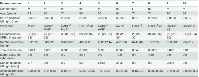
Bilateral MCA monitoring of 10 patients during cardiac surgery yielded over 17 hours of TCD recordings containing 18667 individual embolic signals. A summary of parameters recorded during surgery, and the results of our analysis, are presented inTable 1.
Total numbers of emboli entering the MCAs during a single operation varied from a mini-mum of 371 (patient 8) to a maximini-mum of 6476 (patient 3). Similar total numbers of emboli were observed in the left and right MCAs,Fig 1. A paired t-test confirmed no significant differ-ence in the median number of emboli in the left [491 (IQR: 349 to 950)] or the right sides [599 (IQR: 322 to 886)], (paired t-test,t= -1.19,p= 0.27). Higher average numbers of bubbles tended to be observed during the 5 intra-cardiac procedures (mean 1366, range: 671, 6476) than the 5 CABG procedures (mean 792, range: 371, 1551), though with so few patients this did not reach significance (Wilcoxon rank-sum test,z= -1.15,p= 0.25).
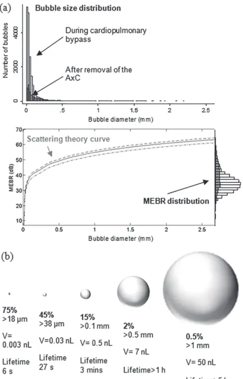
Patient-specific bubble sizing revealed that the majority (85%) of bubbles were less than 100μm and can be expected to dissolve within minutes,Fig 2. The median bubble diameter
from all 18667 signals was 33μm (IQR: 18 to 71μm). Conversion of MEBR values to bubble
di-ameters is illustrated inFig 2(A). Note that the exact form of this curve varies slightly between
Table 1. Summary of patient details, intra-operative haematocrit, and analysis of TCD recordings.
Patient number 1 2 3 4 5 6 7 8 9 10
Gender, (m/f) M m m m m m f m m m
Age, (years) 71 66 74 60 55 64 71 71 76 69
MCAadiameter
left:right, (mm)
2.6:2.7 2.6:2.8 2.9:2.6 2.6:2.9 3.5:2.6 3.3:3.2 3:3.1 2.6:2.8 2.9:2.8 2.6:2.7
Procedure MVRc CABGd
MVRc
CABGd
MVRc
CABGdx3 CABGd
AVRe
MVRc CABGd CABGdx3 CABGd
x3
CABGdx2
Haematocrit on CPBb, % (range)
30 (30–
33)
28 (26–
32)
33 (28–36) 33 (32–34) 28 (27–33) 31 (29–
35)
23 (22–
30)
34 (30–37) 26 (22–
30)
31 (29–34)
Number of emboli, left:right
502:596 349:322 2785:3691 409:383 2080:2144 480:886 674:843 196:175 950:601 284:317
Total volume (mL) 0.021 0.016 0.005 0.0003 0.12 0.003 0.04 0.0002 0.022 0.01
Signal Loss, left: right (mins)
0:6 5:0 0:0 5:11 0:0 12:0 0:4 4:10 0:0 0:0
Curtain duration, left:right (secs)
7:7 0:0 0:0 0:0 65:65 31:31 0:0 0:0 22:15 0:0
Affected arterioles, left:right (%)
0.38:0.30 0.41:0.15 0.1:0.11 0.007:0.005 1.01:2.25 0.04:0.03 0.15:0.79 0.002:0.007 0.46:0.22 0.006:0.034
aMCA
—Middle Cerebral Artery bCPB
—Cardiopulmonary Bypass cMVR
—Mitral Valve Replacement dCABG
—Coronary Artery Bypass Graft eAVR
—Atrial Valve Replacement.
patients depending on MCA diameter and intra-operative haematocrit levels.[11] A summary of the percentage of bubbles above a particular diameter, and their estimated dissolve times, is provided inFig 2(B). Only 2.2% of bubbles were larger than 500μm. Of these, approximately
90 bubbles (0.5%) were estimated to represent clinically significant macrobubbles over 1 mm in diameter.
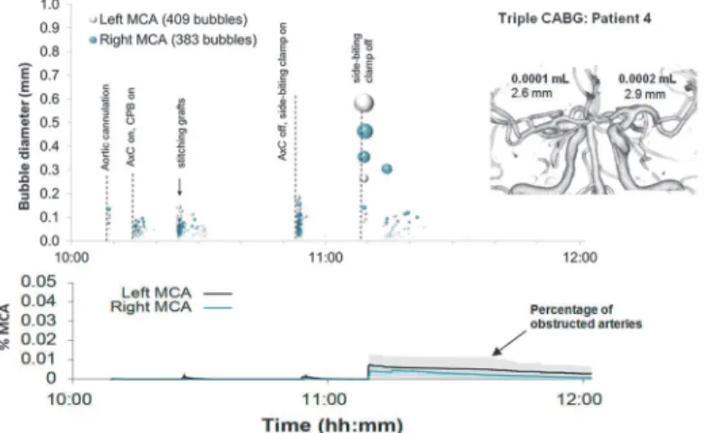
Examples of detailed plots illustrating the timing and estimated sizes of bubbles in individu-al patients are presented in Figs3–6. Each marker in the upper panel represents an individual embolic event. The y-axis and marker width shows estimated bubble diameter. The x-axis dis-plays time during surgery. Closer examination of the timing of embolic signals shows that events during surgery generate distinct showers of emboli seen as vertical stripes in the plot. Showers typically occurred in left and right MCAs simultaneously and contain a broad distri-bution of bubble sizes. Restarting the heart following removal of the aortic cross-clamp consis-tently generated dense showers containing larger bubbles than during bypass. In four patients there were sections of the recording where embolic signals became too dense to analyse indi-vidually. We were unable to include these in our simulations but the durations of these 'cur-tains' of emboli are listed inTable 1.
By manually analysing data from the entire procedure, and maintaining detailed transcripts, we were able to pin-point potential sources of emboli showers (see labels in the upper panels of Figs3–6). Light showers of bubbles were observed to coincide with the introduction and re-moval of cannulas, handling the heart, stitching grafts, and injections. Heavy showers were most often associated with the commencement of CPB, application and removal of the aortic cross-clamp (AxC), and restarting the heart during weaning from bypass. Nearly all patients experienced showers of emboli during commencement of bypass and following removal of the AxC.
Nearly three quarters of bubbles (73%) occurred prior to removal of the AxC during CPB, with an estimated median bubble diameter of 28μm (IQR: 17 to 51μm). Bubbles arising
dur-ing CPB were numerous but considerably smaller than those enterdur-ing the brain durdur-ing the
Fig 1. Total numbers of bubbles detected in the left and right middle cerebral arteries.Similar numbers of bubbles were detected in the left and right MCAs of individual patients. Markers are labelled with
patient identifiers.
Fig 2. Estimated distribution of bubble sizes detected during cardiac surgery.(a) Distribution of bubble sizes estimated based on analysis of the ultrasound backscatter (MEBR values) from 18667 embolic signals. Overall, the median diameter of bubbles was 33μm (IQR: 18 to 69μm). Signals observed following removal of the aortic cross-clamp are shaded. (b) Percentage of emboli, bubble volume, and predicted dissolve times for 18μm, 38μm, 100μm, 500μm and 1 mm diameter air bubbles.
later stages of the surgery. The 28μm median diameter of bubbles detected during CPB is
con-sistent with the use of a 38μm filter positioned in the aortic line of the CPB circuit.
Although the later stages of surgery, following removal of the AxC, contributed only 27% of embolic signals, these tended to include larger bubbles than during CPB, with a median bubble diameter of 72μm (IQR: 28 to 202μm). The majority of larger bubbles were observed as the
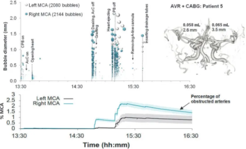
heart began to eject following removal of the AxC. The largest detected bubbles (>1 mm) reached diameters approaching the diameter of the MCA (e.g. ~3.5 mm in patient 5).
In individual patients, the total volume of air entering the MCAs during surgery varied from virtually no air at all [0.0002 mL (patient 8)] to a maximum of 0.12 mL (patient 5),
Table 1. Patient specific Monte-Carlo estimates for the instantaneous percentage of obstructed
Fig 3. Timing and diameters of bubbles in a 60 year old patient during Triple CABG.Markers denote individual embolic events where the y-axis and marker size indicate estimated bubble diameter. The lower panel displays the predicted number of blocked MCA end arterioles estimated by Monte-Carlo simulation. The shaded regions highlight 95% confidence intervals, based on uncertainty in bubble diameter and variations in outcome between simulations. The inset shows a 3D reconstruction of the circle of Willis labelled with estimated total volume of air and MCA diameters.
doi:10.1371/journal.pone.0122166.g003
Fig 4. Timing and diameters of bubbles in a 76 year old patient during Triple CABG.Markers denote individual embolic events where the y-axis and marker size indicate estimated bubble diameter. The lower panel displays the predicted number of blocked MCA end arterioles estimated by Monte-Carlo simulation. The shaded regions highlight 95% confidence intervals, based on uncertainty in bubble diameter and variations in outcome between simulations. The inset shows a 3D reconstruction of the circle of Willis labelled with estimated total volume of air and MCA diameters.
end arterioles, with 95% Confidence Intervals, are plotted as the solid line in the lower panels of Figs3–6. Our simulations suggest that small showers of filtered (<38μm) bubbles occurring during bypass do not impair cerebral blood flow. The greatest predicted threats to perfusion es-timated by our simulations were associated with unfiltered macrobubbles detected following removal of the AxC. However, showers of large emboli following removal of the AxC still only affected a small (<2.2%) percentage of the model vasculature.
Discussion
This study represents a detailed survey of the backscatter intensities of Doppler embolic signals observed during cardiac surgery and estimates the likely size distribution of bubbles and
Fig 5. Timing and diameters of bubbles in a 71 year old patient during combined MVR and CABG. Markers denote individual embolic events where the y-axis and marker size indicates estimated bubble diameter. The lower panel displays the predicted number of blocked end arterioles obtained by Monte-Carlo simulation. The shaded regions highlight 95% confidence intervals, based on uncertainty in bubble diameter and variations in outcome between simulations. The inset shows a 3D reconstruction of the circle of Willis labelled with estimated total volume of air and MCA diameters.
doi:10.1371/journal.pone.0122166.g005
Fig 6. Timing and diameters of bubbles in a 55 year old patient during combined AVR and CABG. Markers denote individual embolic events where the y-axis and marker size indicates estimated bubble diameter. The lower panel displays the predicted number of blocked end arterioles obtained by Monte-Carlo simulation. The shaded regions highlight 95% confidence intervals, based on uncertainty in bubble diameter and variations in outcome between simulations. The inset shows a 3D reconstruction of the circle of Willis labelled with estimated total volume of air and MCA diameters.
volume of air entering the middle cerebral arteries. We confirm that the majority (~85%) of bubbles entering the cerebral circulation are less than 100μm in diameter. The number and
di-mensions of air emboli revealed by our analysis is broadly consistent with previous autopsy studies by Moodyet al., which revealed numerous small capillary arteriolar dilatations post-op-eratively.[15] Moody and colleagues studied 100μm thick slices of the basal ganglia of patients
who died within one week of surgery and observed numerous empty ~40μm dilatations, with a
density of approximately 40 dilatations per cm2of tissue. The median diameter of air emboli estimated by our analysis of 33μm (IQR: 18–71) is similar to Moody’s findings.
A limitation of our study was the difficulty in analysing heavy showers of emboli. During these 'curtains' of emboli some signals could not be analysed and the number of emboli, and volume of air received, would therefore have been underestimated; seeTable 1for curtain dura-tions. This impacted mainly valve and combined procedure patients, where showers were par-ticularly heavy. Based on these limitations, the numbers of emboli reported in our study for patients 1, 5, 6 and 9 should be considered a conservative estimate.
Our findings demonstrate that up to 0.12 mL of air can enter the MCA territories during surgery,Table 1. Although this volume of air is lower than the total volume of microbubbles found to generate acute cerebral injury in animal studies [6], as the dissolve time for an individ-ual bubble depends crucially on surface area, and multiple bubbles have potential to coalesce, the potential for more subtle localised injuries cannot be completely discounted.
The time taken for an air embolus to completely dissolve, and proportion of the vascular tree that becomes obstructed, are both sensitively dependent on bubble size [1]; bubbles less than 38μm clear within seconds, whereas a 1 mm bubble is thought to take over 5 hours,Fig 2
(B). To better understand the relationship between vascular obstruction and bubble size rela-tive to the dimensions of the arterial tree we used a Monte-Carlo simulation of gas bubbles moving through a model MCA vasculature.[9,13,14] Based on the results of these simulations, showers of bubbles with diameters less than ~38μms did not generate any significant
obstruc-tion. Showers of larger (>100μm) macrobubbles following removal of the aortic-cross clamp and during weaning from bypass were predicted to affect up to 2.2% of the model vasculature for several hours. However, a limitation of our model is the absence of a mechanism for allow-ing multiple small bubbles to coalesce. Large bubbles pose a much greater threat to blood flow because they take longer to dissolve and become lodged higher up in the arterial tree. If our simple model can be considered to reflect true levels of microvascular obstruction and rates of embolus clearance, the greatest threat to cerebrovascular perfusion is predicted to occur follow-ing removal of the AxC durfollow-ing weanfollow-ing from bypass. A more sophisticated model would be re-quired to model physiological responses associated with progression from obstruction to cerebral ischaemia by modeling oxygen diffusion in the capillary mesh, cerebral autoregulation, inflammation, and by including a detailed knowledge of the cerebrovasculature.
One of the major limitations of our study is that we have focused entirely on the impact of air emboli. Our current study is therefore limited to conclusions regarding the sizes of bubbles and overlooks the potential contribution to microvascular obstruction of solid particles. The presence of solid emboli would not affect our findings, as these would have been misclassified as very small bubbles, but would provide a significant additional embolic burden, which is not considered in the present study. To predict the impact of both solid and gaseous emboli, would require advances in distinguishing solid and gas emboli based on transcranial Doppler
ultrasound.
benign microbubbles, which pass harmlessly through the circulation, the ability to identify large macrobubbles intra-operatively through analysis of Doppler recordings could provide a valuable clinical monitoring tool during cardiovascular interventions. In future work we plan to use this technique, in combination with neuropsychological testing and MRI, to examine whether the presence of large macrobubbles, or high accumulated volume of air entering the circulation, provides an explanation for new brain injuries observed following heart surgery.
Acknowledgments
We wish to thank the patients, cardiac surgeons and theatre staff who have assisted us with this research.
Author Contributions
Conceived and designed the experiments: EMLC JPH CB DHE. Performed the experiments: CB NP JJ. Analyzed the data: CB NP DM MAH CR JK JPH. Contributed reagents/materials/ analysis tools: CR. Wrote the paper: EMLC CB JPH.
References
1. Barak M, Katz Y (2005) Microbubbles: pathophysiology and clinical implications. Chest 128: 2918–
2932. PMID:16236969
2. Mitchell S, Gorman D (2002) The pathophysiology of cerebral arterial gas embolism. J Extra Corpor Technol 34: 18–23. PMID:11911624
3. Chaudhuri K, Marasco SF (2011) The effect of carbon dioxide insufflation on cognitive function during cardiac surgery. J Card Surg 26: 189–196. doi:10.1111/j.1540-8191.2011.01217.xPMID:21395683 4. Gerriets T, Schwarz N, Sammer G, Baehr J, Stolz E, et al. (2010) Protecting the brain from gaseous
and solid micro-emboli during coronary artery bypass grafting: a randomized controlled trial. Eur Heart J 31: 360–368. doi:10.1093/eurheartj/ehp178PMID:19541675
5. Kruis RW, Vlasveld FA, Van Dijk D (2010) The (un)importance of cerebral microemboli. Semin Cardi-othorac Vasc Anesth 14: 111–118. doi:10.1177/1089253210370903PMID:20478951
6. Haines DE, Stewart MT, Barka ND, Kirchhof N, Lentz LR, et al. (2013) Microembolism and catheter ab-lation II: effects of cerebral microemboli injection in a canine model. Circ Arrhythm Electrophysiol 6: 23–30. doi:10.1161/CIRCEP.112.973461PMID:23275248
7. Chaudhuri K, Storey E, Lee GA, Bailey M, Chan J, et al. (2012) Carbon dioxide insufflation in open-chamber cardiac surgery: a double-blind, randomized clinical trial of neurocognitive effects. J Thorac Cardiovasc Surg 144: 646–653 e641. doi:10.1016/j.jtcvs.2012.04.010PMID:22578685
8. Lynch JE, Riley JB (2008) Microemboli detection on extracorporeal bypass circuits. Perfusion 23: 23–
32. PMID:18788214
9. Hague JP, Banahan C, Chung EM (2013) Modelling of impaired cerebral blood flow due to gaseous emboli. Phys Med Biol 58: 4381–4394. doi:10.1088/0031-9155/58/13/4381PMID:23743635 10. Ringelstein EB, Droste DW, Babikian VL, Evans DH, Grosset DG, et al. (1998) Consensus on
microem-bolus detection by TCD. International Consensus Group on Microemmicroem-bolus Detection. Stroke 29: 725–
729. PMID:9506619
11. Banahan C, Hague JP, Evans DH, Patel R, Ramnarine KV, et al. (2012) Sizing gaseous emboli using Doppler embolic signal intensity. Ultrasound Med Biol 38: 824–833. doi:10.1016/j.ultrasmedbio.2012. 01.008PMID:22402022
12. Moehring MA, Klepper JR (1994) Pulse Doppler ultrasound detection, characterization and size estima-tion of emboli in flowing blood. IEEE Trans Biomed Eng 41: 35–44. PMID:8200666
13. Chung EM, Hague JP, Evans DH (2007) Revealing the mechanisms underlying embolic stroke using computational modelling. Phys Med Biol 52: 7153–7166. PMID:18029999
14. Hague JP, Chung EM (2009) Statistical physics of cerebral embolization leading to stroke. Phys Rev E Stat Nonlin Soft Matter Phys 80: 051912. PMID:20365011