Apoptotic Cell Death Induced by Resveratrol
Is Partially Mediated by the Autophagy
Pathway in Human Ovarian Cancer Cells
Fangfang Lang2, Zhaoyang Qin3, Fang Li4, Huilin Zhang5, Zhenghui Fang2, Enkui Hao1*
1Department of Cardiology, Qianfoshan Hospital, Affiliated with Shandong University, Jinan, China, 2Department of Obstetrics and Gynecology, Jinan Central Hospital, Affiliated with Shandong University, Jinan, China,3Department of General Surgery, Rizhao People’s Hospital, Rizhao, China,4Department of Health, Jinan Central Hospital, Affiliated with Shandong University, Jinan, China,5Central Laboratory, Jinan Central Hospital, Affiliated with Shandong University, Jinan, China
*haoenkui@sdu.edu.cn
Abstract
Resveratrol (trans-3,4,5’ –trihydroxystilbene) is an active compound in food, such as red grapes, peanuts, and berries. Resveratrol exhibits an anticancer effect on various human cancer cells. However, the mechanism of resveratrol-induced anti-cancer effect at the mo-lecular level remains to be elucidated. In this study, the mechanism underlying the anti-can-cer effect of resveratrol in human ovarian cananti-can-cer cells (OVCAR-3 and Caov-3) was
investigated using various molecular biology techniques, such as flow cytometry, western blotting, and RNA interference, with a major focus on the potential role of autophagy in res-veratrol-induced apoptotic cell death. We demonstrated that resveratrol induced reactive oxygen species (ROS) generation, which triggers autophagy and subsequent apoptotic cell death. Resveratrol induced ATG5 expression and promoted LC3 cleavage. The apoptotic cell death induced by resveratrol was attenuated by both pharmacological and genetic inhi-bition of autophagy. The autophagy inhibitor chloroquine, which functions at the late stage of autophagy, significantly reduced resveratrol-induced cell death and caspase 3 activity in human ovarian cancer cells. We also demonstrated that targeting ATG5 by siRNA also sup-pressed resveratrol-induced apoptotic cell death. Thus, we concluded that a common path-way between autophagy and apoptosis exists in resveratrol-induced cell death in OVCAR-3 human ovarian cancer cells.
Introduction
Ovarian cancer is one of the major leading causes of cancer-related death for females and a high rate of recurrence after surgery [1] [2]. In most cases, the diagnosis is made at late stages of the cancer, and it becomes challenging for surgical resection and recovery [2]. Thus, studies on the active ingredients of food products might provide useful alternative therapeutic ap-proaches for this malignancy.
OPEN ACCESS
Citation:Lang F, Qin Z, Li F, Zhang H, Fang Z, Hao E (2015) Apoptotic Cell Death Induced by Resveratrol Is Partially Mediated by the Autophagy Pathway in Human Ovarian Cancer Cells. PLoS ONE 10(6): e0129196. doi:10.1371/journal.pone.0129196
Academic Editor:Dominique Delmas, UMR INSERM U866, FRANCE
Received:October 31, 2014
Accepted:May 7, 2015
Published:June 11, 2015
Copyright:© 2015 Lang et al. This is an open access article distributed under the terms of the
Creative Commons Attribution License, which permits unrestricted use, distribution, and reproduction in any medium, provided the original author and source are credited.
Data Availability Statement:All data are provided in the manuscript.
Funding:This study was supported by the promotive research fund for young and middle-aged scientists of Shandong Province (Grant No. 07-21). The funders had no role in study design, data collection and analysis, decision to publish, or preparation of the manuscript.
Resveratrol is an active ingredient from our food sources, such as grapes, peanuts, and ber-ries, which has long been used in traditional Chinese medicine. Numerous studies have demon-strated the beneficial effects of resveratrol in cardiovascular diseases, neural diseases, obesity, and inflammatory disorders [3–5]. One of the major areas of resveratrol research is at the
fore-front of cancer research [6,7]. It is well known that a high dose of resveratrol results in
apopto-tic cell death of ovarian cancer cells [8–10]. Several mechanisms of ovarian cancer cell death have been proposed. Phosphorylation of Cdc2-tyr15 by resveratrol treatment result in cell cycle arrest of OVCAR-3 [9]. Down-regulation of Akt/GSK and ERK signaling pathways has been shown to be critical for resveratrol-mediated cell death [10]. Recently, Lin et al. described the important role of ceramide and COX-2 in apoptotic cell death by resveratrol in OVCAR-3 [8].
Autophagy is a conservative self-degradation pathway in which cytosolic components are sequestered to lysosomes for degradation and recycling [11]. In healthy tissues, this is a process of clearing of damaged organelles. However, it is a complex process in cancer cells where it can either suppress or induce the growth of cancer cells depending on the cellular microenviron-ment [12].
In the present study, we investigated the potential role of autophagy in resveratrol-induced apoptotic cell death in OVCAR-3 cancer cells. We found that resveratrol treatment induced ROS generation and apoptosis, as well as activation of the autophagy pathway in OVCAR-3 cells. Inhibition of autophagy by a pharmacological inhibitor or a siRNA against ATG5 signifi-cantly attenuated resveratrol-mediated apoptotic cell death. Thus, our study established an im-portant role of autophagy in resveratrol-induced apoptosis in human ovarian cancer cells.
Materials and Methods
Reagents
Resveratrol, NAC (N acetyl cysteine), chloroquine, caspase 3 assay kit, and LC3 antibody were purchased from Sigma (USA). Resveratrol was dissolved in DMSO (Sigma, USA) and was freshly prepared every time prior to cell treatment. Anti-ATG5 antibody was purchased from Beijing Biosynthesis Biotechnology, ATG5-ATG12 Complex Antibody was from AbD Serotec, and anti-cleaved caspase 3 antibody was ordered from Cell Signal technologies. siRNA against ATG5 were obtained from Shanghai GenePharma Co. Ltd. Z-VAD-FMK was purchased from R&D.
Cell culture
OVCAR-3 and Caov-3 human ovarian cancer cell lines were obtained from ATCC (USA). The cells were cultured in RPMI 1640 (Life Technology, USA) supplemented with 10% fetal bovine
serum, insulin, and penicillin–streptomycin. The cell line was grown in a CO2incubator at
37°C. Human Ovarian Surface Epithelial Cells (HOSEpiC) cells were grown with Ovarian Epi-thelial Cell Medium provided from manufacturer (ScienCell Research Laboratories). Resvera-trol treatment were described in text and figures.
Cell viability assays
Apoptosis Assay using flow cytometry
Apoptotic cell death were measured by staining with Sytox Green and Annexin V–APC (Life Technology, USA) as previously described [13].
Cellular ROS assays
Total intracellular ROS were measured by flow cytometry using the H2DCFDA dye according
to the manufacturer’s recommendation (Life Technology, USA).
Determination of mitochondrial electron potential change
Mitochondrial membrane potential was assessed using a live cell assay with the fluorescent li-pophilic cationic dye TMRE (Sigma)). This dye is a cell permeant dye that readily accumulates in active mitochondria due to their relative negative charge. After treatment OVCAR-3 cells were stained with 500 nM TMRE for 30 min at 37°C, then washed twice in medium and re-sus-pended in PBS. The cells were analyzed in a flowcytometer at FL2 channel and the mean inten-sity is plotted. itochondrial potential also analyzed by another dye JC-1. After treatment as described in the text, the fluorescent images were immediately taken using a Zeiss AX10 in-verted scope (Carl Zeiss Microimaging Inc., Germany).
Determination of 4HNE Protein adducts
The 4-HNE level in cell lysates was measured using the OxiSelect HNE Adduct ELISA Kit (Cell Biolabs) according to the manufacturer's instructions. Western blot was also performed using an anti-HNE antibody (Abcam).
Western blotting analyses
Cells were washed with cold PBS and lysed with RIPA buffer (Sigma, USA) containing a prote-ase inhibitor cocktail (Roche, Germany). Protein concentrations were determined using a
pro-tein assay reagent (Bio Rad, USA), and equal amounts of propro-tein (50μg) were loaded in each
well of 4–20% or 12% SDS-PAGE. After the proteins were transferred onto a nitrocellulose membrane, the blots were blocked with 5% milk, followed by incubation with the appropriate diluted primary antibody overnight. After three washes with TBS-T buffer, the blots were incu-bated with appropriately diluted HRP-conjugated secondary antibodies for 2 h. The immuno-blots were developed using the SuperSignal West Femto kit (Fisher Scientific, USA).
RNA Interference
OVCAR cells were transfected with either nonspecific or ATG5 siRNA using lipofectamine (Life technologies, USA) according to manufacturer’s instruction. Cells were incubated 48 h prior to treatment with resveratrol for maximum blocking of ATG5 expression.
Caspase 3 activity
PARP activity
PARP activity, we used the HT Universal Colorimetric PARP assay kit from Trevigen. PARP activity is determined by the amount of PAR deposited onto immobilized histone proteins. The procedure was performed according to the recommendations of the manufacturer.
Live cell imaging with autophagy indicator dye
Autophagic flux also was determined using Cyto-ID Autophagy Detection Kit (Enzo Life Sci-ences) according to manufacturer's instructions.
Live cell imaging with fluroscent caspase substrate
Caspase activation was determined using CellEvent Caspase-3/7 Green Detection Reagent (Life technologies,USA) according to manufacturer’s instruction.
Statistical analyses
All data were expressed as the mean with standard error of mean. Experiments were repeated four times in duplicate for all experiments. Student’s t-test or one-way ANOVA were per-formed as appropriate to determine the statistical significance. The significance was established at p<0.05.
Results
Resveratrol induced apoptotic cell death of human ovarian cancer cells
in a dose-dependent manner
OVCAR-3 is a chemo-resistant cancer cell line established from an ovarian adenocarcinoma
patient. We utilized OVCAR-3 cells as anin vitrocancer cell model to test the anticancer effects
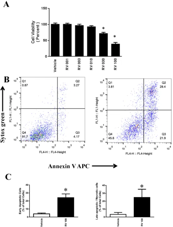
of resveratrol. As shown inFig 1A, treatment with resveratrol at 1μM to 100μM for 48 h
re-sulted in a dose-dependent loss of cell viability as determined using MTT assays. Treatment
with 30μM and 100μM resveratrol showed a statistically significant loss of cell viability at
48 h.
The effects of resveratrol on the apoptotic cell death of OVCAR-3 cells were examined by
flow cytometry using Sytox Green (FL1H) and Annexin V–Allophycocyanin (FL4H).
Treat-ment with 100μM of resveratrol for 48 h induced significant early apoptotic and late
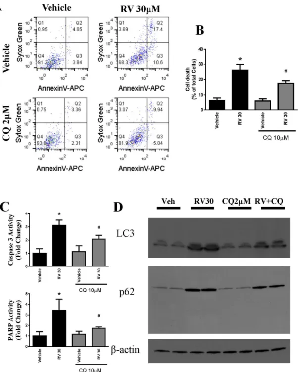
apopto-tic/necrotic cell death (Fig 1B and 1C). Treatment with 30μM of resveratrol for 48 h also
induced significant early apoptotic and late apoptotic/necrotic cell death (Fig 2A and 2B) but
the effect was less compared to 100μM.
Resveratrol induced autophagy in human ovarian cancer cells
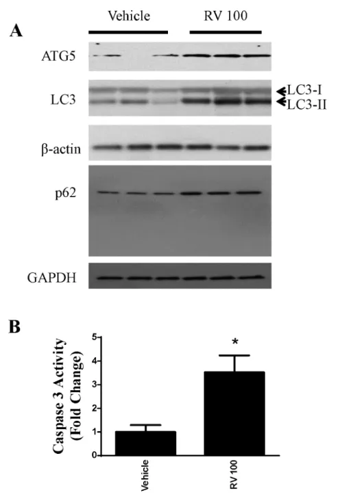
We also evaluated the effect of resveratrol on autophagy. Key markers of autophagy were
as-sessed. As shown inFig 2CandFig 3A, resveratrol ainduced LC3-II and p62 in OVCAR-3
cells. Western blotting analyses revealed an increase in the specific autophagic marker ATG5,
which is essential for autophagosome formation(Figs2Aand3A). In addition, the activity of
caspase 3 was significantly increased in resveratrol-treated OVCAR-3 cells (Figs2Dand3B).
Resveratrol induced oxidative stress in OVCAR-3 human ovarian cancer
cells
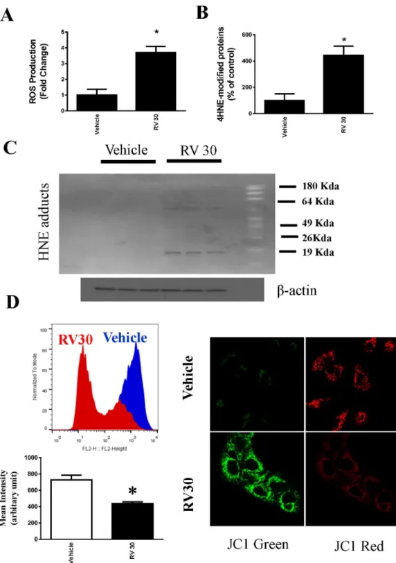
diacetate (H2DCFDA) dye to measure intracellular ROS, and found that resveratrol at 30μM
resulted in significantincrease in intracellular ROS production in OVCAR3 cells (Fig 4A). To further confirm this result, we also used a complimentary method to measure the oxidative
Fig 1. Effect of resveratrol in the cell death of ovarian cancer cells OVCAR-3. A.OVCAR3 cells were treated with 1 to 100μM resveratrol for 48 h. The cell viability was determined using MTT assays. The data were obtained from an average of four independent experiments.*P<0.05 compared with vehicle control, n = 4/group.B.Representative flow cytometric dot plots for the measurement of apoptosis in OVCAR-3 cells. Resveratrol-induced apoptosis was measured by staining with Sytox Green dye (Y-axis, FL1) and Annexin V-APC (X-axis, FL4).C. Quantitative determination of early apoptotic cell death as the number of annexin V-positive cells with Sytox green-negative cells, and late apoptotic/necrotic cell death as the number of annexin V-positive and Sytox green-positive cells.
footprint by measuring HNE protein adducts (4-Hydroxynonenal). Consistent with the intracellular ROS data, HNE protein adducts were increased significantly compared with
Fig 2. Role of autophagy in resveratrol-induced cytotoxicity in ovarian cancer cells OVCAR-3. A. Representative western blotting analyses of OVCAR-3 total cell lysates with ATG5, LC3, and p62 antibodies.
β-actin and GAPDH were used as loading controls. Three replicate samples were loaded for each treatment group.B.Caspase 3 activation was measured from cell lysates and expressed as the fold change.*P<0.05
compared with the vehicle control, n = 4/group.
vehicle-treated OVCAR cells (Fig 4B and 4C). Furthermore, we showed that resveratrol also significantly decreased mitochondrial electron potential (Fig 4D).
Resveratrol similarly induced apoptosis and autophagy in human
ovarian cancer cells Caov-3
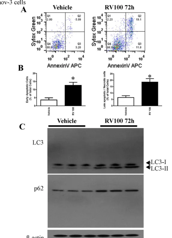
To determine whether the resveratrol effect is cell line specific, we utilized another human ovarian cancer cell line Caov-3. Consistent with the effect observed in OVCAR-3 cells,
Fig 3. Resveratrol induced apoptosis and autophagy in OVCAR-3 cells at a lower dose.A. OVCAR-3 cells were treated with 30μM resveratrol for 48 h, and cell death was determined by flow cytometry. B. Quantitative result of the flow cytometry. C. Representative western blotting analyses of OVCAR-3 total cell lysates with ATG5, LC3, and p62 antibodies withβ-actin as a loading control.
Fig 4. Generation of ROS by resveratrol (30μM) in ovarian cancer cells OVCAR-3.A.Intracellular ROS of OVCAR-3 were measured by flow cytometry using H2DCFDA and expressed as the fold change. Resveratrol at 30μM induced statistically significant ROS.*P<0.05 compared with vehicle control, n = 4/ group.B.Oxidative stress marker 4HNE modified proteins were determined using ELISA from the cell lysates of vehicle and Resveratrol-treated (30μM) OVCAR-3 cells. The quantified modified proteins were expressed as the % of vehicle control samples.*P<0.05 compared with vehicle control, n = 4/group. C. Representative Western blotting showing resveratrol induced elevation of HNE adducts.D. Mitochondrial electron potential change following resveratrol treatment was determined by quantitative flow cytometry and immunofluorescence.
resveratrol also induced apoptosis and autophagy in Caov-3 cells (Fig 5). However, resveratrol at the same dose and time frame did not induce early apoptosis in primary human ovarian sur-face epithelial cells HOSEpiC although there is slightly increase in late apoptotic/necrotic cell death (S1 Fig).
Pharmacological inhibition of autophagy attenuated resveratrol-induced
cell death in OVCAR-3 cells
To test whether autophagy is a link to apoptotic cell death, we treated OVCAR-3 cells with chloroquine (CQ), a pharmacological inhibitor of autophagy, prior to resveratrol treatment. Apoptosis and cell death were determined using Sytox Green and AnnexinV–Allophycocyanin.
As shown inFig 6A, cell death by resveratrol at 30μM was significantly attenuated by
pretreat-ment with 2μM of CQ. Resveratrol significantly reduced the total cellular death by autophagy
inhibition. also analyzed resveratrol induced apoptosis markers caspase 3 activity and PARP activity and both were attenuated by CQ treatment (Fig 6C). In addition, we observed that res-veratrol-induced autophagosome markers LC3II and p62 were also attenuated by CQ treat-ment (Fig 6D).
Inhibition of autophagy by ATG siRNA ameliorated resveratrol-induced
cell death in OVCAR-3 cells
ATG5 is the hallmark of autophagy and a key player in the formation of autophagosomes. In addition to the pharmacological inhibitor, we used a genetic approach by knocking down ATG5 expression using a specific siRNA and examined its effect on resveratrol-induced
can-cer cell death. As shown inFig 7, cell death by resveratrol (30μM) was reduced from 27.9%
to 16.22% by ATG5 siRNA. A similar pattern was also observed in caspase 3 activity and LC3 western blot (Fig 7B and 7C). The efficacy of siRNA technology was confirmed using western blotting analyses, which indicated a significant reduction of ATG5 protein levels (Fig 7A). Interestingly, we found that the cells treated with ATG5 siRNA in combination
with Z-VAD-FMK(50μM) are more significantly protected from resveratrol-induced cell
death compared to those treated with either inhibitor alone (Fig 7E). We also employed new live cell imaging technology using Cyto-ID dye for autophagy flux and the above results were confirmed.
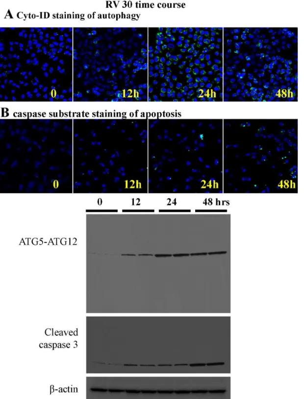
We used live cell imaging for autophagic flux and caspase 3 substrate dye (marker for early apoptosis) using microscopy (Fig 8A and 8B). It was evident that autophagic flux was initiated earlier (12h) than apoptosis (24h) when treated with 30M resveratrol, which is in agreement of the aforementioned ATG5 siRNA data. We also performed a time course study with Western blot analyses using two sets of samples from independent experiments at each time points (Fig 8C) and found that autophagy was activated earlier than apoptosis, suggesting that autop-hagy is upstream of apoptosis in the ovarian cancer cells treated with resveratrol. However there are overlap between the two processes which is a typical pattern of any cancer cell population.
Discussion
chloroquine attenuated resveratrol-induced cell death in OVCAR-3, and similar results were obtained by blocking ATG5 expression using siRNA.
Fig 5. Resveratrol induced apoptosis and autophagy in Caov-3 cells.A. Caov-3 cells were treated with 30μM resveratrol for 72 h, and cell death was determined by flow cytometry. B. Quantitative result of the flow cytometry. C. Representative western blotting analyses of Caov-3 total cell lysates with LC3, and p62 antibodies.
Several recent studies have shown some intriguing pathways that mediate resveratrol-in-duced apoptotic cell death. Vergara et al. described the role ERK signaling and AKT/GSK path-ways in apoptotic cell death using a proteomic approach [10]. In another elegant study, Lin
Fig 6. Pharmacological inhibition of autophagy protects ovarian cancer cells OVCAR-3 against resveratrol-induced cell death.A. Representative flow cytometry dot diagrams representing each OVCAR-3 cells. Cellular apoptosis was measured in the same method as inFig 1. Chloroquine reduced the cell killing effect of resveratrol. B. Quantitative determination of total cell death, which was expressed as the percentage of total cells.*P<0.05 compared with vehicle control; # P<0.05 compared with resveratrol-treated samples. n = 4/group. C. Caspase 3 and PARP activities were assayed and plotted as fold
change. D. Representative western blotting analyses of OVCAR-3 total cell lysates with LC3, and p62 antibodies.
Fig 7. RNA interference of ATG5 attenuated resveratrol-induced cell death and caspase activity.A. ATG5 protein expression was determined from cellular lysates obtained from OVCAR-3 cells loaded with either control random siRNA or ATG5 siRNA. B. Caspase 3 activities were determined from cellular lysates obtained from OVCAR-3 cells loaded with either control random siRNA or ATG5 siRNA. The activities were expressed as the fold change.*P<0.05 compared with vehicle control, # P<0.05 compared with resveratrol-treated samples. n = 4/group. C. Representative western blotting analyses of OVCAR-3
treated with vehicle (Veh) or Resveratrol at 30μM (RV) with and without siATG5 and total cell lysates were anlyzedwith LC3 andβ-actin D. Live cell image from confocal microscope(with 20X objective) using Cyto-Id dye loaded for 15 minutes after treatment with resveratrol at 30μM in OVCAR-3 cells
transformed with random or ATG5 siRNA. E. Quantitative determination of cell death in OVCAR cells using flow cytometry. Resveratrol-induced cell death is attenuated in siATG5-treated and siATG5 plus Z-VAD-FMK (50μM) treated OVCAR-3 cells.*P<0.05 compared with vehicle control; # P<0.05 compared
with resveratrol-treated samples. n = 4/group.
Fig 8. The time course of autophagy and apoptosis induction by 30μM resveratrol treatment.A. Live cell imaging by confocal microscopy (with 10X
objective) with Cyto-ID dye in OVCAR-3 cells treated with 30μM resveratrol at 0, 12, 24 and 48 hour time points. B. Live cell imaging by confocal microscopy (with 10X objective) with caspase 3 green detection dye in OVCAR-3 cells at 0, 12, 24 and 48 hour time points. C. OVCAR-3 cells were treated with 30μM of resveratrol for 12 h, 24 h, and 48 h. Western blotting of total cell lysates were performed with anti-ATG5-ATG12, LC3, and caspase 3 antibodies. No cleavage of ATG5 was observed. The results shown represent an experiment, in which each time-point of treatment was duplicated.
et al. demonstrated that resveratrol-induced apoptosis shared similar pathways with ceramide-induced COX-2-dependent apoptosis in ovarian cancer cells [8]. ere, we have shown that the apoptotic cell death of OVCAR-3 induced by resveratrol is far more complex and autophagy plays a critical role in this process.
One of our key observations was the production of intracellular ROS and oxidative stress in resveratrol-induced cell death in OVCAR-3 cells. ROS is involved in modulating various path-ways, including the ERK signaling pathway [14–16]. Intriguingly, ROS could induce autophagy in resveratrol-treated OVCAR-3 cells [17]. Resveratrol is known to have beneficial antioxidant effects on healthy cells [18], but these antioxidant activities have different consequences in highly proliferating cancer cells. Antioxidants, including resveratrol, have been shown to
con-tribute to ROS-induced cell death in other cancer cell types [19,20]. Our data suggest that ROS
production by resveratrol in OVCAR-3 cells is associated with cancer cell apoptosis, which is consistent to the findings obtained in previous studies focused on colorectal and colon carcino-ma cells.
In general, autophagy inhibits the induction of apoptosis, and apoptosis-associated caspase activation turns off the autophagy process. However, there are many cases where both of these processes occur simultaneously or co-activates to ROS production induced both autophagy and apoptosis in cancer cells [21]. Autophagy itself can induce cell death, a process known as autophagic cell death [22]. It has also been reported that induction of autophagy facilitates the activation of apoptosis [23]. Puissant et al found that both autophagy and apoptosis are in-volved in resveratrol-induced cell death in a myelogenous leukemia cell line [24–26]. In MCF7 breast cancer cells, resveratrol induced beclin-1 dependent and independent autophagy [27]. Here, we also observed both caspase dependent and independent pathway exists for resveratrol induced cell death in ovarian cancer cells. Either caspase or autophagy inhibitors were unable to restore resveratrol induced cell death. One of the key proteins involved in this process is the tumor suppressor protein p53 and its protein levels in the cytosol modulate activation or
inhi-bition of autophagy[28,29]. Nuclear translocation of p53 is also important for apoptotic
induc-tion. It has been shown that p53 is crucial for resveratrol-mediated cell death in OVCAR-3 cells [8]. Other common mediators of both autophagy and apoptosis are several BH3-only (BCL-3 homology 3) proteins. During apoptosis, BH3-only proteins directly interact with the BCL-2 family, thereby silencing the anti-apoptotic role and stimulating apoptosis. Several BH3-only proteins, such as BAD and BNIP3 can also promote autophagy by competitive inhi-bition between Beclin1 and apoptotic proteins BCL-2 [30–32]. Interestingly, both BCL2 and Bad have been shown to be dysregulated in OVCAR-3 cells [8].
The involvement p62 in resveratrol induced cell death in leukemia cell line is critical[25]. We also observed induction of p62 in ovarian cancer cells in response to resveratrol treatment.
In our study, we found that inhibition of autophagy attenuates (but not completely abol-ishes) resveratrol-induced ovarian cancer cell death. We also performed a time course study, and found that resveratrol activates autophagy earlier than apoptosis, suggesting that autop-hagy is upstream of apoptosis. Together, these data support that resveratrol induces apoptosis partially through autophagy. We agree that some more classic mechanisms are likely also in-volved in resveratrol induced apoptosis.
effect. This would be an interesting and important topic worth further study. Importantly, pre-vious studies in esophageal squamous cell carcinoma demonstrated the opposite effect by inhi-bition of autophagy in resveratrol-induced cell death [34]. This might be partially due to the different dosages and types of cancer cells used in this study, and autophagy is a complex pro-cess that functions like a double-edged sword when harnessed as a way to fight against cancer [35–39].
Supporting Information
S1 Fig. Resveratrol did not induce apoptosis in healthy ovarian surface epithelial cells.Flow cytometric analyses of Human Ovarian Surface Epithelial Cells (HOSEpiC) cells, which were grown with Ovarian Epithelial Cell Medium provided from manufacturer (ScienCell Research
Laboratories). Resveratrol treatment was performed at 100μM for 48 hours. Cells were then
stained with AnnexinV and sytox green for early apoptosis and late apoptosis/necrotic cell death markers respectively. Quantitative determination of flow cytometry data demonstrated no significant difference in early apoptic cell death markers but little significant increase in late
apoptosis/necrotic cell death markers.P<0.05 compared with vehicle control; n = 3/group.
(TIFF)
Author Contributions
Conceived and designed the experiments: EH F. Lang. Performed the experiments: F. Lang ZQ F. Li HZ ZF EH. Analyzed the data: F. Lang ZQ FLi HZ ZF EH. Contributed reagents/materi-als/analysis tools: F. Lang ZQ FLi HZ ZF EH. Wrote the paper: F. Lang EH.
References
1. Davis M, Rauh-Hain JA, Andrade C, Boruta DM 2nd, Schorge JO, Horowitz NS, et al. Comparison of clinical outcomes of patients with clear cell and endometrioid ovarian cancer associated with endome-triosis to papillary serous carcinoma of the ovary. Gynecol Oncol. 2014; 132(3):760–6. Epub 2014/01/ 21. doi:10.1016/j.ygyno.2014.01.012S0090-8258(14)00023-7 [pii]. PMID:24440832.
2. Jayson GC, Kohn EC, Kitchener HC, Ledermann JA. Ovarian cancer. Lancet. 2014. Epub 2014/04/29. doi: S0140-6736(13)62146-7 [pii] doi:10.1016/S0140-6736(13)62146-7PMID:24767708.
3. Hao E, Lang F, Chen Y, Zhang H, Cong X, Shen X, et al. Resveratrol alleviates endotoxin-induced myo-cardial toxicity via the Nrf2 transcription factor. PLoS One. 2013; 8(7):e69452. Epub 2013/07/31. doi:
10.1371/journal.pone.0069452 PONE-D-13-17928[pii]. PMID:23894482; PubMed Central PMCID: PMC3718737.
4. Mukhopadhyay P, Mukherjee S, Ahsan K, Bagchi A, Pacher P, Das DK. Restoration of altered micro-RNA expression in the ischemic heart with resveratrol. PLoS One. 2010; 5(12):e15705. Epub 2011/01/ 05. doi:10.1371/journal.pone.0015705PMID:21203465; PubMed Central PMCID: PMC3009730. 5. Vang O, Ahmad N, Baile CA, Baur JA, Brown K, Csiszar A, et al. What is new for an old molecule?
Sys-tematic review and recommendations on the use of resveratrol. PLoS One. 2011; 6(6):e19881. Epub 2011/06/24. doi:10.1371/journal.pone.0019881 PONE-D-11-01755[pii]. PMID:21698226; PubMed Central PMCID: PMC3116821.
6. Delmas D, Lancon A, Colin D, Jannin B, Latruffe N. Resveratrol as a chemopreventive agent: a promis-ing molecule for fightpromis-ing cancer. Curr Drug Targets. 2006; 7(4):423–42. Epub 2006/04/14. PMID:
16611030.
7. Le Corre L, Chalabi N, Delort L, Bignon YJ, Bernard-Gallon DJ. Resveratrol and breast cancer chemo-prevention: molecular mechanisms. Mol Nutr Food Res. 2005; 49(5):462–71. Epub 2005/03/24. doi:
10.1002/mnfr.200400094PMID:15786518.
8. Lin HY, Delmas D, Vang O, Hsieh TC, Lin S, Cheng GY, et al. Mechanisms of ceramide-induced COX-2-dependent apoptosis in human ovarian cancer OVCAR-3 cells partially overlapped with resveratrol. J Cell Biochem. 2013; 114(8):1940–54. Epub 2013/03/16. doi:10.1002/jcb.24539PMID:23495037. 9. Tyagi A, Singh RP, Agarwal C, Siriwardana S, Sclafani RA, Agarwal R. Resveratrol causes Cdc2-tyr15
human ovarian carcinoma Ovcar-3 cells. Carcinogenesis. 2005; 26(11):1978–87. Epub 2005/06/25. doi: bgi165 [pii] doi:10.1093/carcin/bgi165PMID:15975956.
10. Vergara D, Simeone P, Toraldo D, Del Boccio P, Vergaro V, Leporatti S, et al. Resveratrol downregu-lates Akt/GSK and ERK signalling pathways in OVCAR-3 ovarian cancer cells. Mol Biosyst. 2012; 8 (4):1078–87. Epub 2012/01/12. doi:10.1039/c2mb05486hPMID:22234583.
11. Duan WJ, Liu FL, He RR, Yuan WL, Li YF, Tsoi B, et al. Autophagy is involved in the effects of resvera-trol on prevention of splenocyte apoptosis caused by oxidative stress in restrained mice. Mol Nutr Food Res. 2013; 57(7):1145–57. Epub 2013/03/19. doi:10.1002/mnfr.201200662PMID:23505001. 12. Cheong H, Lu C, Lindsten T, Thompson CB. Therapeutic targets in cancer cell metabolism and
autop-hagy. Nat Biotechnol. 2012; 30(7):671–8. Epub 2012/07/12. doi:10.1038/nbt.2285 nbt.2285[pii]. PMID:22781696; PubMed Central PMCID: PMC3876738.
13. Mukhopadhyay P, Rajesh M, Hasko G, Hawkins BJ, Madesh M, Pacher P. Simultaneous detection of apoptosis and mitochondrial superoxide production in live cells by flow cytometry and confocal micros-copy. Nat Protoc. 2007; 2(9):2295–301. Epub 2007/09/15. doi: nprot.2007.327 [pii] doi:10.1038/nprot. 2007.327PMID:17853886; PubMed Central PMCID: PMC2225540.
14. Tan BJ, Chiu GN. Role of oxidative stress, endoplasmic reticulum stress and ERK activation in tripto-lide-induced apoptosis. Int J Oncol. 2013; 42(5):1605–12. Epub 2013/03/08. doi:10.3892/ijo.2013. 1843PMID:23467622.
15. Zhang Y, Zheng S, Zheng JS, Wong KH, Huang Z, Ngai SM, et al. Synergistic Induction of Apoptosis by Methylseleninic Acid and Cisplatin, The Role of ROS-ERK/AKT-p53 Pathway. Mol Pharm. 2014. Epub 2014/02/22. doi:10.1021/mp400749fPMID:24555485.
16. Gupta SC, Francis SK, Nair MS, Mo YY, Aggarwal BB. Azadirone, a limonoid tetranortriterpene, in-duces death receptors and sensitizes human cancer cells to tumor necrosis factor-related apoptosis-in-ducing ligand (TRAIL) through a p53 protein-independent mechanism: evidence for the role of the ROS-ERK-CHOP-death receptor pathway. J Biol Chem. 2013; 288(45):32343–56. Epub 2013/10/01. doi:10.1074/jbc.M113.455188 M113.455188[pii]. PMID:24078627; PubMed Central PMCID: PMC3820870.
17. Li L, Ishdorj G, Gibson SB. Reactive oxygen species regulation of autophagy in cancer: implications for cancer treatment. Free Radic Biol Med. 2012; 53(7):1399–410. Epub 2012/07/24. doi:10.1016/j. freeradbiomed.2012.07.011 S0891-5849(12)00406-6[pii]. PMID:22820461.
18. Santos JA, de Carvaho GS, Oliveira V, Raposo NR, da Silva AD. Resveratrol and analogues: a review of antioxidant activity and applications to human health. Recent Pat Food Nutr Agric. 2013; 5(2):144–
53. Epub 2013/05/22. doi: PFNA-EPUB-20130513-1 [pii]. PMID:23688141.
19. Wenzel U, Nickel A, Daniel H. alpha-Lipoic acid induces apoptosis in human colon cancer cells by in-creasing mitochondrial respiration with a concomitant O2-*-generation. Apoptosis. 2005; 10(2):359–
68. Epub 2005/04/22. doi:10.1007/s10495-005-0810-xPMID:15843897.
20. Juan ME, Wenzel U, Daniel H, Planas JM. Resveratrol induces apoptosis through ROS-dependent mi-tochondria pathway in HT-29 human colorectal carcinoma cells. J Agric Food Chem. 2008; 56 (12):4813–8. Epub 2008/06/05. doi:10.1021/jf800175aPMID:18522405.
21. Wong CH, Iskandar KB, Yadav SK, Hirpara JL, Loh T, Pervaiz S. Simultaneous induction of non-canon-ical autophagy and apoptosis in cancer cells by ROS-dependent ERK and JNK activation. PLoS One. 2010; 5(4):e9996. Epub 2010/04/07. doi:10.1371/journal.pone.0009996PMID:20368806; PubMed Central PMCID: PMC2848860.
22. Lamy L, Ngo VN, Emre NC, Shaffer AL 3rd, Yang Y, Tian E, et al. Control of autophagic cell death by caspase-10 in multiple myeloma. Cancer Cell. 2013; 23(4):435–49. Epub 2013/04/02. doi:10.1016/j. ccr.2013.02.017 S1535-6108(13)00073-1[pii]. PMID:23541952.
23. Marino G, Niso-Santano M, Baehrecke EH, Kroemer G. Self-consumption: the interplay of autophagy and apoptosis. Nat Rev Mol Cell Biol. 2014; 15(2):81–94. Epub 2014/01/10. doi:10.1038/nrm3735 nrm3735[pii]. PMID:24401948; PubMed Central PMCID: PMC3970201.
24. Puissant A, Auberger P. AMPK- and p62/SQSTM1-dependent autophagy mediate resveratrol-induced cell death in chronic myelogenous leukemia. Autophagy. 2010; 6(5):655–7. doi:10.4161/auto.6.5. 12126PMID:20458181.
25. Puissant A, Robert G, Fenouille N, Luciano F, Cassuto JP, Raynaud S, et al. Resveratrol promotes autophagic cell death in chronic myelogenous leukemia cells via JNK-mediated p62/SQSTM1 expres-sion and AMPK activation. Cancer research. 2010; 70(3):1042–52. doi: 10.1158/0008-5472.CAN-09-3537PMID:20103647.
27. Scarlatti F, Maffei R, Beau I, Codogno P, Ghidoni R. Role of non-canonical Beclin 1-independent autop-hagy in cell death induced by resveratrol in human breast cancer cells. Cell death and differentiation. 2008; 15(8):1318–29. doi:10.1038/cdd.2008.51PMID:18421301.
28. Delmas D, Solary E, Latruffe N. Resveratrol, a phytochemical inducer of multiple cell death pathways: apoptosis, autophagy and mitotic catastrophe. Current medicinal chemistry. 2011; 18(8):1100–21. PMID:21291372.
29. Tasdemir E, Maiuri MC, Galluzzi L, Vitale I, Djavaheri-Mergny M, D'Amelio M, et al. Regulation of autop-hagy by cytoplasmic p53. Nat Cell Biol. 2008; 10(6):676–87. Epub 2008/05/06. doi:10.1038/ncb1730 ncb1730[pii]. PMID:18454141; PubMed Central PMCID: PMC2676564.
30. Andreu-Fernandez V, Genoves A, Messeguer A, Orzaez M, Sancho M, Perez-Paya E. BH3-mimetics-and cisplatin-induced cell death proceeds through different pathways depending on the availability of death-related cellular components. PLoS One. 2013; 8(2):e56881. Epub 2013/02/26. doi:10.1371/ journal.pone.0056881 PONE-D-12-33808[pii]. PMID:23437261; PubMed Central PMCID: PMC3578861.
31. Malik SA, Orhon I, Morselli E, Criollo A, Shen S, Marino G, et al. BH3 mimetics activate multiple pro-autophagic pathways. Oncogene. 2011; 30(37):3918–29. Epub 2011/04/05. doi:10.1038/onc.2011. 104 onc2011104[pii]. PMID:21460857.
32. Malik SA, Shen S, Marino G, BenYounes A, Maiuri MC, Kroemer G. BH3 mimetics reveal the network properties of autophagy-regulatory signaling cascades. Autophagy. 2011; 7(8):914–6. Epub 2011/04/ 22. doi: 15785 [pii]. PMID:21508685.
33. Wu Z, Chang PC, Yang JC, Chu CY, Wang LY, Chen NT, et al. Autophagy Blockade Sensitizes Pros-tate Cancer Cells towards Src Family Kinase Inhibitors. Genes & cancer. 2010; 1(1):40–9. doi:10. 1177/1947601909358324PMID:20811583; PubMed Central PMCID: PMC2930266.
34. Tang Q, Li G, Wei X, Zhang J, Chiu JF, Hasenmayer D, et al. Resveratrol-induced apoptosis is en-hanced by inhibition of autophagy in esophageal squamous cell carcinoma. Cancer letters. 2013; 336 (2):325–37. doi:10.1016/j.canlet.2013.03.023PMID:23541682.
35. Kimura T, Takabatake Y, Takahashi A, Isaka Y. Chloroquine in cancer therapy: a double-edged sword of autophagy. Cancer research. 2013; 73(1):3–7. doi:10.1158/0008-5472.CAN-12-2464PMID:
23288916.
36. Kubisch J, Turei D, Foldvari-Nagy L, Dunai ZA, Zsakai L, Varga M, et al. Complex regulation of autop-hagy in cancer—integrated approaches to discover the networks that hold a double-edged sword. Sem-inars in cancer biology. 2013; 23(4):252–61. doi:10.1016/j.semcancer.2013.06.009PMID:23810837. 37. Martinet W, Agostinis P, Vanhoecke B, Dewaele M, De Meyer GR. Autophagy in disease: a
double-edged sword with therapeutic potential. Clinical science. 2009; 116(9):697–712. doi:10.1042/ CS20080508PMID:19323652.
38. Shintani T, Klionsky DJ. Autophagy in health and disease: a double-edged sword. Science. 2004; 306 (5698):990–5. doi:10.1126/science.1099993PMID:15528435; PubMed Central PMCID:
PMC1705980.