Methodology/Principal Findings:Our results showed that Rhabdastrellic acid-A, an isomalabaricane triterpenoid isolated from the sponge Rhabdastrella globostellata, inhibited proliferation of human cancer cell lines Hep3B and A549 and induced caspase-independent cell death in both the cell lines. Further investigation showed that Rhabdastrellic acid-A induced autophagy of cancer cells determined by YFP-LC3 punctation and increased LC3-II. The pretreatment with autophagy inhibitor 3-MA inhibited Rhabdastrellic acid-A-induced cell death. Knockdown of autophagy-related gene Atg5 inhibited Rhabdastrellic acid-A-induced cell death in A549 cells. Also, phospho-Akt and its downstream targets significantly decreased after treatment with Rhabdastrellic acid-A in both cancer cell lines. Transfection of constitutive active Akt plasmid abrogated autophagy and cell death induced by Rhabdastrellic acid-A.
Conclusions/Significance:These results suggest that Rhabdastrellic acid-A could induce autophagy-associated cell death through blocking Akt pathway in cancer cells. It also provides the evidence that Rhabdastrellic acid-A deserves further investigation as a potential anticancer or cancer preventive agent.
Citation:Li D-D, Guo J-F, Huang J-J, Wang L-L, Deng R, et al. (2010) Rhabdastrellic Acid-A Induced Autophagy-Associated Cell Death through Blocking Akt Pathway in Human Cancer Cells. PLoS ONE 5(8): e12176. doi:10.1371/journal.pone.0012176
Editor:Gian Maria Fimia, INMI, Italy
ReceivedFebruary 19, 2010;AcceptedJuly 21, 2010;PublishedAugust 17, 2010
Copyright:ß2010 Li et al. This is an open-access article distributed under the terms of the Creative Commons Attribution License, which permits unrestricted use, distribution, and reproduction in any medium, provided the original author and source are credited.
Funding:This study is supported by grants from the National Nature Science Foundation of China (30873085, 30972882) (http://www.nsfc.gov.cn/Portal0/ default124.htm). The funders had no role in study design, data collection and analysis, decision to publish, or preparation of the manuscript.
Competing Interests:The authors have declared that no competing interests exist.
* E-mail: [email protected]
.These authors contributed equally to this work.
Introduction
Marine sponges have been proven to be a particularly fruitful source of unusual terpenoids[1,2]. Rhabdastrellic acid-A (Fig. 1), an isomalabaricane triterpenoid, was isolated from the yellow sponge Rhabdastrella globostellata (Carter) collected from the South China Sea near Hainan Island, People’s Republic of China. Its structure was established on the basis of UV, IR, MS, 1 H-NMR,13C-NMR, and 2D NMR spectrometry [3,4]. The relative
and absolute stereochemistries were solved by NOESY and CD studies, respectively. It has been reported that Rhabdastrellic acid-A can inhibit growth of cancer cell line HCT-116. Tasdemir et al. found that stellettin B and E, two terpenoids from Rhabdastrella globostellata with structure similar to Rhabdastrellic acid-A, preferentially inhibit p212/2 HCT-116 cell growth [5]. Other isomalabaricane triterpenoids were found to induce reactive oxygen species (ROS), decrease mitochondrial membrane poten-tial, increase the levels of Bax and cytochrome c, decrease the levels of Bcl-2 and mediate a caspases-3 apoptotic pathway, but the molecular mechanisms responsible for triterpenoids-induced
cell death have not been elucidated yet[5]. Also, our investigation showed that Rhabdastrellic acid-A induced apoptosis in HL-60 cells [4], but the development of apoptotic features in Hep3B and A549 cells following exposure to Rhabdastrellic acid-A was not observed.
The role of autophagy in mediating the cellular response to stress has been examined by interferencing the expression of mammalian orthologues of the ATG gene [10]. For example, Beclin 1 (BECN1; also known as Atg6) is one component of a complex including the class III phosphotidylinositol-3-kinase that is stimulatory for autophagy. Beclin 1 (ATG6) is also known to interact with Bcl-2 family members, Bcl-2 and Bcl-xL[11,12]. The induction of Beclin 1 expression can be found during autophagy in various cell types[13]; and ATG5 is required for autophagy but can also induce cell death via its interaction with Fas associated protein through death domain[14]; Therefore, the results of these important experiments may not at the moment be taken as a final evidence for the role of autophagy as either a perpetrator or a damage-ameliorating agent during stress, but more as a demon-stration of the important roles its regulators are playing in cellular homeostasis and tumorigenesis[15].
A number of signaling pathways are involved in regulation of autophagy. One of the central regulators of autophagy is the target of rapamycin, TOR kinase, which is the major inhibitory signal that suppresses autophagy in the presence of growth factors and abundant nutrients. The class I PI3K/Akt signaling molecules link receptor tyrosine kinases to TOR activation and thereby repress autophagy in response to insulin-like and other growth factor signals[16]. Some of the other regulatory molecules that control autophagy include 59-AMP-activated protein kinase (AMPK), which responds to low energy; the eukaryotic initiation factor 2a (eIF2a), which responds to nutrient starvation, double-stranded RNA, and endoplasmic reticulum (ER) stress[17,18]. The suppression of autophagic cell death by caspase-8 in mammalian cells indicated that caspases can regulate both apoptotic and non-apoptotic cell death[19].
For the past several decades, researchers have focused on the central role of Akt in many human cancers[20,21,22,23,24,25]. Akt signaling pathway has emerged as a central route for regulating multiple cellular processes, including survival and proliferation of many cell types. To date, many human cancers show hyperactivation of Akt kinases, thereby inhibition of Akt pathway is considered as a promising strategy for cancer treatment[26,27,28,29,30,31,32].
In the present study, we explored that the possibility that Rhabdastrellic acid-A killed human cancer cells via the induction of autophagy. We found that Hep3B and A549 cells treated with Rhabdastrellic acid-A underwent morphologic and biochemical changes consistent with the induction of autophagy and cell death. The mechanism of autophagy induction by Rhabdastrellic acid-A is associated with inhibition of Akt pathway.
Results
1. Rhabdastrellic acid-A induced caspase-independent cell death in Hep3B and A549 cells
The treatment of Hep3B and A549 cells for 3 d with various concentrations of Rhabdastrellic acid-A resulted in inhibition of cell growth in a dose-dependent manner. IC50value was 0.42mg/mL and 2.50mg/mL (Fig. 2A). Inhibition of cell growth could be the results of the induction of apoptosis, autophagy and/or cell cycle arrest. Hep3B and A549 cells treated with various concentrations of Rhabdastrellic acid-A for 48 h and analyzed for the cell cycle by flow cytometry. Fig. 2B showed that Rhabdastrellic acid-A did not induce cell cycle arrest in Hep3B and A549 cells within these concentration ranges at 48 h. Thereby, we investigated whether Rhabdastrellic acid-A could induce apoptosis or autophagy-associated cell death in Hep3B and A549 cells.
Many kinds of anticancer agents function to induce apoptosis of cancer cells which is characterized by caspase-3 and PARP cleavage. To determine whether the cell death was caspase-dependent, we first detected the cleavage of caspase-3. Using immunoblotting analysis, the cleavage of caspase-3 in both cancer cells was not observed following treatment with Rhabdastrellic acid-A. The cleavage of PARP was also undetectable (Fig. 2A). The cleavage of caspase-3 and PARP was also detected in both cell lines treated with adriamycin. The results showed that pro-caspase-3 was cleaved to yield a 17 KDa fragmentation and PARP was cleaved into 89 KDa fragmentation following adriamycin treatment (Fig. 3A). Furthermore, the pan-caspase inhibitor, z-VAD-fmk, did not inhibit Rhabdastrellic acid-A induced cell death (Fig. 3B). To further confirm that Rhabdastrellic acid-A mainly induced non-apoptotic cell death, Hep3B and A549 cells exposed to various concentrations of Rhabdastrellic acid-A were analyzed at 48 h intervals by Annexin V-FITC/PI staining assay. As shown in Fig. 3C, the highest apoptotic rates were 4.4% in Hep3B cells and 0.2% in A549 cells after treatment. These results strongly indicated that Rhabdastrellic acid-A induced a caspase-indepen-dent cell death in Hep3B and A549 cells.
2. Autophagy-associated cell death induced by Rhabdastrellic acid-A in cancer cells
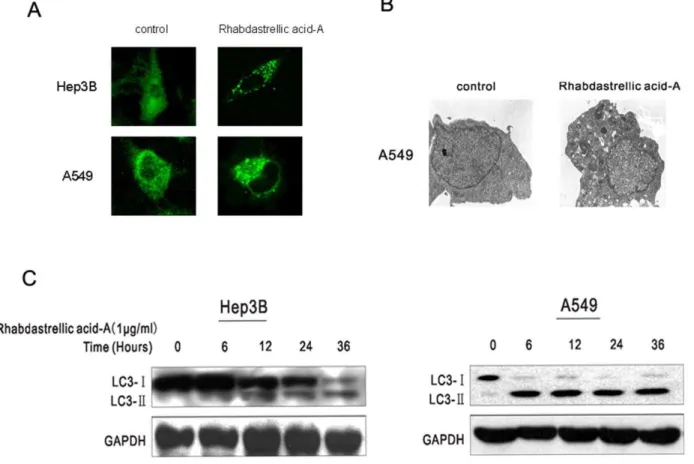
Rhabdastrellic acid-A induces the formation of autoph-agosomes. The membrane-associated light chain 3 protein, MAP LC3 (Atg8), localizes to the elongation membrane and stays in the membrane until autophagosome maturation. It is a key marker for autophagy. Upon induction of autophagy, LC3-I is
Figure 1. Molecular structure of Rhabdastrellic acid-A. doi:10.1371/journal.pone.0012176.g001
converted into LC3-II, which is most likely conjugated to phosphatidylethanolamine (PE) and tightly bound to the autophagosomal membranes forming ring-shaped structures in the cytoplasm. To measure autophagy, Hep3B and A549 cells were transfected with an expression construct for LC3 fused to yellow fluorescent protein (YFP-LC3). In DMSO-treated control cells YFP-LC3 was evenly distributed in the entire cytoplasm. After Rhabdastrellic acid-A treatment, ring-shaped structures were detectable in the cytosol, indicating the association of YFP-LC3 with autophagosomal membranes following an induction of autophagy (Fig 4A). Ultrastructural analysis of Rhabdastrellic acid-A-treated A549 cells by electron microscopy showed large autophagic vacuoles in contrast to control cells where no such vacuoles could be detected (Fig. 4B).
LC3-II expression induced by Rhabdastrellic acid-A in cancer cells. The amount of PE-conjugated form of LC3 (LC3-II) correlates well with the number of autophagosomes. After Rhabdastrellic acid-A treatment for the indicated times, LC3 was detected by immunoblotting analysis. The results indicated that the expression of LC3-II was gradually upregulated (Fig 4C). This confirmed the results of YFP-LC3 transfection and indicated that Rhabdastrellic acid-A could induce autophagy in Hep3B and A549 cells.
Autophagy-associated cell death induced by Rhabda-strellic acid-A in cancer cells. To confirm that Rhabdastrellic acid-A induced autophagy-associated cell death,
Hep3B cells were treated with Rhabdastrellic acid-A and/or 3-methyladenine (an inhibitor of PI3K-C3 commonly used for specific inhibition of autophagy). In contrast to control cells, after Rhabdastrellic acid-A treatment, most of the Hep3B cells appeared round, some were detached from the surface of wells and the number of cells was reduced (Fig 5A). However, the effects by Rhabdastrellic acid-A treatment were reversed by 3-MA (Fig 5A). It was suggested that the induction of cell death by Rhabdastrellic acid-A treatment was blocked when the cells were in parallel treated with autophagy inhibitor 3-MA.
shRNA-based depletion of an essential autophagy protein Atg5, effectively blocked Rhabdastrellic acid-A-induced LC3-II accu-mulation (Fig 5B). And the inhibition of autophagy by depletion of Atg5 inhibited Rhabdastrellic acid-A-induced cell death in A549 cells within the concentration ranges from 0.05 to 3.2mg/mL (Fig 5C).
A549 cells exposed to 4mM Rhabdastrellic acid-A and/or 3-MA were analyzed for cell death at 24 h by flow cytometry. Propidium iodide (PI) positive were counted as ‘dead’ cells. The cell death induced by Rhabdastrellic acid-A was suppressed when the cells were treated in combination with 3-MA (Fig. 5D).
3. Rhabdastrellic acid-A inhibited Akt pathway in Hep3B and A549 cells
Rhabdastrellic acid-A could inhibit Akt pathway in HL-60 cells[4]. Therefore, it was interesting to test whether it was the
Figure 2. Growth inhibition of Rhabdastrellic acid-A on Hep3B and A549 cells.A. Cancer cells were plated at the density of 8000 cells/well in a 96-well plate. The stock of Rhabdastrellic acid-A was added to wells. Cells were cultured for 3 days. MTT assay was conducted. B. Hep3B and A549 cells were treated with indicated concentrations of Rhabdastrellic acid-A for 48 h and analyzed for the DNA content by flow cytometry. The results were representative of 3 experiments.
Figure 3. Rhabdastrellic acid-A induced caspase-independent cell death in Hep3B and A549 cells.A. in Hep3B and A549 cells, Cell lysates were prepared from 0.1% DMSO, 1mg/mL Rhabdastrellic acid-A or 4mM adriamycin for 24 h and 48 h. Western blot analysis was conducted with caspase-3 and PARP antibodies. B. Cells were incubated with various concentrations of Rhabdastrellic acid-A in the presence or absence of 20mM z-VAD-fmk for 3 d. MTT assay was conducted. Results (mean6S.E.) represent the average of three experiments. C. Hep3B and A549 cells were treated with 0 to 4mg/mL Rhabdastrellic acid-A for 48 h. After treatment, the cells were collected sequentially and stained with Annexin V and PI and analysed by the flow cytometry assay. doi:10.1371/journal.pone.0012176.g003
same in other cancer cells. As shown in Fig 6A, After Rhabdastrellic acid-A treatment for the indicated times or various concentrations, Rhabdastrellic acid-A inhibited the phosphoryla-tion of Akt in Hep3B and A549 cells.
mTOR, FKHR and STAT3 are three important downstream targets of Akt pathway and play key roles in autophagy and apoptosis[33]. As shown in the results (Fig 6B), the phosphoryla-tion of mTOR, FKHR and STAT3 had dramatic decrease after Rhabdastrellic acid-A treatment in a time-dependent manner in both cell lines. These results indicated that AKT pathway was inhibited by Rhabdastrellic aicd-A in Hep3B and A549 cells.
4. The induction of autophagy by Rhabdastrellic acid-A was abrogated by constitutive active Akt ectopic expression
Rhabdastrellic acid-A could inhibit Akt pathway and induce autophagy in Hep3B cells. In order to further confirm an intrinsic role of Akt in autophagy induced by Rhabdastrellic acid-A, we transfected the Hep3B cells with myr-Akt1 plasmids (activated) and selected the positive clone using G418. Compared with the control cells (Hep3B cells transfected with the vector plasmid), the exogenous Akt expression significantly increased in Hep3B/myr-Akt1 cells (Fig. 7A). Furthermore, the marker of autophagy was determined while constitutive active Akt expression in Hep3B cells. Compared with the control cells, massive induction of autophagy was observed in Hep3B cells, indicated by increase of LC3-II.
However, expressing constitutive active Akt blocked increase of LC3-II (Fig 7B). Consistent with the notion that some population of LC3-II is degraded in lysosomes [34], treatment with lysosomal enzyme inhibitors Pepstatin A resulted in the accumulation of LC3-II in cancer cells (Fig 7C), this phenomenon was also impaired while constitutive active Akt ectopic expression. Furthermore, constitutive active Akt expression could rescue Hep3B cells from Rhabdasterllic acid-A-mediated growth inhibition under treatment of Rhabdas-trellic acid-A (Fig. 7D). These results shown in Fig 7 indicated that constitutive active Akt expression could inhibit Rhabdastrellic acid-A-mediated autophagy.
To investigate whether other pathways are involved in autophagy induced by Rhabdastrellic acid-A, we analysed the levels of Bip, CHOP, phospho-EIF2a and phospho-AMPK in Hep3B cells following Rhabdastrellic acid-A treatment. The results showed that phosphorylation of AMPK, EIF2a and CHOP increased after Rhabdastrellic acid-A treatment in a dose-dependent manner in Hep3B cells. However, an increase of Bip was not detected (shown in Fig. S1). These results indicated that multiple pathways might be involved in Rhabdastrellic acid-A-induced autophagy.
Discussion
The results described here demonstrate that Rhabdastrellic acid-A could induce autophagy in the human cancer cell lines.
Figure 4. Rhabdastrellic acid-A induced autophagy in Hep3B and A549 cells.A. Hep3B and A549 cells were transfected with an expression construct for LC3 fused to yellow fluorescent protein (YFP-LC3) for 24 h. Thereafter, cells were treated with or without 1mg/mL or 4mg/mL Rhabdastrellic acid-A for 36 h, and visualized under a confocal microscope. B. Electron micrograph showing autophagic vacuole of A549 cells following 4mg/mL Rhabdastrellic acid-A treatment. C. Rhabdastrellic acid-A time-dependently induced the formation of LC3-II, a marker for autophagy. Hep3B and A549 cells were treated with 1mg/mL Rhabdastrellic acid-A for the indicated times. Lysates were analyzed by immunoblotting with LC3 antibody.
Rhabdastrellic acid-A could block Akt pathway in Hep3B and A549 cells, and constitutive active Akt expression could inhibit Rhabdastrellic acid-A-mediated autophagy. These results indicat-ed that inhibition of Akt/mTOR pathway is involvindicat-ed in Rhabdastrellic acid-A induced autophagy and cell death.
Autophagy has been reported to be a process whereby double membrane vacuoles termed autophagosomes form around, engulf, cytosol and organelles. Though autophagy was reported to effect as a prosurvival response, in some instance, the cytotoxicity of arsenic trioxide, imatinib, ionizing radiation etc. to some cell types is mediated by the induction of autophagy. In those circumstances, autophagy constitutes a nonapoptotic pathway for programmed cell death. Many reports show that triterpenoids have potential cytotoxicity to cancer cell lines [35,36]. Rhabdastrellic acid-A is one of the triterpenoids that induced non-apoptotic cell death in Hep3B and A549 cells. Therefore, we explored whether the induction of autophagy was required for Rhabdastrellic acidA -mediated cell death in these two cancer cell lines. Autophagic cells
enlarge without permeabilization of the plasma membrane, convert LC3-I to LC-II, show punctate cytoplasmic LC3 translocation and develop autophagosome. In this study, Hep3B and A549 cells exhibited morphologic and biochemical features that are characteristic of cells undergoing autophagy, not apoptosis following Rhadastrellic acid-A treatment. Also, growth inhibition of Rhabdasterllic acid-A in A549 cells was weaker than that in Hep3B cells. But Rhabdasterllic acid-A increased more LC3-II expression. Conversion of LC3-I to LC3-II can be observed in autophagy initiation. But partial LC3-II can be degraded when autophagy maturation. Only LC3-II expression could not indicate the sensitivity to autophagy.
Previous studies on mechanism underlying the regulation of autophagy in cancer cells showed that autophagy was regulated by multiple signaling pathways as diverse as the class III PI 3-kinase, and the protein kinases mTOR, ERK, and p38. Aberrant activation of Akt/mTOR signaling pathway was implicated in a variety of human malignances [20,21,22,23,24,25,37].
Phosphor-Figure 5. Requirement of autophagy induction for Rhabdastrellic acid-A-induced cell death.A. Hep3B cells were treated with 1mg/mL Rhabdastrellic acid-A and/or 10 mM 3-MA for 36 h, and then examined by inverted microscope. *, p,0.05 vs. Rhabdastrellic acid-A2/3-MA-. **, p,0.05 vs. Rhabdastrellic acid-A+/3-MA-. B. Lysates from A549-Vector and A549-shAtg5 cells were analyzed by immunoblotting with Atg5 and LC3 antibodies. C. A549 vector cells and A549-shAtg5 cells were cultured at 6000 cells per well in a 96-well plate, exposed to different concentrations of Rhabdastrellic acid-A from 0.05 to 3.2mg/mL for 72 h. The growth inhibition was detected using MTT assay. Reported values are mean6SD of triplicate samples from a representative experiment. *P-vlaue,0.05 as compared with cells treated in the same way, but transfected without shRNA. D. A549 Cells were incubated with 4mg/mL Rhabdastrellic acid-A and/or 10 mM 3-MA for 24 h. Cell death was quantified using flow cytometry as PI staining assay. The experiment was repeated 3 times.
doi:10.1371/journal.pone.0012176.g005
ylated Akt plays central roles in some fundamental processes, including cell proliferation, cell death, cell motility/adhesion, cell transformation, and neovascularization. When activated, Akt acts as a survival factor by preventing release of cytochrome c from mitochondria and inactivating FKHR which is known to induce expression of pro-apoptotic factors such as Fas ligand [38]. Inhibition of mTOR induces apoptosis in some types of tumor cells, whereas they trigger autophagy in other settings. Rapamycin, an inhibitor of mTOR, was reported to induce the classic autophagic cell death in cancer cells. Our previous study also showed that Rhabdastrellic acid-A inhibited PI3K/Akt pathway and induced apoptosis in human leukemia HL-60 cells[4]. So inhibition of Akt may activate some autophagy-related proteins and induce autophagy. In keeping with this finding, we observed that Rhabdastrellic acid-A could inhibit the phosphorylation of Akt in Hep3B and A549 cells. The phosphorylation of mTOR, FKHR and STAT3 also dramatically decreased after Rhabdas-trellic acid-A treatment. Furthermore, ectopic constitutive active Akt expression could block autophagy induced by Rhadastrellic acid-A. Taken together, Rhadastrellic acid-A induced autophagy through inhibition of Akt/mTOR pathway.
In summary, the current studies show that Rhabdastrellic acid-A kills Hep3B and A549 cells via the induction of autophagy. Inhibition of the Akt/mTOR pathway plays a key role in Rhabdastrellic acid-A-mediated autophagy. As tumor cells frequently acquire defects in autophagy compared with normal cells, pharmacological activation of autophagy through inhibition of Akt/mTOR pathway may kill cancer cells and limit tumorigenesis, especially in cancers with defects in apoptosis. Our study also provides the evidence that Rhabdastrellic acid-A deserves further investigation as a potential anticancer agent or cancer preventive agent.
Materials and Methods
Drugs and reagents
Rhabdastrellic acid-A was isolated from the sponge Rhabdas-trella globostellata and initially dissolved in 100% Dimethylsul-foxid (DMSO) and stored at 220uC. Methylthiazolyldiphenyl-tetrazolium bromide (MTT) and 3-methyladenine (3-MA) were purchased from Sigma Co. (Sigma Aldrich, M2128). RPMI medium 1640 were purchased from invitrogen (Invitrogen, 11875).
Figure 6. The effect of Rhabdastrellic acid-A on Akt pathway in Hep3B and A549 cells. Hep3B and A549 cells were treated with Rhabdastrellic acid-A in the indicated concentrations for 36 h or treated with 1mg/mL or 4mg/mL Rhabdastrellic acid-A for the indicated times. A. Cells were sequentially harvested and lysed. cell lysates were analyzed by immunoblotting with Akt or Akt antibodies. B. mTOR, phospho-mTOR, FKHR, phospho-FKHR, STAT3 and phospho-STAT3 were analyzed by immunoblotting. The experiment was performed 3 times.
Cell lines and Cell culture
Human hepatocellular carcinoma cell line Hep3B[39] and human lung adenocarcinoma epithelial cell line A549[40] were cultured in RPMI-1640 supplemented with 10% heat-inactivated fetal bovine serum, penicillin (50U/mL), and streptomycin (50mg/ mL). The cells were incubated at 37uC in humidified 5% CO2.
MTT assay
Cells were plated in 96-well plate. The stock of Rhabdastrellic acid-A was diluted, added to the wells for the desired final assay concentration. After 3 days exposure to Rhabdastrellic acid-A, 10mL of MTT (5 mg/L) was added to each well and incubated for four more hours, then liquid in the wells was evaporated. 100mL of DMSO was added to each well. The absorbance was detected in the microplate reader 550 model with 565 nm wave length (Bio-rad Co., 17024). Growth inhibition was calculated and IC50value
was determined using the Bliss Software.
Annexin V-FITC/PI staining assay
Cells with different treatments were collected, resuspended with 10% 1640 medium, adjusted the cell suspension concentration to approximately 16106 cells/ml, and transferred 0.5 ml of cell suspension to a microfuge tube. After added 10ml media binding reagent and 1.25ml Annexin V-FITC, cells were incubated at room temperature for 15 min in the dark, then centrifuged and
removed media. When resuspended in 0.5 ml cold 16binding Buffer (10 mM HEPES, 150 mM NaCl, 2.5 mM CaCl2, 1 mM MgCl2, 20% BSA pH 7.4), cells were added 10ml propidium iodide (30mg/ml), placing samples on ice and away from light. Apoptosis was analysed by flow cytometry ((Beckman Coulter, Fullerton, CA) at the wavelength of 488 nm immediately.
Cell cycle detection
Cells were collected, resuspended with 1 ml precooled 70% ethanol, and fixed in 4uC overnight. After removed ethanol and added 0.5 ml staining solution (50mg/ml PI, 100mg/ml RNaseA, 0.2% Triton-100), cells were incubated in 37uC for 30 min in the dark. Cell cycle distribution was analysed by flow cytometry ((Beckman Coulter, Fullerton, CA) at the wavelength of 488 nm immediately.
PI staining assay
Cells were trypsinized with 0.5 ml 0.25% trypsin for 3 min, collected and resuspended with 1 ml precooled PBS. After added 0.5 ml staining solution (50mg/ml PI, 100mg/ml RNaseA, 0.2% Triton-100), cells were incubated in 37uC for 30 min in the dark. Cell death was detected by flow cytometry ((Beckman Coulter, Fullerton, CA).
Confocal microscopy and indirect immunofluorescence Cells were grown on glass coverslips and transfected with pYFP-LC3 for Hep3B and A549 cells. 36 h after transfection the cells
Figure 7. Constitutive active Akt expression could inhibit Rhabdastrellic acid-A-mediated autophagy.A. A. the positive clone stably expressed myr-Akt1 was analyzed by immunoblotting with Akt antibody. B. Hep3B cells were treated with 1mg/mL Rhabdastrellic acid-A for 36 h in the absence or presence of constitutive active Akt ectopic expression. Then LC3 protein expression was analyzed. C. Cancer cells were treated with 1mg/mL Rhabdastrellic acid-A and/or 10mg/mL pepstatin A (pepA) for 36 h, Then LC3 protein expression was analyzed. D. After treatment, viable cells of two cell lines were measured. *, p,0.05 vs. Rhabdastrellic acid-A (vector).
doi:10.1371/journal.pone.0012176.g007
citrate, and were examined in an electron microscope (Philip, CN10).
Western blot analysis
Lysates were prepared from 46105cells by dissolving cell pellets in 100mL of lysis buffer (20 mM Na2PO4 (pH 7.4), 150 mM
NaCl, 1% Triton X-100, 1% aprotinin, 1 mM phenymethysulfo-nyl fluoride, 10 mg/mL leupeptin, 100 mM NaF, and 2 mM Na3VO4). Lysates were centrifuged at 12,000 rpm for 10 min. The
supernatant was collected. The protein content was determined using the Bio-Rad protein assay. SDS-PAGE sample buffer (10 mM Tris-HCl, pH 6.8, 2% SDS, 10% Glycerol, 0.2 M DTT) was added to lysates. Lysates were heated to 100uC for 5 min, and 40mg of protein was loaded in each well of a 4–20% SDS-PAGE gel. Resolved proteins were electrophoretically transferred to nitrocellulose and incubated sequentially with primary antibody and horseradish peroxidase-conjugated goat anti-mouse IgG (Santa Cruz, sc-2005) or goat anti-rabbit-IgG (Santa Cruz, sc-2004). After washing, the bound antibody complex was detected using LumiGLO reagent (Cell Signaling Technology, #7003) and XAR film (Kodak, XBT-1) as described by the manufactures. The following primary antibodies were used: caspase-3 antibody (Santa Cruz, sc-7272), PARP antibody (Santa Cruz, sc-7150), glyceraldehyde 3-phosphate dehydrogenase anti-body (Santa Cruz, sc-47724), LC3 antianti-body (Novus Biologicals, NB100-2220), Atg5 antibody (Cell Signaling Technology,#2630), Phospho-Akt1/2/3(ser473) antibody (Santa Cruz, sc-7985-R), Akt1 antibody (Santa Cruz, sc-1618), Phospho-mTOR(ser2448) antibody(Cell Signaling Technology, #2971), mTOR antibody (Cell Signaling Technology, #2972), Phospho-FKHR(ser256) antibody (Cell Signaling Technology, #9461), FKHR antibody
For stable ATG5 siRNA expression, the retroviral vector (pSUPER. puro, a gift of Professor Musheng Zeng, Cancer Center, Sun Yat-sen University, Guangzhou, China) encoding hairpin RNA sequences was constructed. A distinct short hairpin RN (shRNA) sequences against ATG5 (shATG5) were generated and cloned into the expression vector. The ATG5 sense primers were: 59-GGC ATT ATC CAA TTG GTT TA-39, Vesicular stomatitis virus–pseudotyped vectors were produced by transfec-tion of the VSV-GPG producer cell line with 5mg DNA using lipofectamine 2000 (Invitrogen) in a six-well plate. Retrovirus-containing supernatants were collected at days 5 to 7 after transfection. Then A549 cells were infected with retrovirus-containing supernatants thrice. The positive cells stably expressing shATG5 were selected under puromycine (1mg/mL).
Supporting Information
Figure S1 Rhadbstrellic acid-A up-regulated p-AMPK, p-EIF2a, CHOP proteins in Hep3B cells. Hep3B cells were treated with 0– 2mg/mL Rhadbstrellic acid-A for 36 h, then the cells were collected and lysed. Cell lysates were analyzed by immunoblotting with phospho-AMPK, phospho-EIF2a, Bip and CHOP antibodies. Found at: doi:10.1371/journal.pone.0012176.s001 (2.04 MB TIF)
Author Contributions
Conceived and designed the experiments: XSZ XFZ. Performed the experiments: DDL JFG JJH LLW RD. Analyzed the data: DDL JNL GKF. Contributed reagents/materials/analysis tools: DJX SZD. Wrote the paper: DDL.
References
1. Liu Y, Mansoor TA, Hong J, Lee CO, Sim CJ, et al. (2003) New cytotoxic sesterterpenoids and norsesterterpenoids from two sponges of the genus Sarcotragus. J Nat Prod 66: 1451–1456.
2. Carroll J, Jonsson EN, Ebel R, Hartman MS, Holman TR, et al. (2001) Probing sponge-derived terpenoids for human 15-lipoxygenase inhibitors. J Org Chem 66: 6847–6851.
3. Rao Z, Deng S, Wu H, Jiang S (1997) Rhabdastrellic Acid-A, a Novel Triterpenoid from the Marine Sponge Rhabdastrella globostellata. J Nat Prod 60: 1163–1164.
4. Guo JF, Zhou JM, Zhang Y, Deng R, Liu JN, et al. (2008) Rhabdastrellic acid-A inhibited PI3K/Akt pathway and induced apoptosis in human leukemia HL-60 cells. Cell Biol Int 32: 48–54.
5. Tasdemir D, Mangalindan GC, Concepcion GP, Verbitski SM, Rabindran S, et al. (2002) Bioactive isomalabaricane triterpenes from the marine sponge Rhabdastrella globostellata. J Nat Prod 65: 210–214.
6. Levine B, Klionsky DJ (2004) Development by self-digestion: molecular mechanisms and biological functions of autophagy. Dev Cell 6: 463–477. 7. Furuya N, Yu J, Byfield M, Pattingre S, Levine B (2005) The evolutionarily
conserved domain of Beclin 1 is required for Vps34 binding, autophagy and tumor suppressor function. Autophagy 1: 46–52.
8. Yuan J, Lipinski M, Degterev A (2003) Diversity in the mechanisms of neuronal cell death. Neuron 40: 401–413.
9. Cuervo AM (2004) Autophagy: in sickness and in health. Trends Cell Biol 14: 70–77.
10. Levine B, Yuan J (2005) Autophagy in cell death: an innocent convict? J Clin Invest 115: 2679–2688.
11. Liang XH, Jackson S, Seaman M, Brown K, Kempkes B, et al. (1999) Induction of autophagy and inhibition of tumorigenesis by beclin 1. Nature 402: 672– 676.
12. Liang XH, Yu J, Brown K, Levine B (2001) Beclin 1 contains a leucine-rich nuclear export signal that is required for its autophagy and tumor suppressor function. Cancer Res 61: 3443–3449.
13. Chu CT, Zhu J, Dagda R (2007) Beclin 1-independent pathway of damage-induced mitophagy and autophagic stress: implications for neurodegeneration and cell death. Autophagy 3: 663–666.
14. Liang XH, Kleeman LK, Jiang HH, Gordon G, Goldman JE, et al. (1998) Protection against fatal Sindbis virus encephalitis by beclin, a novel Bcl-2-interacting protein. J Virol 72: 8586–8596.
16. Lum JJ, Bauer DE, Kong M, Harris MH, Li C, et al. (2005) Growth factor regulation of autophagy and cell survival in the absence of apoptosis. Cell 120: 237–248.
17. Levine B (2007) Cell biology: autophagy and cancer. Nature 446: 745–747. 18. Mizushima N (2007) Autophagy: process and function. Genes Dev 21:
2861–2873.
19. Yu L, Alva A, Su H, Dutt P, Freundt E, et al. (2004) Regulation of an ATG7-beclin 1 program of autophagic cell death by caspase-8. Science 304: 1500–1502.
20. Abdul-Ghani R, Serra V, Gyorffy B, Jurchott K, Solf A, et al. (2006) The PI3K inhibitor LY294002 blocks drug export from resistant colon carcinoma cells overexpressing MRP1. Oncogene 25: 1743–1752.
21. Stitt TN, Drujan D, Clarke BA, Panaro F, Timofeyva Y, et al. (2004) The IGF-1/PI3K/Akt pathway prevents expression of muscle atrophy-induced ubiquitin ligases by inhibiting FOXO transcription factors. Mol Cell 14: 395–403. 22. Cichy SB, Uddin S, Danilkovich A, Guo S, Klippel A, et al. (1998) Protein
kinase B/Akt mediates effects of insulin on hepatic insulin-like growth factor-binding protein-1 gene expression through a conserved insulin response sequence. J Biol Chem 273: 6482–6487.
23. Parry R, Smith G, Reif K, Sansom DM, Ward S (1997) Activation of the PI3K effector protein kinase B following ligation of CD28 or Fas. Biochem Soc Trans 25: S589.
24. Franke TF, Kaplan DR, Cantley LC (1997) PI3K: downstream AKTion blocks apoptosis. Cell 88: 435–437.
25. Theodoropoulou M, Zhang J, Laupheimer S, Paez-Pereda M, Erneux C, et al. (2006) Octreotide, a somatostatin analogue, mediates its antiproliferative action in pituitary tumor cells by altering phosphatidylinositol 3-kinase signaling and inducing Zac1 expression. Cancer Res 66: 1576–1582.
26. Chang F, Lee JT, Navolanic PM, Steelman LS, Shelton JG, et al. (2003) Involvement of PI3K/Akt pathway in cell cycle progression, apoptosis, and neoplastic transformation: a target for cancer chemotherapy. Leukemia 17: 590–603.
27. Liang J, Slingerland JM (2003) Multiple roles of the PI3K/PKB (Akt) pathway in cell cycle progression. Cell Cycle 2: 339–345.
28. Stephenson I, Nicholson KG, Gluck R, Mischler R, Newman RW, et al. (2003) Safety and antigenicity of whole virus and subunit influenza A/Hong Kong/
1073/99 (H9N2) vaccine in healthy adults: phase I randomised trial. Lancet 362: 1959–1966.
29. Chen YL, Law PY, Loh HH (2005) Inhibition of PI3K/Akt signaling: an emerging paradigm for targeted cancer therapy. Curr Med Chem Anticancer Agents 5: 575–589.
30. Hennessy BT, Smith DL, Ram PT, Lu Y, Mills GB (2005) Exploiting the PI3K/ AKT pathway for cancer drug discovery. Nat Rev Drug Discov 4: 988–1004. 31. Kim D, Cheng GZ, Lindsley CW, Yang H, Cheng JQ (2005) Targeting the
phosphatidylinositol-3 kinase/Akt pathway for the treatment of cancer. Curr Opin Investig Drugs 6: 1250–1258.
32. Tan J, Hallahan DE (2003) Growth factor-independent activation of protein kinase B contributes to the inherent resistance of vascular endothelium to radiation-induced apoptotic response. Cancer Res 63: 7663–7667.
33. Levine B, Kroemer G (2008) Autophagy in the pathogenesis of disease. Cell 132: 27–42.
34. Kabeya Y, Mizushima N, Ueno T, Yamamoto A, Kirisako T, et al. (2000) LC3, a mammalian homologue of yeast Apg8p, is localized in autophagosome membranes after processing. Embo J 19: 5720–5728.
35. Feng XZ, Dong M, Gao ZJ, Xu SX (2003) Three new triterpenoid saponins from Ixeris sonchifolia and their cytotoxic activity. Planta Med 69: 1036–1040. 36. Fu GM, Wang YH, Gao S, Tang MJ, Yu SS (2005) Five new cytotoxic triterpenoid saponins from the roots of Symplocos chinensis. Planta Med 71: 666–672.
37. Bockbrader KM, Tan M, Sun Y (2005) A small molecule Smac-mimic compound induces apoptosis and sensitizes TRAIL- and etoposide-induced apoptosis in breast cancer cells. Oncogene 24: 7381–7388.
38. Datta SR, Brunet A, Greenberg ME (1999) Cellular survival: a play in three Akts. Genes Dev 13: 2905–2927.
39. Hiron M, Daveau M, Arnaud P, Bauer J, Lebreton JP (1992) The human hepatoma Hep3B cell line as an experimental model in the study of the long-term regulation of acute-phase proteins by cytokines. Biochem J 287 (Pt 1): 255–259.
40. Foster KA, Oster CG, Mayer MM, Avery ML, Audus KL (1998) Character-ization of the A549 cell line as a type II pulmonary epithelial cell model for drug metabolism. Exp Cell Res 243: 359–366.