Volume 2012, Article ID 347848,6pages doi:10.1155/2012/347848
Research Article
Bleaching Gels Containing Calcium and Fluoride:
Effect on Enamel Erosion Susceptibility
Alessandra B. Borges,
1Carlos R. G. Torres,
1Paulo A. B. de Souza,
1Taciana M. F. Caneppele,
1Luciana F. T. F. Santos,
1and Ana Carolina Magalh˜aes
21Department of Restorative Dentistry, School of Dentistry, Universidade Estadual Paulista (UNESP), S˜ao Jos´e dos Campos, Av. Francisco Jos´e Longo, 777, 12245-000 S˜ao Jos´e dos Campos, SP, Brazil
2Department of Biological Sciences, Bauru School of Dentistry, University of S˜ao Paulo (USP), Alameda Octavio Pinheiro Brisola, 9-75, 17012-901 Bauru, SP, Brazil
Correspondence should be addressed to Alessandra B. Borges,[email protected]
Received 24 May 2012; Revised 23 July 2012; Accepted 13 August 2012
Academic Editor: Alix Young
Copyright © 2012 Alessandra B. Borges et al. This is an open access article distributed under the Creative Commons Attribution License, which permits unrestricted use, distribution, and reproduction in any medium, provided the original work is properly cited.
Thisin vitrostudy evaluated the effect of 35% hydrogen peroxide (HP) bleaching gel modified or not by the addition of calcium and
fluoride on enamel susceptibility to erosion. Bovine enamel samples (3 mm in diameter) were divided into four groups (n=15)
according to the bleaching agent: control—without bleaching (C); 35% hydrogen peroxide (HP); 35% HP with the addition of 2% calcium gluconate (HP + Ca); 35% HP with the addition of 0.6% sodium fluoride (HP + F). The bleaching gels were applied on the enamel surface for 40 min, and the specimens were subjected to erosive challenge with Sprite Zero and remineralization with
artificial saliva for 5 days. Enamel wear was assessed using profilometry. The data were analyzed by ANOVA/ Tukey’s test (P <0.05).
There were significant differences among the groups (P=0.009). The most enamel wear was seen for C (3.37±0.80µm), followed
by HP (2.89±0.98µm) and HP + F (2.72±0.64µm). HP + Ca (2.31±0.92µm) was the only group able to significantly reduce
enamel erosion compared to C. The application of HP bleaching agent did not increase the enamel susceptibility to erosion. However, the addition of calcium gluconate to the HP gel resulted in reduced susceptibility of the enamel to erosion.
1. Introduction
Tooth whitening is a highly desirable esthetic treatment, as the tooth color is one of the most important factors related
to the patients’ satisfaction with their appearance [1].
Dental bleaching treatments are mainly based on the action of hydrogen peroxide, which is able to penetrate the tooth structure and release free radicals, oxidizing the chromophore molecules. Such molecules are mainly organic,
although inorganic molecules can also be affected by these
reactions [2]. Nevertheless, the free radical reaction is not
specific and it may also alter the organic component of
enamel [3]. Since the organic content contributes to the
integrity of enamel, different adverse effects on both mineral
and organic parts of bleached enamel have been observed [3].
Alterations in enamel surface morphology [3–6],
chemi-cal composition [7–10], and microhardness values [3,11,12]
after bleaching were previously reported. Furthermore, some changes in bleached enamel were also described as slight
erosive effects promoted by the bleaching agent [6, 13].
Nevertheless, some authors claim that the erosive pattern on the surface of bleached enamel only occurs when bleaching
gels with low pH are used [14,15].
Attempts to minimize the adverse effects of
bleach-ing treatments by increasbleach-ing enamel remineralization have been conducted, however, the results are contradictory. De
Oliveira et al. [16] observed no significant increase of
bleached enamel microhardness when calcium and fluoride were added to 10% carbamide peroxide gel. On the other
hand, Borges et al. [12] observed a significant increase of
enamel microhardness after bleaching with 35% hydrogen peroxide agent with the addition of calcium and fluoride.
Chen et al. [6] also reported a less distinct erosion pattern
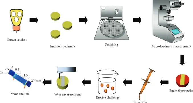
Crown section
Enamel specimens Polishing Microhardness measurement
Wear analysis Wear measurement
Erosive challenge
Bleaching
Enamel protection
Y(mm)
7.5 0 0.5
1
2 1.5
X(mm)
Figure1: Schematic representation of sample preparation and analysis.
The association between the bleached enamel surface alterations and the subsequent susceptibility to erosive lesions resulting from the contact of bleached enamel with demineralizing solutions has been discussed. In a previous study, the application of carbamide peroxide gel rendered
enamel more susceptible to demineralization [17]. In other
studies, at-home bleaching technique did not increase the
susceptibility of enamel to erosion [18, 19]. However, the
effect of at-office bleaching agent (35% HP) on the enamel
susceptibility to erosion has not been properly discussed. Considering the possibility that bleaching gels with high concentration of HP could increase the susceptibility of enamel to erosion, the addition of remineralizing ions into bleaching gels would be beneficial for preventing a further
enamel demineralization [6].
Therefore, the objective of this in vitro study was to
evaluate the effect of bleaching agents based on 35%
hydro-gen peroxide modified or not by the addition of calcium or fluoride on the susceptibility of enamel to erosion. The null hypotheses tested were (1) 35% HP bleaching agent does not increase the susceptibility of enamel to erosive challenges and
(2) there is no difference in erosion susceptibility of enamel
that has been treated with hydrogen peroxide compared to enamel treated with hydrogen peroxide supplemented with calcium or fluoride.
2. Materials and Methods
2.1. Preparation of the Samples. Freshly extracted
nondam-aged and intact bovine incisors were stored in a 0.1% thymol
solution and refrigerated at 4◦C, until required. Cylindrical
enamel samples (3 mm in diameter and 1 mm height) were prepared from the labial surface of the tooth using a
trephine mill (Dentoflex, S˜ao Paulo, SP, Brazil). The enamel surface was ground flat and polished with water-cooled silicon carbide (SiC) paper discs (1200, 2500, and 4000 grit; Fepa-P, Panambra, S˜ao Paulo, SP, Brazil), thereby removing
approximately 200µm of the outermost layer as verified
with a micrometer (Micromar 40EXL, Mahr-Goettingen, Germany).
The specimens were immersed in deionized water and placed in an ultrasonic bath for 10 min (Ultrasonic Cleaner, Odontobras, Ribeirao Preto, Brazil) for the removal of all waste, and then stored in thymol solution at 0.1% for rehydration.
After polishing, the enamel specimens were selected from the average of the surface microhardness measured using a Microhardness tester (FM-700, Future-Tech, Tokyo, Japan-Knoop tip, average of three indentations, under 25 g-load for 10 s) with an allowable variation within 20% of the mean. Microhardness average of each specimen was used
for stratified allocation among 4 groups (n = 15), so that
the average microhardness for each group was similar. In order to maintain the reference surfaces for lesion-depth determination (profilometry), 2 layers of nail varnish were applied on 2/3 of the surface of each sample (1/3 of each side)
exposing a central band area (see Figure1).
2.2. Bleaching Procedures. The groups were divided as
fol-lows: C (control)-nonbleached, HP-bleached with 35% hydrogen peroxide gel without addition of remineralizing
agents (pH=6.35), HP + Ca-bleached with 35% hydrogen
peroxide gel with the addition of 2% calcium gluconate
(pH = 7.99), and HP + F-bleached with 35% hydrogen
peroxide gel modified by the addition of 0.6% sodium
The experimental groups were subjected to 35% hydro-gen peroxide-based bleaching ahydro-gents and modified by the manufacturer (based on Whiteness HP Blue formulation-FGM Dental Products, Joinville, SC, Brazil). The pH of the agents was measured using a pH meter (Digimed DM-20-Digicrom Anal´ıtica Ltda., S˜ao Paulo, Brazil) fitted with an electrode (DME-Digimed CV8) that was calibrated using solutions with pH 4.01 and 6.86.
A layer of approximately 2 mm of the bleaching gel was applied on the enamel surface for 40 minutes. The gel was periodically shaken to remove the air bubbles formed during the procedure. After this period, the specimens were rinsed with deionized water to remove the bleaching agent (20 s).
2.3. Erosive Challenge. After the bleaching procedures, the
enamel samples were immersed in artificial saliva for 2 h and then subjected to erosive challenges for 5 days. The erosive cycles consisted of immersion in unstirred soft drink (20 mL/sample, Sprite Zero, Companhia Fluminense de Refrigerantes, Porto Real, RJ, Brazil), pH 2.8, four times per
day for 2 min each time [20], followed by a remineralizing
period of 2 h between erosive challenges (immersion in unstirred artificial saliva, pH 7.0, 20 mL/sample), at room temperature in small containers. The samples were kept in artificial saliva overnight. The composition of the artificial
saliva was the same as used by G¨ohring et al. [21]:
hydro-gen carbonate (22.1 mmol/L), potassium (16.1 mmol/L), sodium (14.5 mmol/L), hydrogen phosphate (2.6 mmol/L), boric acid (0.8 mmol/L), calcium (0.7 mmol/L), thiocyanate (0.4 mmol/L), and magnesium (0.2 mmol/L). The pH was adjusted to 7.0 using concentrated HCl.
The beverage was replaced at the end of each challenge, and artificial saliva was renewed daily.
2.4. Surface Wear Assessment. Profiles were obtained from
the enamel surfaces with a profilometer (MaxSurf XT 20, Mahr-Goettingen, Germany) after the erosive challenge. To determine the change in the surface profile after the experi-ment, the nail varnish was carefully removed with a spatula and a solution of acetone (1 : 1-acetone : water) to clean the surface. The diamond stylus moved from the first reference to the exposed area and then over to the other reference area (2.5 mm long and 1.0 mm wide). The vertical distance between the horizontal line drawn on the reference areas and the horizontal line drawn on experimental area was defined as tooth wear using the software (Software Mahr Surf XT20, 2009). Five profile measurements were performed for each specimen at intervals of 0.25 mm and the values were averaged (µm).
A schematic representation of the sample preparation
and analysis is presented in Figure1.
2.5. Statistical Analysis. Assumptions of normal distribution
of error and equality of variance (Kolmogorov-Smirnov and Bartlett’s tests) were checked for all the variables tested. Since the assumptions were justified, one-way ANOVA followed
by post hoc Tukey’s test were used to compare the different
bleaching agents in relation to the susceptibility of enamel to erosive wear. Statistical analysis was performed with the
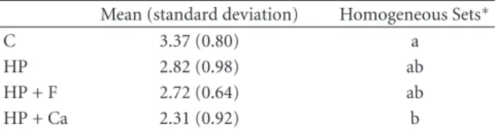
Table1: Mean (standard deviation) of the erosive enamel wear
(µm) for the different tested groups.
Mean (standard deviation) Homogeneous Sets∗
C 3.37 (0.80) a
HP 2.82 (0.98) ab
HP + F 2.72 (0.64) ab
HP + Ca 2.31 (0.92) b
∗
Groups with different letters showed significant differences between them (P <0.05).
software Statistica for Windows (Statsoft, Tulsa, OK, USA), with a significance level of 5%.
3. Results
One-way ANOVA revealed significant differences between
the groups (P=0.009). Table1shows the results of post hoc
Tukey’s test. The bleaching groups HP, HP + F, and HP + Ca, presented similar enamel wear values among them (n.s.). However, specimens from group HP + Ca had significantly less mean enamel wear compared to the control after the erosive challenges, while the groups HP and HP + F did not
differ from the control.
4. Discussion
Although in-office bleaching is considered a short-term
treatment, it should not represent an additional risk for subjects susceptible to erosion. In the present study, the exposure of 35% HP-bleached enamel to the acid beverage did not cause higher enamel surface wear compared to control group (unbleached). Therefore, the first null hypoth-esis tested was accepted. It may be suggested that possible microstructural changes caused by the bleaching treatment did not predispose the enamel to greater erosive wear. These microstructural changes may have been repaired by the adsorption and precipitation of salivary calcium and phosphate, when immersed in artificial saliva for 2 h before
the erosive challenge [17]. The ability of saliva to reharden
and replenish lost minerals from bleached enamel has been
demonstrated previously [22,23].
Although the adverse effects of bleaching agents on
enamel are mainly attributed to the concentration and pH
of the bleaching gel [14,24], some surface alterations are
reported even with low-concentrated and nonacidic agents
[4,5,9]. In fact, the demineralization of bleached enamel can
occur as a result of mineral and organic content alterations
[4,5,25]. Even though these factors are considered reversible
with exposure to saliva [22], it might be speculated that
when the bleached enamel is exposed to acid, these minimal changes could promote a greater spread of erosive agents inside enamel, leading it to a more pronounced
demineral-ization [17].
Previous studies have shown that bleached enamel exposed to 37% phosphoric acid, which is applied before bonding procedures, presented a higher acid dissolution,
an uneven etched surface, compared to the unbleached
enamel [26,27]. Nevertheless, other authors only reported
this increased decalcified effect when high-concentrated
hydrogen peroxide was used [28].
The association between carbamide peroxide bleaching and erosion and abrasion were previously investigated.
Pretty et al. [18] applied 20 cycles of bleaching (2 h) using
carbamide peroxide gels, from 10 to 22% concentration, and brushing (2 min), on enamel surface. The specimens were then immersed in 0.1% citric acid for a total of 14 h. The authors found no increased risk of enamel to acid
dis-solution. Engle et al. [19] also failed to demonstrate a
significant increase in the susceptibility of bleached enamel to erosion/abrasion after a 5 days-bleaching treatment with 10% carbamide peroxide (10 h/day) associated with erosive and abrasive challenges. Although the bleaching agent tested in this study was more concentrated, which could increase
the adverse effects, the unique application time was shorter
(40 min) and, thus, similar results were achieved compared to control.
This study simulated only one session of bleaching treatment, however, multiple appointments are needed for
obtaining optimal bleaching outcomes [29]. Therefore, the
subsequent erosive wear of bleached enamel could be more evident after successive bleaching, as more severe chemical alterations have been observed in enamel bleached with high-concentrated hydrogen peroxide for long periods of
exposure [30]. On the other hand, it might be speculated
that the alterations provoked by the bleaching agents could be relevant only for the susceptibility of the enamel to initial erosive lesions (“erosion” phase), in which enamel softening,
but not wear, is seen, as observed previously [17].
This experiment was conducted to simulate the effect of
the intake of soft drinks (Sprite Zero that has no potential
to stain the bleached enamel) after a session of in-office
bleaching treatment using a high-concentrated hydrogen peroxide gel. The use of bovine enamel is considered a convenient substitute for human enamel in erosion studies, as they are easier to obtain, to handle, and standardize. Nevertheless, it has to be considered that these two substrates
can behave in different physical and chemical manners and it
was shown that the demineralization occurs faster in bovine
enamel, due to its greater porosity [31–33].
The addition of potentially remineralizing agents in bleaching gels was also investigated in this study. The calcium and fluoride were added to the thickener phase of the gel, resulting in a concentration of 2% and 0.6%, respectively, after the final mixture, as these concentrations were com-patible with the chemical formulation of the gel, without impairing its thickness. It would be conceivable that the bleaching gels containing remineralizing agents could act not only as whitening agents but also as remineralizing agents
[34]. Supplements of fluoride and calcium in the bleaching
gels have been shown to minimize the deleterious effects on
enamel. It is supposed that the saturation of these ions in the gel allow their incorporation into the enamel apatite,
increasing the resistance to demineralization [12,35].
It was previously reported that the application of fluoride gel after bleaching resulted in increased resistance of the
enamel against erosive attacks, compared to
bleached/unflu-oridated enamel [36]. However, when fluoride was added
to the bleaching gel, the protection against demineralization was reported to be limited compared to fluoride only, since peroxide reduced the fluoride uptake by bleached enamel
[34].
In the present study, the erosive challenges tested were frequent (4 cycles of 2 min each per day, for 5 days); therefore, the presence of fluoride into gel might have not been able to provide protection against enamel wear. As a reduced formation of KOH-soluble fluoride was observed in enamel
bleached with fluoridated agents [34], it can be speculated
that any “calcium fluoride-like” precipitation was readily
dissolved by the subsequent acid challenges [37].
In addition, it has to be considered that fluoride den-tifrices are frequently used in the daily practice and this additional source of fluoride can eventually contribute rem-ineralizing the enamel surface after the bleaching procedures and erosive challenges. Nevertheless, the fluoride dentifrice was not included in this experimental model as this study
attempted to analyze the effect of remineralizing agents
added to the bleaching gel. Furthermore, fluoride dentifrice application is usually associated with a brushing procedure,
which may increase the enamel erosive wear [38]. Due to
the fact that the focus of this study was only on enamel susceptibility to erosion, brushing with fluoride dentifrice was not included in the pH cycling model.
In contrast to the effect of adding fluoride into the
bleaching gel, the addition of calcium gluconate to the
bleaching gel resulted in a protective effect against the
ero-sion; thus, the second null hypothesis was rejected. In a previous study, deposits of calcium on the surface of the enamel bleached with calcium-added agent were shown
using scanning electron microscope analysis [23]. These
deposits may have acted as a physical barrier, minimizing the contact of the acid to enamel, or providing additional mineral to be dissolved during the acid challenge before the underlying enamel was attacked. Another forms of cal-cium combined with bleaching agents have been previously investigated, such as the casein phosphopeptide-amorphous
calcium phosphate (CPP-ACP) [39,40] and calcium
chlo-ride [12], resulting in increased mechanical properties of
bleached enamel.
Nevertheless, future studies should be conducted to
investigate the solubility of different presentation forms of
calcium salts, as well as their interaction with enamel surface, so that the mechanism of action of calcium in improving the resistance of bleached enamel to erosion can be established
in different phases of its development (erosion and erosive
wear).
5. Conclusion
Acknowledgments
The authors wish to thank FGM Produtos Odontologicos for providing the bleaching gels. This study was supported by the State of S˜ao Paulo Research Foundation Grants no. (FAPESP 09/18588-9 and 12/16307-5).
References
[1] G. R. Samorodnitzky-Naveh, S. B. Geiger, and L. Levin,
“Patients’ satisfaction with dental esthetics,” Journal of the
American Dental Association, vol. 138, no. 6, pp. 805–808, 2007.
[2] M. A. Sulieman, “An overview of tooth-bleaching techniques:
chemistry, safety and efficacy,”Periodontology 2000, vol. 48, no.
1, pp. 148–169, 2008.
[3] T. Jiang, X. Ma, Y. Wang et al., “Investigation of the effects of
30% hydrogen peroxide on human tooth enamel by Raman
scattering and laser-induced fluorescence,”Journal of
biomed-ical optics, vol. 13, no. 1, p. 014019, 2008.
[4] C. Heged¨us, T. Bistey, E. Fl ´ora-Nagy, G. Keszthelyi, and A.
Jenei, “An atomic force microscopy study on the effect of
bleaching agents on enamel surface,”Journal of Dentistry, vol.
27, no. 7, pp. 509–515, 1999.
[5] M. T¨urk¨un, F. Sevgican, Y. Pehlivan, and B. O. Aktener,
“Effects of 10% carbamide peroxide on the enamel surface
morphology: a scanning electron microscopy study,”Journal
of Esthetic and Restorative Dentistry, vol. 14, no. 4, pp. 238– 244, 2002.
[6] H. P. Chen, C. H. Chang, J. K. Liu, S. F. Chuang, and J. Y. Yang,
“Effect of fluoride containing bleaching agents on enamel
surface properties,”Journal of Dentistry, vol. 36, no. 9, pp. 718–
725, 2008.
[7] M. S. McCracken and V. B. Haywood, “Demineralization
effects of 10 percent carbamide peroxide,”Journal of Dentistry,
vol. 24, no. 6, pp. 395–398, 1996.
[8] I. Rotstein, E. Dankner, A. Goldman, I. Heling, A. Stabholz, and M. Zalkind, “Histochemical analysis of dental hard tissues
following bleaching,”Journal of Endodontics, vol. 22, no. 1, pp.
23–26, 1996.
[9] N. Efeoglu, D. J. Wood, and C. Efeoglu, “Thirty-five percent carbamide peroxide application causes in vitro
demineraliza-tion of enamel,”Dental Materials, vol. 23, no. 7, pp. 900–904,
2007.
[10] I. Potoˇcnik, “Effect of 10% carbamide peroxide bleaching
gel on enamel microhardness, microstructure, and mineral
content,”Journal of Endodontics, vol. 26, no. 4, pp. 203–206,
2000.
[11] I. Lewinstein, N. Fuhrer, N. Churaru, and H. Cardash, “Effect
of different peroxide bleaching regimens and subsequent
fluoridation on the hardness of human enamel and dentin,”
Journal of Prosthetic Dentistry, vol. 92, no. 4, pp. 337–342, 2004.
[12] A. B. Borges, L. Y. Samezima, L. P. Fonseca, K. C. K. Yui, A. L. S. Borges, and C. R. G. Torres, “Influence of potentially
reminer-alizing agents on bleached enamel microhardness,”Operative
Dentistry, vol. 34, no. 5, pp. 593–597, 2009.
[13] T. Ushigome, S. Takemoto, M. Hattori, M. Yoshinari, E. Kawada, and Y. Oda, “Influence of peroxide treatment on
bovine enamel surface—Cross-sectional analysis,” Dental
Materials Journal, vol. 28, no. 3, pp. 315–323, 2009.
[14] M. Sulieman, M. Addy, E. Macdonald, and J. S. Rees, “A safety
study in vitro for the effects of an in-office bleaching system
on the integrity of enamel and dentine,”Journal of Dentistry,
vol. 32, no. 7, pp. 581–590, 2004.
[15] B. Xu, Q. Li, and Y. Wang, “Effects of pH values of hydrogen
peroxide bleaching agents on enamel surface properties,”
Operative Dentistry, vol. 36, no. 5, pp. 554–562, 2011.
[16] R. De Oliveira, A. F. Paes Leme, and M. Giannini, “Effect
of a carbamide peroxide bleaching gel containing calcium or
fluoride on human enamel surface microhardness,”Brazilian
Dental Journal, vol. 16, no. 2, pp. 103–106, 2005.
[17] T. Attin, M. Kocabiyik, W. Buchalla, C. Hannig, and K. Becker, “Susceptibility of enamel surfaces to demineralization after
application of fluoridated carbamide peroxide gels,” Caries
Research, vol. 37, no. 2, pp. 93–99, 2003.
[18] I. A. Pretty, W. M. Edgar, and S. M. Higham, “The effect of
bleaching on enamel susceptibility to acid erosion and
dem-ineralisation,”British Dental Journal, vol. 198, no. 5, pp. 285–
290, 2005.
[19] K. Engle, A. T. Hara, B. Matis, G. J. Eckert, and D. T. Zero, “Erosion and abrasion of enamel and dentin associated with
at-home bleaching,”Journal of the American Dental
Associa-tion, vol. 141, no. 5, pp. 546–551, 2010.
[20] A. C. Magalh˜aes, F. H. Stancari, D. Rios, and M. A. R. Buzalaf,
“Effect of an experimental 4% titanium tetrafluoride varnish
on dental erosion by a soft drink,”Journal of Dentistry, vol. 35,
no. 11, pp. 858–861, 2007.
[21] T. N. G¨ohring, M. Zehnder, B. Sener, and P. R. Schmidlin, “In vitro microleakage of adhesive-sealed dentin with lactic
acid and saliva exposure: a radio-isotope analysis,”Journal of
Dentistry, vol. 32, no. 3, pp. 235–240, 2004.
[22] L. M. Justino, D. R. Tames, and F. F. Demarco, “In situ and in
vitro effects of bleaching with carbamide peroxide on human
enamel,”Operative Dentistry, vol. 29, no. 2, pp. 219–225, 2004.
[23] M. U. Da Costa Soares, N. C. Araujo, B. C. Borges, W. D. Sales, and A. P. Sobral, “Impact of remineralizing agents on
enamel microhardness recovery after in-office tooth bleaching
therapies,”Acta Odontologica Scandinavica. In press.
[24] A. B. Borges, K. C. K. Yui, T. C. D’Avila, C. L. Takahashi, C. R. G. Torres, and A. L. S. Borges, “Influence of remineralizing gels
on bleached enamel microhardness in different time intervals,”
Operative Dentistry, vol. 35, no. 2, pp. 180–186, 2010. [25] S. T. Yeh, Y. Su, Y. C. Lu, and S. Y. Lee, “Surface changes and
acid dissolution of enamel after carbamide peroxide bleach
treatment,”Operative Dentistry, vol. 30, no. 4, pp. 507–515,
2005.
[26] B. Yurdukoru, A. C. Ak¨oren, and M. K. ¨Unsal, “Alterations in
human enamel surface morphology following the use of an
office bleaching agent and consecutive application of 37%
phosphoric acid in vivo,”Journal of Clinical Dentistry, vol. 14,
no. 4, pp. 103–107, 2003.
[27] C. L. S. G. De Medeiros, S. Gonz´alez-L ´opez, M. V. Bola˜nos-Carmona, P. Sanchez-Sanchez, and J. Bola˜nos-Bola˜nos-Carmona,
“Effects of phosphoric acid on bovine enamel bleached with
carbamide peroxide,”European Journal of Oral Sciences, vol.
116, no. 1, pp. 66–71, 2008.
[28] C. Torres-Rodr´ıguez, S. Gonz´alez-L ´opez, V. Bola˜nos-Carmo-na, P. S´anchez-S´anchez, A. Rodr´ıguez-Navarro, and T. Attin,
“Demineralization effects of phosphoric acid on surface and
subsurface bovine enamel bleached with in-office hydrogen
peroxide,”The Journal of Adhesive Dentistry, vol. 13, no. 4, pp.
315–321, 2011.
[29] W. Buchalla and T. Attin, “External bleaching therapy with
activation by heat, light or laser—a systematic review,”Dental
[30] T. Bistey, I. P. Nagy, A. Sim ´o, and C. Hegedus, “In vitro FT-IR
study of the effects of hydrogen peroxide on superficial tooth
enamel,”Journal of Dentistry, vol. 35, no. 4, pp. 325–330, 2007.
[31] T. Attin, F. Wegehaupt, D. Gries, and A. Wiegand, “The poten-tial of deciduous and permanent bovine enamel as substi-tute for deciduous and permanent human enamel:
erosion-abrasion experiments,”Journal of Dentistry, vol. 35, no. 10, pp.
773–777, 2007.
[32] A. J. White, C. Yorath, V. Ten Hengel, S. D. Leary, M. C. Huysmans, and M. E. Barbour, “Human and bovine enamel
erosion under ’single-drink’ conditions,”European Journal of
Oral Sciences, vol. 118, no. 6, pp. 604–609, 2010.
[33] P. Laurance-Young, L. Bozec, L. Gracia et al., “A review of the structure of human and bovine dental hard tissues and their physicochemical behaviour in relation to erosive challenge and
remineralisation,”Journal of Dentistry, vol. 39, no. 4, pp. 266–
272, 2011.
[34] T. Attin, K. Albrecht, K. Becker, C. Hannig, and A. Wiegand, “Influence of carbamide peroxide on enamel fluoride uptake,”
Journal of Dentistry, vol. 34, no. 9, pp. 668–675, 2006.
[35] V. Cavalli, L. K. Rodrigues, A. F. Paes-Leme et al., “Effects of
bleaching agents containing fluoride and calcium on human
enamel,”Quintessence International, vol. 41, no. 8, pp. e157–
165, 2010.
[36] G. M. Burgmaier, I. M. Schulze, and T. Attin, “Fluoride uptake and development of artificial erosions in bleached and
fluori-dated enamel in vitro,”Journal of Oral Rehabilitation, vol. 29,
no. 9, pp. 799–804, 2002.
[37] A. C. Magalh˜aes, A. Wiegand, D. Rios, M. A. R. Buzalaf, and A.
Lussi, “Fluoride in dental erosion,”Monographs in Oral
Sci-ence, vol. 22, pp. 158–170, 2011.
[38] A. C. Magalh˜aes, D. Rios, A. C. B. Delbem, M. A. R. Buzalaf, and M. A. A. M. Machado, “Influence of fluoride dentifrice on brushing abrasion of eroded human enamel: an in situ/ex vivo
study,”Caries Research, vol. 41, no. 1, pp. 77–79, 2006.
[39] B. C. D. Borges, J. S. Borges, C. D. De Melo et al., “Efficacy of a
novel at-home bleaching technique with carbamide peroxides
modified by cpp-acp and its effect on the microhardness of
bleached enamel,”Operative Dentistry, vol. 36, no. 5, pp. 521–
528, 2011.
[40] A. G. Gama Cunha, A. A. Meira De Vasconcelos, B. C. Dutra
Borges et al., “Efficacy of in-office bleaching techniques
com-bined with the application of a casein
phosphopeptide-amor-phous calcium phosphate paste at different moments and its
influence on enamel surface properties,”Microscopy Research
Submit your manuscripts at
http://www.hindawi.com
Hindawi Publishing Corporation
http://www.hindawi.com Volume 2014
Oral Oncology
Journal ofDentistry
International Journal ofHindawi Publishing Corporation
http://www.hindawi.com Volume 2014
International Journal of
Biomaterials
Hindawi Publishing Corporationhttp://www.hindawi.com Volume 2014
BioMed Research International
Hindawi Publishing Corporation
http://www.hindawi.com Volume 2014
Hindawi Publishing Corporation
http://www.hindawi.com Volume 2014 Case Reports in Dentistry
Hindawi Publishing Corporation
http://www.hindawi.com Volume 2014
Oral Implants
Journal ofHindawi Publishing Corporation
http://www.hindawi.com Volume 2014
Anesthesiology
Research and Practice
Hindawi Publishing Corporation
http://www.hindawi.com Volume 2014 Radiology
Research and Practice
Environmental and Public Health
Journal of
Hindawi Publishing Corporation
http://www.hindawi.com Volume 2014
The Scientiic
World Journal
Hindawi Publishing Corporation
http://www.hindawi.com Volume 2014
Hindawi Publishing Corporation
http://www.hindawi.com Volume 2014
Dental Surgery
Journal ofDrug Delivery
Journal ofHindawi Publishing Corporation
http://www.hindawi.com Volume 2014
Hindawi Publishing Corporation
http://www.hindawi.com Volume 2014
Oral Diseases
Journal ofHindawi Publishing Corporation
http://www.hindawi.com Volume 2014
Computational and Mathematical Methods in Medicine
Scientiica
Hindawi Publishing Corporationhttp://www.hindawi.com Volume 2014Pain
Research and Treatment
Hindawi Publishing Corporation
http://www.hindawi.com Volume 2014
Hindawi Publishing Corporation
http://www.hindawi.com Volume 2014
International Journal of
Endocrinology
Hindawi Publishing Corporation
http://www.hindawi.com Volume 2014
Hindawi Publishing Corporation
http://www.hindawi.com Volume 2014