Effect of 0.02% NaF solution on enamel demineralization and
fluoride uptake by deciduous teeth
in vitro
Efeito da solução de NaF a 0,02% na desmineralização e
incorporação de fluoreto pelo esmalte de dentes decíduos
in vitro
Silvia José Chedid* Jaime Aparecido Cury**
INTRODUCTION
Fluoride dentifrice is considered an important factor in explaining the decline in dental caries observed either in developed or in developing coun-tries at the end of the last century3,14. However,
fluoride dentifrice is also a risk factor with respect to dental fluorosis13 because young children ingest
a large amount of toothpaste during brushing16.
In addition, there is evidence that dental fluorosis is increasing in developed countries irrespective of whether the areas concerned have water fluorida-tion or not12. These findings in developed countries
are an alert to developing countries that have car-ies control programs based on fluoride use17.
In order to reduce the risk of dental fluoro-sis some recommendations have been made for young children: (i) do not use dentifrice, or use a non-fluoride dentifrice7; however, the presence of
fluoride during toothbrushing is considered
in-dispensable21; (ii) use a dentifrice with low
fluo-ride concentration9; however, the anticaries effect
of dentifrice with less than 1,100 µg F/g is not well-established15,19; (iii) use a small amount of
dentifrice18; however, the anticaries effect of this
procedure has not yet been established.
In addition to these international recommen-dations, the use of 0.02% NaF solution for children under 3 years of age in Brazil has been suggested, instead of fluoride dentifrice23. Parents or
guard-ians should apply this solution to the children’s teeth daily, using a cotton swab. This alternative is safer than the use of fluoride dentifrice with regard to dental fluorosis because the child is subjected daily to a low amount of fluoride (approximately 0.01 mg F/cotton swab). However, its anti-caries potential is unknown.
Thus, the aim of this study was to evaluate
* Associate Professor, Pediatric Dentistry, São Leopoldo Mandic Centre of Research in Dentistry. ** Professor, Biochemistry, School of Dentistry of Piracicaba, State University of Campinas.
ABSTRACT: The application of 0.02% NaF solution on teeth with a cotton swab instead of brushing with fluoride dentifrice has been suggested for young children to reduce the risk of dental fluorosis, but its anticariogenic effect has not been evaluated. Thus, we studied the in vitro effect of 0.02% NaF solution on enamel demineralization and fluoride uptake in deciduous teeth; non-fluoride dentifrice and fluoride dentifrice (1,100 µg F/g) were used, respectively, as negative and positive controls. The treatment with fluoride dentifrice was more effective in reducing enamel demineralization (p < 0.05) and on fluoride uptake by the enamel (p < 0.05) than the non-fluoride dentifrice and the 0.02% NaF solution. Data suggest that the alternative use of 0.02% NaF solution instead of fluoride den-tifrice should be reevaluated especially if dental caries are to be controlled.
DESCRIPTORS: Fluorides; Dentifrices; Dental enamel; Tooth, deciduous; Fluorosis, dental.
RESUMO: A aplicação da solução de NaF a 0,02%, no lugar de dentifrício fluoretado, tem sido sugerida para ser aplicada com cotonete nos dentes de bebês para reduzir o risco de fluorose dental. Como o efeito anticariogênico dessa recomendação não tem sido estudado, avaliou-se in vitro seu efeito na redução da desmineralização e in-corporação de fluoreto no esmalte de dentes decíduos; dentifrício não fluoretado e fluoretado (1.100 µg F/g) foram utilizados como controles negativo e positivo, respectivamente. O dentifrício fluoretado foi mais efetivo que a solu-ção de NaF a 0,02% na redusolu-ção de desmineralizasolu-ção e na incorporasolu-ção de fluoreto no esmalte (p < 0,05). Os dados sugerem que uso alternativo de NaF a 0,02% ao invés de dentifrício fluoretado para reduzir o risco de fluorose dental deve ser reavaliado, especialmente se a cárie dental precisa ser controlada.
the in vitro effect of 0.02% NaF solution on enamel demineralization and fluoride uptake in decidu-ous teeth.
MATERIAL AND METHODS
Experimental design
Seventy-five enamel blocks (3 x 3 mm) were prepared from human upper incisor deciduous teeth provided by the Human Tooth Bank, School of Dentistry, University of São Paulo (USP). The enamel surface of the blocks was then sequentially polished and those with a hardness of 43 ± 3 KHN (knoop hardness number) units were random-ized into three groups of 15 specimens each. The groups of blocks, presenting known baseline enamel surface microhardness, were submitted to a pH-cycling model simulating a high cario-genic challenge. During pH-cycling, one group was treated with a non-fluoride dentifrice (nega-tive control); the experimental group was treated with 0.02% NaF solution and the positive control with fluoride dentifrice (1,100 µg F/g, w/w). After pH-cycling, surface enamel microhardness was again determined in the blocks to evaluate its loss. Enamel cross-sectional microhardness was also determined to evaluate the caries lesion. In addi-tion, fluoride in enamel was assessed to evaluate fluoride uptake in each treatment. The use of hu-man teeth was ethically conducted according to the Brazilian guidelines (Resolution No. 196 of the National Health Council, Health Ministry, Brasília, DF, 10/03/1996).
pH-cycling
The enamel blocks were submitted to a pH-cycling model for 10 days, simulating a high caries challenge, essentially according to Featherstone
et al.5. The blocks were kept in a demineralizing
solution (2.0 mM calcium, 2.0 mM phosphate in 0.075 M acetate buffer, pH 4.3) at 37oC for 3 h
(60 ml per block), and in a remineralizing solution (1.5 mM calcium, 0.9 mM phosphate, 150 mM of KCl in 0.1 M Tris buffer, pH 7.0) for 21 h (30 ml per block). During the weekend, the enamel blocks were stored in the remineralizing solution and both solutions were changed before starting another cycle of 5 days. During pH-cycling, twice a day, the enamel blocks were treated with the dentifrices or the 0.02% NaF solution.
Fluoride treatment
The treatments were applied to the enamel blocks before and after the time that these were
im-mersed in the demineralizing solution. The nega-tive control group was treated with a non-fluoride dentifrice (placebo) and the positive one with fluo-ride dentifrice (Tandy, Colgate-Palmolive Ind. Com. Ltda., São Bernardo do Campo, SP, Brazil) contain-ing 1,100 µg F/g (w/w as NaF). Both dentifrices were silica-based. The dentifrices were applied to the enamel surface for 20 s, using a toothbrush, but without pressure. The amount of dentifrice applied was 0.1 ± 0.02 g and it was standardized by collecting the dentifrice set in the tip-top cap of the dentifrice tube. In the experimental group of enamel blocks, 0.02% NaF solution (w/v) was applied to the enamel surface with a cotton swab for 20 s. After the treatments the blocks were im-mersed in artificial saliva2 (1.5 mM Ca, 3.0 mM P,
20.0 mM NaHCO3, pH 7.0, 1.0 ml per block). The blocks were briefly washed with deionized water and the artificial saliva was changed for a fresh amount every time treatment was applied. After the pH-cycling, the enamel of the blocks was evalu-ated.
Microhardness determination
After pH-cycling, the surface microhardness of the enamel block was measured again. Five in-dentations spaced 100 µm from each other and from the baseline ones were made. A Shimadzu HMV-2000 (Shimadzu Corp., Kyoto, Japan) mi-crohardness tester with a Knoop diamond with 50 g load was used for 5 s. The percentage of surface microhardness change (%SMC) was cal-culated (%SMHC = hardness after pH-cycling −
baseline × 100/baseline). Surface microhardness (SMH) was evaluated because this method is high-ly sensitive and reproducible to evaluate enamel demineralization24. After surface microhardness
analysis, the blocks were longitudinally sectioned in the centre. Half of each block was used for cross-sectional microhardness determination and the other half for fluoride enamel analysis.
con-sidering that there is a high correlation (r = 0.91) between enamel microhardness and percentage of mineral in caries lesions, and the values of Knoop hardness number (KHN) were converted to min-eral contents (%vol) using the relation: minmin-eral content = 4.3 ( ) + 11.3, according to Feath-erstone et al.6.
Fluoride concentration in enamel
Five enamel layers were sequentially removed from half of each block by immersion in 0.25 ml of 0.5 M HCl for 30, 30, 30, 60 and 60 s under agita-tion10. An equal volume of TISAB II pH 5.0
modi-fied with 20 g NaOH/l was added to each solution containing the dissolved enamel layer. Fluoride measurements were performed using an ion-se-lective electrode Orion 96-09 and an ion analyzer Orion EA-940 (Orion Research Inc., Boston, MA, USA), previously calibrated with various standard fluoride solutions from 0.1 to 5.0 µg F/ml. The thickness of enamel layer removed during the acid biopsy was calculated from the inorganic phospho-rus concentration, determined colorimetrically8.
Phosphorus content in enamel of 17.4% was esti-mated to calculate the amount of enamel removed and enamel density of 2.92 was considered to cal-culate the depth of each enamel layer11.
Statistical analysis
The SMH and fluoride uptake data were ana-lyzed by ANOVA, after transformation by exponen-tial and square root respectively. The difference among the treatments was analyzed by Newman-Keuls test. The results of cross-sectional micro-hardness were analyzed by the non-parametric Kruskal-Wallis test. The software BioEstat 2.0 was used1 for all the analyses, and the significance level
was established at 5%.
RESULTS
None of the treatments were able to prevent
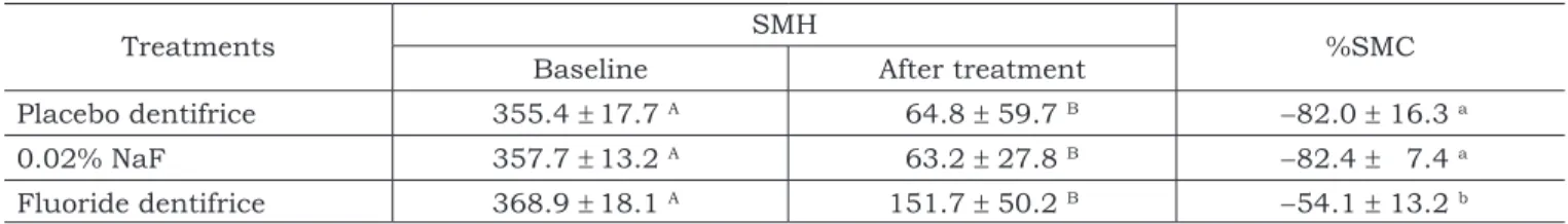
the reduction of surface microhardness (Table 1), because the difference (baseline values vs. after treatment) was statistically significant (p < 0.0001). However, the effect of the treatments was found (p < 0.0001) by ANOVA regarding the percentage of SMH change (%SMC). The treatment with fluo-ride dentifrice was more effective than those with placebo dentifrice and with 0.02% NaF regarding reduction of the percentage of SMC (p < 0.01). The effect of 0.02% NaF treatment was not statistically significant in comparison with placebo dentifrice (p > 0.05).
Graph 1 shows that only the treatment with fluoride dentifrice (FD) was statistically more ef-fective than the placebo (PD) in reducing the loss of mineral at all the distances from the enamel surface (p < 0.05). The results observed with the 0.02% NaF solution were not statistically different to those observed with the non-fluoride dentifrice treatment (PD) regarding caries lesions.
With regard to fluoride uptake (Graph 2), data collected from the first layer of enamel analyzed show that the 0.02% NaF solution was able to increase concentrations of F in enamel in compari-son with the non-fluoride dentifrice (PD). However, the fluoride dentifrice (FD) was more effective than the other two treatments (p < 0.05).
DISCUSSION AND CONCLUSION
Some recommendations have been made to re-duce the risks of dental fluorosis, considering that pre-school children involuntarily swallow a consid-erable amount of dentifrice during toothbrushing. However, the effect of these recommendations on the anticariogenic properties of fluoride has not been evaluated. In the present study we evaluated the in vitro use of 0.02% NaF solution, in terms of its effect on the anticariogenic potential of fluoride. In this in vitro study we tried to simulate clinical situations regarding toothbrushing by children and caries: (i) as substrate we used deciduous
TABLE 1 - Surface microhardness analysis of enamel blocks according to the treatments (means ± SD; n = 15).
Treatments SMH %SMC
Baseline After treatment
Placebo dentifrice 355.4 ± 17.7 A 64.8 ± 59.7 B −82.0 ± 16.3 a
0.02% NaF 357.7 ± 13.2 A 63.2 ± 27.8 B −82.4 ± 7.4 a
Fluoride dentifrice 368.9 ± 18.1 A 151.7 ± 50.2 B −54.1 ± 13.2 b Means followed by different letters are statistically significant (p < 0.05). Capital letters show difference between baseline and after treatment, SMH for each treatment and lower case among the treatments. SMH = surface microhardness; SMC = surface micro-hardness change. SD = standard deviation.
teeth, (ii) a way to use a small amount of dentifrice was idealized, (iii) a pH-cycling model was used to mimic caries lesion development.
The results clearly showed that the use of a small amount of fluoride dentifrice was as effec-tive in reducing enamel demineralization (Table 1 and Graph 1) as in forming fluoride in enamel (Graph 2). However, the findings with the use of 0.02% NaF solution were not conclusive because it did not show better effects than the negative control used (non-fluoride dentifrice) for all the evaluations made.
The positive effect of fluoride dentifrice and the lack of effect of 0.02% NaF solution are compre-hensible because the concentration of fluoride in dentifrice (1,100 µg F/g) is 12 times higher than that in the 0.02% NaF solution (90 µg F/ml). It is recognized that loosely-bound fluoride, like CaF2, is the product formed in the enamel, which is responsible for the anticariogenic effect of topi-cal fluoride22. Considering that the formation of a
CaF2-like product is dependent on the concentra-tion of fluoride applied to the enamel20, the result
found is not surprising. However, some effects of the 0.02% NaF solution were expected, because 90 µg F/ml of fluoride is reacting on the enamel surface twice a day. We did not find any effect of this fluoride solution in reducing enamel surface demineralization (Table 1), and a very small effect was observed in the reduction of mineral loss in the caries lesion (Graph 1) and in fluoride uptake (Graph 2). These findings suggest that 0.02% NaF
solution applied with a cotton swab in enamel has doubtful anticariogenic potential. Thus, this al-ternative to fluoride dentifrice can be safer with regard to dental fluorosis risk, but it must not be chosen, especially for a child with caries risk or activity. The use of a small amount of dentifrice would be a better solution considering the benefits and risks of fluoride use17.
With regard to the effect of fluoride denti-frice, during toothbrushing we did not simulate the dilution of the dentifrice by saliva, which is approximately 3-4 times. We tried to simulate the dilution after toothbrushing, by immersing the enamel blocks in artificial saliva for 30 min. The dilution of the dentifrice by saliva is important, as shown by DenBesten, Ko4 (1996).The authors
suggest that the reduction in the amount of fluo-ride toothpaste to a pea-sized amount should be limited only to young children, who are at risk of ingesting toothpaste. However, these authors did the study with 4-5 year old children and the risk of dental fluorosis occurs at an earlier age.
In conclusion, the results of this laboratory study suggest that the use of 0.02% NaF solution in a clinical basis has very little anticariogenic po-tential, and the recommendation to use it instead of fluoride dentifrice should be reevaluated.
ACKNOWLEDGEMENTS
The authors thank Professor Rosana C. Paren-te, University of Amazonas, for assistance in statis-tical analysis; Miss Mariza de Jesus Carlos Soares
GRAPH 1 - Enamel volume mineral percentage according to the treatments and the distance (µm) from the surface. Different letters show significant difference (p < 0.05) among treatments at each distance from the enamel surface; bars denote standard errors (n = 15). PD: non-fluoride dentifrice; FD: fluoride dentifrice.
GRAPH 2 - Fluoride concentration in enamel (mg/kg) according to the groups/treatments and the distance (µm) from the surface. Different letters show significant difference (p < 0.05) among groups/treatments at each distance from the enamel surface; bars denote standard errors (n = 15). PD: non-fluoride dentifrice; FD: fluoride dentifrice.
0 500 1,000 1,500 2,000 2,500 3,000
0 10 20 30 40 50 60 70 80 90 100 110
Depth (�m)
Fl uo rid e co nce nt ra tio n in e na me l (mg F/ kg ) a b c a a
a aa
a a a a a a a PD 0.02% NaF FD 0 20 40 60 80 100 120
0 20 40 60 80 100 120
%vo l mi ne ra l a b b a a,b b a a b a b c a b b
Depth (�m)
PD
and Mr. Waldomiro Vieira Filho, School of Den-tistry of Piracicaba, State University of Campinas, for technical assistance; Miss Adriana Franco Paes
Leme, Doctorate Student in Cariology, School of Dentistry of Piracicaba, State University of Campi-nas, for helping with the graphs.
REFERENCES
1. Ayres M, Ayres Jr M, Ayres DL, Santos AS. BioEstat 2.0: statistical applications in biological sciences and medi-cine. Belém, Sociedade Civil Mamirauá, Brasília, CNPq; 2000.
2. Birkeland JM. The effect of pH on the interaction of fluo-ride and salivary ions. Caries Res 1973;7:11-8.
3. Clarkson JJ. International collaborative research on fluo-ride. J Dent Res 2000;79:893-904.
4. DenBesten P, Ko HS. Fluoride levels in whole saliva of preschool children after brushing with 0.25 g (pea-sized) as compared to 1.0 g (full-brush) of a fluoride dentifrice. Pediatr Dent 1996;18:277-80.
5. Featherstone JDB, O’Reilly MM, Shariati M, Brugler S. Enhancement of remineralization in vitro and in vivo. In: Leach SA (editor). Factors relating to demineralization and remineralization of the teeth. Oxford: IRL Press; 1986. p. 23-34.
6. Featherstone JDB, ten Cate JM, Shariati M, Arends J. Comparison of artificial caries-like lesions by quantitative microradiography and microhardness profiles. Caries Res 1983;17:385-91.
7. Feigal RJ. Recent modifications in the use of fluorides for children. Northwest Dent 1983;62:19-21.
8. Fiske CH, Subarrow Y. The colorimetric determination of phosphorous. J Biol Chem 1925;66:375-400.
9. Horowitz HS. The need for toothpastes with lower than conventional fluoride concentrations for preschool-aged children. J Public Health Dent 1992;52:216-21.
10. Koo H, Cury JA. Soluble calcium/SMFP dentifrice: effect on enamel fluoride uptake and remineralization. Am J Dent 1998;11:173-6.
11. Lazzari EP. Dental biochemistry. 2nd ed. London: Lea &
Febiger; 1976.
12. Levy SM, Kohout FJ, Kiritsy MC, Heilman JR, Wefel JS. Infants’ fluoride ingestion from water, supplements and dentifrice. J Am Dent Assoc 1995;126:1625-32.
13. Mascarenhas AK. Risk factors for dental fluorosis: a re-view of the recent literature. Pediatr Dent 2000;22:269- 77.
14. Narvai PC, Frazão P, Castellanos RA. Decline in caries experience in permanent teeth of Brazilian scholars at the end of the twentieth century. Odontol Soc 1999;1:25-9. 15. Newbrun E. Current regulations and recommendations
concerning water fluoridation, fluoride supplements and topical fluoride agents. J Dent Res 1992;71:1255-65. 16. Paiva SM, Cury JA. Fluoride dentifrice and risk of dental
fluorosis. RPG Rev Pós Grad 2001;8:322-8.
17. Paiva SM, Lima YBO, Cury JA. Fluoride intake by Brazil-ian children from two communities with fluoridated water. Community Dent Oral Epidemiol 2003;31:184-91. 18. Pang DT, Vann WF Jr. The use of fluoride-containing
toothpastes in young children: the scientific evidence for recommending a small quantity. Pediatr Dent 1992;14: 384-7.
19. Richards A, Banting DW. Fluoride toothpastes. In: Fejer-skov O, Ekstrand J, Burt BA. Fluoride in dentistry. 2nd ed.
Copenhagen: Munksgaard; 1996. p. 328-43.
20. Saxegaard E, Rölla G. Fluoride acquisition on and in hu-man enamel during topical application in vitro. Scand J Dent Res 1988;96:523-35.
21. Scheie AA. Dentifrices in the control of dental caries. In: Embery G, Rolla G (editors). Clinical and biological as-pects of dentifrices. New York: Oxford University Press; 1992.
22. ten Cate JM. Review on fluoride, with special emphasis on calcium fluoride mechanisms in caries prevention. Eur J Oral Sci 1997;105:461-5.
23. Walter LRF. Dentistry for toddler. São Paulo: Artes Médi-cas; 1996.
24. Zero DT. In situ caries models. Adv Dent Res 1995;9:214-30.