Anupriyaa Mukherjeeet al. Int. Journal of Engineering Research and Applications www.ijera.com
ISSN : 2248-9622,Vol. 5, Issue 2, ( Part -4) February 2015, pp.21-24
www.ijera.com 21|P a g e
Diagnosis of Diabetic Retinopathy
Anupriyaa Mukherjee, Diksha Rathore, Supriya Shree, Asst Prof. Shaik Jameel*
ABSTRACT
Diabetic retinopathy is a disease, caused by alternation in the retinal blood vessels. It is a strong sign of early blindness and if it is not treated may tend to complete blindness and the vision lost once cannot be restored once again. In this paper different image processing techniques are used to differentiate between the normal and the diseased image. The attempt is made to see where the problem actually lies so that proper diagnosis of patient can be done. Pre processing of an image, optic disk detection, Blood vessels extraction, Exudates detection are some of the methods that are applied here. Other algorithms are designed to obtain the desired result. A large number of populations are affected by this disease around the world.
KEYWORDS : Diabetic retinopathy; retinal image; morphology; exudates; image processing
I.
INTRODUCTION
Diabetes is one of the most common causes of blindness among the people of working groups. It causes cataract, glaucoma and damage of the blood vessels inside the eye, such condition is called
“Diabetic Retinopathy’. Diabetic Retinopathy is an
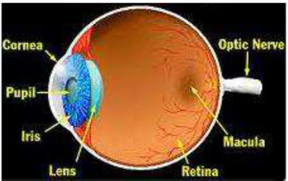
acute retinal disorder which causes manifestation of diabetes on the retina. About 210 million people all over the world have Diabetes Mellitus; among which 10-18% of people are suffering from Diabetic Retinopathy. So for the prevention of Diabetic Retinopathy and gradual vision loss, early detection and diagnosis of Diabetic Retinopathy is required. Human Eye is like a camera. Light enters into the eye through cornea and iris and it is focused to the retina through lens which is present between iris and retina. Retina interprets the light and transmitted it to the brain through optical disc. Figure 1 shows the anatomy image of the human eye,
In our paper, retina is the most important part of the work. By examining the retina we can determine diabetic retinopathy which mainly occurs due to the vascular changes of the eye.
Figure 1 Anatomy of Human Eye
The symptoms of diabetic retinopathy are blurred vision, seeing black spots in the center of the eye,
difficulty to see at night. If it is left untreated it causes complete blindness to the patients.
Figure 2 Normal Vision and Vision with Diabetic Retinopathy
Diabetic Retinopathy is of two types: a) NPDR (Non-proliferative diabetic retinopathy) b) PDR (Proliferative Diabetic Retinopathy). In NPDR, blood vessels got damaged in the retina and retina got swollen up due to the leakage of blood or fluid whereas in case of PRD, new blood vessels form in the retina which is fragile and susceptible to leakage of blood and fluids within the retina. Diabetic Retinopathy is mainly identified due to the development of microaneuryms, hemorrhages and exudates. Microaneuryms are mainly tiny swelling in the venous end of the retinal capillaries which lead to hemorrhages. It can be identified as small red spots in the retinal image. Exudates are mainly occurs when fat or lipids leaks from the ruptured blood vessels or aneuryms,it appears as yellow lesions. The degree of disease can be determined from the severity of the development of microaneuryms and exudates. If the exudates move to the macular region of the eye it may lead to total vision loss.
Anupriyaa Mukherjeeet al. Int. Journal of Engineering Research and Applications www.ijera.com
ISSN : 2248-9622,Vol. 5, Issue 2, ( Part -4) February 2015, pp.21-24
www.ijera.com 22|P a g e Figure 3: A typical Fundus Retinal Image
This paper proposes that pre processing of the image is done by separating the optical disk from the retinal image then by using morphological operations,detection of the blood vessels is done. Exudates detection technique is done by mathematical morphology on the given retinal image.
II.
OBJECTIVE
The idea behind this topic is to detect the diabetic retinopathy in the eye among the diabetic patients. The condition can cause complete blindness to the patients so the diagnosis is required to detect it in the early stage of it. By taking the image of the retina we can distinguish the diseased retina and normal retina. Swelling of the blood vessels in the eye and rupturing causes blurred vision then if it is not treated it causes blindness. By using MATLAB, the fundus retinal image is processed by using different operations and ruptured blood vessels and exudates is detected and made visually cleared for the ophthalmologists to study. The degree and severity of the disease can also be determined by this method.
III.
LITERATURE SURVEY
Nearly 50% of the patient who suffers from diabetes for more than ten years is affected by diabetic retinopathy. For the first time in history in the year of 1856 the diabetic macular changes in the form of yellowish spots was observed by Edward Jaeger. In the year of 1876 the proliferative changes that occurs in diabetic retinopathy and the importance of fractional detachment of retina was described by Wilhelm Manz. Diabetic retinopathy ocular complication is a leading cause of vision loss around the world. Approximately 5000 cases of blindness caused by this disease is reportedly annually in United States. It is expected that almost 366 million people may be affected by this disease by 2030 or the number may be increase by more than this. This disease also initiates moderate, severe
vision loss and new onset of blindness. The symptoms of diabetic retinopathy are not known in actual who do not lead to the proper treatment and finally the patients tend to new onset of blindness. Therefore proper diagnosis of this disease is very important.
IV.
PROBLEM DEFINATION
The patients with diabetes are at risk, they should check up their eyes in a regular interval for the diagnosis of diabetic retinopathy. Since Proliferative diabetic retinopathy does not show any symptoms until they are in last stage of vision loss. By image processing we can determine whether exudates and microaneuryms is developed in the retina or not which will detect the patient shown be taken care of. The retinal image of the eye and processing it by different operations is the only method to diagnose diabetic retinopathy and it also determines the degree of risk.
V.
METHODOLOGY
Morphological image processing is a type of processing and a collection of non linear operations which is used to determine the structure and morphology of the image. It has two mathematical morphology operations: erosion and dilation where erosion is used to shrink the image whereas dilation is used to expand the given image. Based on erosion and dilation, opening and closing operation is done in the morphological image processing. In this process we are using these operations to detect blood vessels and exudates.
Diabetes Retinopathy detection methodology follows from the elimination of optic disk then detection of blood vessels exudates in the fundus retinal image. The image which is used here is in JPEG image format i.e .jpg format and the image dimension is 400x273 with 24 bit depth. The overall procedure is demonstrated here:
a) Pre-processing
Anupriyaa Mukherjeeet al. Int. Journal of Engineering Research and Applications www.ijera.com
ISSN : 2248-9622,Vol. 5, Issue 2, ( Part -4) February 2015, pp.21-24
www.ijera.com 23|P a g e b) Optic disk detection
In this method, segmentation of the image is done by thresholding as it divides the image
into its constituent’s objects or region. In a gray
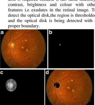
scale image, thresholding is done to convert it into a binary image. It divides the image into foreground and background. Detection of optic disk is required because it has same intensity, contrast, brightness and colour with other features i.e exudates in the retinal image. To detect the optical disk,the region is thresholded and the optical disk is being detected with a proper boundary.
Figure 4: a) fundus retinal image; b) optic disk localization; c)optic disk; d) optic disk detection
c)Blood vessel extraction
Detection of blood vessel is very important in this image processing approach. We use green channel of the input retinal image because blue channel is very low in contrast whereas the red channel is always saturated. Closing of the morphological operation is now done on the green channel retinal image with two different sizes of the structuring element. Then subtraction of these two images is done which gives the blood vessels present in the image. For better contrast, thresholding is done and then to remove the small artifacts, a median filter is applied.
Figure 5: a)input fundus image; b)gradient image ; c) thresholded gradient image; d) blood vessel extraction
d) Exudate Detection
Dilation of the morphological operation is used for the detection of the exudate edges. Edge detectors like canny and sobel adds a lot of noises in the image and may misses out key edges so they are avoided. Dilation causes bright regions to be highlighted whereas dark regions are avoided. Now dilation is performed on the green channel of the retinal image on two different sizes which are different from the sizes uses during blood vessel extraction. Subtraction is done between two sizes to get the edge detection of the exudates. Thresholding is done for better contract. On thresholded image, morphological filling is done to get optical disk region. While detecting exudates, optical disk is also detected so the optical disk co-ordinates is removed to get the final exudates.
Figure 3:a) fundus retinal image; b) dilation gradient Image; c) thresholded and filled image; d) exudates detected
Anupriyaa Mukherjeeet al. Int. Journal of Engineering Research and Applications www.ijera.com
ISSN : 2248-9622,Vol. 5, Issue 2, ( Part -4) February 2015, pp.21-24
www.ijera.com 24|P a g e
The features such as pre processing, optic disk detection, blood vessel extraction and exudates detection is performed by using the proposed algorithms. Forty eight images are tested by using the software MATLAB. The pre processing of the image is mainly done to increase the contrast of the image, then optic disk detection is done because it same intensity with the exudates, since exudates detects the presence of diabetic retinopathy. Detection of blood vessels is done to detect hemorrhages, Microaneuryms or swelling up is present or not. The detection of exudates by removing the optic disk co-ordinates is done which gives the idea of the severity of the disease.
VI.
CONCLUSION
Here we have described a proposed low cost retinal diagnosis system which can be used to detect or diagnose the various stages of diabetic retinopathy. It will help the ophthalmologists to know the severity of the disease and it can be detected at the early stage. The main advantage of our proposed algorithm is the accuracy of the result is very high; it gives proper detection of the exudates. The proposed algorithm technique can also work on a poor computing system.
REFERENCES
[1] AkaraSopharak a, BunyaritUyyanonvara a, , Sarah Barmanb, Thomas H.Williamson;
“Automatic detection of diabetic retinopathy
exudates from nondilatedretinal images using mathematical morphology
methods”Elsevier2008.
[2] Berrichi Fatima Zohra, Benyettou Mohamed;
“Automatic diagnosis of retinal images using
the support vector machine (SVM)”
[3] JagadishNayak& P Subbanna Bhat &Rajendra Acharya U & C. M.
Lim&ManjunathKagathi “Automated
Identification of Diabetic Retinopathy [4] Sinthanayothin C, Boyce JF, Williamson TH,
Cook HL, Mensah E, LalS. Automated detection of diabetic retinopathy on digital fundus image. JDiabet Med 2002; 19:105–12. [5] Niemeijer M, van Ginneken B, Staal J,

