David C. Wyld et al. (Eds) : COSIT, DMIN, SIGL, CYBI, NMCT, AIAPP - 2014
pp. 233–246, 2014. © CS & IT-CSCP 2014 DOI : 10.5121/csit.2014.4923
S.Sukanya, S.Abinaya and Dr.D.Tamilselvi
Department of CSE, Thiagarajar College of Engineering Madurai.
sabinayavasan@tce.edu, dtamilselvi@tce.edu
A
BSTRACTThe proposed method measures the retinal blood vessel diameter to identify arteriolar narrowing, arteriovenous (AV) nicking, branching coefficients to detect early diabetic retinopathy. It utilizes the vessel centerline and edge information to measure the width for a vessel segment. From the input retinal image, the vascular network is extracted using the local entropy thresholding method. The vessel boundaries are extracted using sobel edge detection method. The skeletonization operation is applied to the vascular network and mapping the vessel boundaries and the skeleton image. The branching point detection method is then performed to localize all crossing locations. A rotational invariant mask to search the pixel pairs from the edge image, and calculate the shortest distance pair which provides the vessel width (or diameter) for that cross-section. Variation in the width measurement identifies the diabetic retinopathy.
I
NDEXT
ERMSComputer Vision, Image Processing, Edge Detection, Mapping, Diabetic Retinopathy.
1.
I
NTRODUCTIONKlemen et al. 1980) [4]. Normally the circulating blood is separated from the extravascular compartment of the retina by tight encircling functional complexes between contiguous endothelial cells in the case of the intra retinal vessels (inner blood-retinal barrier) and between cells of the retinal pigment epithelium (outer blood-retinal barrier) [5]. A number of multi- centered clinical trials during the last ten years have contributed substantially to the understanding of the natural history of diabetic retinopathy and have established the value of intensive glycemic control in reducing both the risk of onset and the progression of diabetic retinopathy [5].Despite this intimidating statistics, research indicates that at least 90% of these new cases could be reduced if there was proper and vigilant treatment and monitoring of the eyes[7]. Diabetic Retinopathy often has no early warning signs, which may cause vision loss more rapidly. Diabetic Retinopathy tends to appear and progress in stages beginning with Mild Non-Proliferative Diabetic Retinopathy, progressing to Moderate Non Proliferative Diabetic Retinopathy, further advancing to Severe Non-Proliferative Diabetic Retinopathy and without proper attention developing in to the most severe stage, Proliferative Diabetic Retinopathy . Mild Non-Proliferative Diabetic Retinopathy is characterized by the presence of dot and blot hemorrhages [11] and micro aneurysms[10] in the Retina during your eye examination[8][9]. In Moderate Non-Proliferative Diabetic Retinopathy some of the small blood vessels in the Retina may actually become blocked. The blockage of these tiny blood vessels causes a decrease in the supply of nutrients and oxygen to certain areas of the Retina[8]. Severe Non-Proliferative Diabetic Retinopathy is characterized by a significant number of small blood vessels in the Retina actually becoming blocked, which results in areas of the Retina being deprived of nourishment and oxygen. A lack of sufficient oxygen supply to the Retina results in a condition called Retinal Ischemia[12][8]. Proliferative Retinopathy is the most severe stage of Diabetic Retinopathy and carries a significant risk of vision loss. The Retina responds to a lack of oxygen, by attempting to compensate for the reduced circulation by growing new, but abnormal blood vessels-a process called neovascularization [13][8].
2. S
YMPTOMS
OF
D
IABETIC
R
ETINOPATHY
A symptom is something the patient senses and describes, while a sign is something other people, such as the doctor notice. For example, rowsiness may be a symptom while dilated pupils may be a sign. Diabetic retinopathy typically has no symptoms during the early stages. Unfortunately, when symptoms become noticeable the condition is often at an advanced stage. Sometimes the only detectable symptom is a sudden and complete loss of vision. The only way patients with diabetes can protect themselves is attend every eye examination their doctor tells them to go to. Based on the research done by the Professors Tapp RJ, Shaw JE, Harper CA et al it is identified that patient can be prevented from the loss of vision if Diabetic Retinopathy is detected earlier. The blood vessel in the retina bulges called microneurysms causes the vessel width to vary (when the vessel bulges) compare to the other vessels or the other eye of the human. This symptom will be taken as earlier sign for the Diabetic Retinopathy and sometimes burst in the blood vessels causes tiny blood spots (hemorrhages) in the retina. Therefore the methods are proposed below to measure the blood vessel width more accurately to identify diabetic Retinopathy earlier.
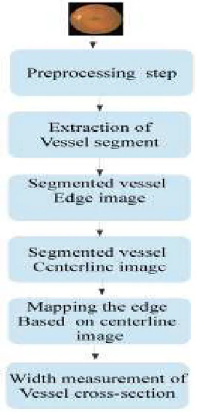
3. P
ROPOSED
A
LGORITHM
algorithm is composed of four steps. Since blood vessels usually have lower reflectance compared with the background, we apply the matched filter to enhance blood vessels with the generation of a MFR image. Secondly, an entropy-based thresholding scheme can be used to distinguish between vessel segments and the background in the MFR image. A length filtering technique is used to remove mis-classified pixels. Vascular intersection detection is performed by a branch point detection method. Then the vessel width is measured by proposed width measurement technique.
A. Preprocessing
The color components are considered separately because green channel exhibits the best vessel/background contrast while the red and blue ones tend to be very noisy in case of RGB. The proposed method work on the inverted green channel images, where vessels appear brighter than the back-ground. The two dimensional matched filter kernel is designe d to convolve with the original image in order to enhance the blood vessels.
B. Matched Filter
In [15], This concept is used to detect piecewise linear segments of blood vessels in retinal images. Blood vessels usually have poor local contrast. The two dimensional matched filter kernel is designed to convolve with the original image in order to enhance the blood vessels. A prototype matched filter kernel is expressed as
f (x, y) = −exp( −x2 ), for |y| L/2 (1)
where L is the length of the segment for which the vessel is assumed to have a fixed orientation. Here the direction of the vessel is assumed to be aligned along the y-axis. Because a vessel may be oriented at any angles, the kernel needs to be rotated for all possible angles. A set of twelve 16x15 pixel kernels is applied by convolving to a fund us image and at each pixel only the maximum of their responses is retained. The operator generates a template of values that are then applied to groups of pixels in the image.
C. Local Entrophy Thresholding
the probability of co-occurrence of gray levels i and j can therefore be written as
if s, 0 s L − 1, is a threshold. Then s can partition the co-occurrence matrix into 4 quadrants, namely A, B, C, and D
Fig.1.Quadrants of co-occurrence matrix [16].
Let us define the following quantities
Normalizing the probabilities within each individual quadrant, such that the sum of the probabilities of each quadrant equals one. The second-order entropy of the object can be defined as.
Hence, the total second-order local entropy of the object and the background can be written as
The gray level gives the optimal threshold for object back ground classification.
D. Vessel Edge Detection
The Sobel operator is used for edge detection algorithms. Technically, it is a discrete differentiation operator computing an approximation of the gradient of the image intensity function. At each point in the image, the result of the Sobel operator is either the corresponding gradient vector or the norm of this vector. The operator uses two 33 kernels which are convolved with the original image to calculate approximations of the derivatives - one for horizontal changes, and one for vertical.
E. Vessel Centerline Detection
Vessel skeleton is obtained by applying mathematical morphology reducing the vessel to a centerline of single pixel width. The perifoveolar capillaries were detected by the authors to locate candidate pixels(central part of vessel).Selection of vessel centerline candidates are done by using directional information provided from a set of four directional difference of Offset Gaussians filters. Connection of the candidate points are obtained in the previous step, by a region growing process guided by some image statistics. Validation of centerline segment candidates is based on the characteristics of the line segments; this operation is applied in each one of the four directions and finally combined, resulting in the map of the detected vessel centerlines.
F. Vessel Branch Point Detection
The vessel skeletons have to be converted into vessel segments separated by interruptions at the branching points. Segment start and end positions are determined as follows.Each of the centerline pixels on the vessel skeleton is analyzed within its 33 neighborhood, and branching points are detected as centerline pixels with more than 2 neighbors. The detection of vessel end points is required for the graph search and they are determined as the centerline pixels with only one neighbor.
G. Vessel Width Measurement
mask and at the same time it rotate the position from 0 to 180 degrees. For increasing the rotation
angle we use the step size (depending on the size of the mask) less than . Hence, the method access every cell inthe mask using this angle.
Fig. 2. Finding the mirror of an edge pixel(left) and width or minimum distance from potential pairs of pixels (right).
For each obtained position gray scale value is searched to check whether it is an edge pixel or not. Once we find an edge pixel then find its mirror by shifting the angle to 180 degree and increasing the distance from one to the maximum size of the mask (fig. 2) In this way the method produce a rotational invariant mask and pick all the potential pixel pairs to find the width or diameter of that cross sectional area.
where (x , y ) is the vessel centerline pixel position, r =1, 2, .. and = 0, .., 180. For any pixel position, if the gray scale value in the edge image is 255 (white or edge pixel) then we find the pixel (x2, y2) in the opposite edge(mirror of this pixel) considering = 0 + 180 and varying r this can be described in [17],[18]. After applying this operation we obtain the pairs of pixels which are on the opposite edges (at line end points) giving imaginary lines passing through the centerline pixels. From these pixels pairs, find the minimum Euclidian distance
Fig. 3. The overall system for proposed vessel width measurement method
4. E
XPERIMENTAL
R
ESULTS
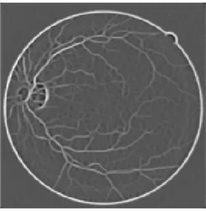
Fig.4. Original Fundus Image
Fig.4. shows the Green Channel Conversion. The green channel conversion is helps to reduce the contrast of the image and used to separate the background and vessel segment.
Fig 5 shows the Gaussian filtering, to remove Gaussian noise and is a realistic model of defocused lens. Large values
Fig. 5. Green Channel conversion
for sigma will only give large blurring for larger template sizes. Noise can be added using the sliders. The radius slider is used to control how template size.
Fig. 7. Matched Filtering Image
The concept of matched filter detection is used to detect piecewise linear segments of blood vessels in retinal images. Blood vessels usually have poor local contrast as shown in Fig. 6. The two-dimensional matched filter kernel is designed to convolve with the original image in order to enhance the blood vessels.
Fig. 8. Local Entropy Thresholding Image
Fig.8. shows the length filtering which is used to produce a clean and complete vascular tree structure by removing misclassified pixels. It isolates the individual objects by using the eight-connected neighborhood and label propagation.
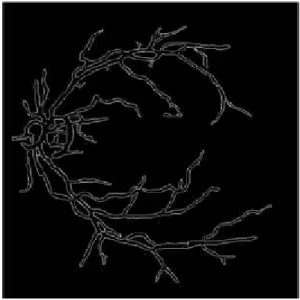
Fig. 9. Vascular Tree Structure
Fig.9. shows the edges of the detected vasculature tree structure (vessel boundaries).The edges of vessels are detectedusing the sobel edge detection method.
Fig. 10. Boundary of the vessel segment
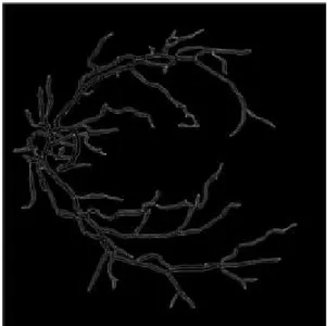
Fig. 11. Skeletonization of vessel segment (Centerline)
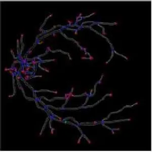
Fig.11. shows the mapping of vessel boundaries and centerline of retinal vessel. This is used to find the vessel width for a particular vessel centerline pixel position. The branching point of each vessel segements are detected and displayed in the Fig.12.The branching point are plotted in green color circle point. This points helps to determine the cross-section point of each vessel segment.
Fig. 13. Branching Point of each vessel segment
Fig.13. shows the branching points and ending points of each vessel segment. The branch points are plotted in blue color point and ending point of each vessel are plotted in red color point. And then the green color line segment shows the width or diameter of the particular vessel.
Fig. 14. Branch point, End point of vessel and Vessel width
Fig. 15. Vessel width
With the width calculated above, comparison based on vessel width calculation will takes place between manual width calculation and proposed method and the error(%) is displayed. The result is analyzed below based on the output obtained.
5. C
ONCLUSION
A
ND
F
UTURE
W
ORK
R
EFERENCES[1] Derek Hoiem, David Forsyth, TA: Varsha Hedau Computer Vision University of Illinosis. [2] Dibetic Retinopathy- http : //en.wikipedia.org/wiki/Diabetic retinopathy
[3] Wolfensberger TJ1, Hamilton AM.”DiabeticRetinopath–an Historical Review” Semin Ophthamill.2001 Mar; 16(1):2-7.
[4] Kalantzis G1, Angelou M, Poulakou-RebelakouE.”Diabetic retinopathy: an historical assessment”.Hormones (Athens). 2006 Jan-Mar;5(1):72-5.
[5] Alec Garner MD FRCpath-Department of Pathology, Institute of Ophthalmology,London EC] V 9A T ”Developments in the pathology of diabetic retinopathy: a review” in Journal of the Royal Society of Medicine Volwne 74 June 1981 427.
[6] Kertes PJ, Johnson TM, ed. (2007). Evidence Based Eye Care. Philadelphia, PA: Lippincott Williams & Wilkins. ISBN 0-7817-6964-7.
[7] Tapp RJ, Shaw JE, Harper CA et al. (June 2003). ”The prevalence of and factors associated with diabetic retinopathy in the Australian population”.Diabetes Care 26 (6): 17317. doi:10.2337/diacare.26.6.1731. PMID 12766102.
[8] T he Eye Center of Colorado -http://www.eyecarecolorado.com/diabeticretinopathy-denver.html [9] A. Hoover, V. Kouznetsova, and M. Goldbaum, Locating blood vessels in retinal images by
piecewise threshold probing of a matched filter response, IEEE Transaction Medical Imaging, Mar.2000.
[10] Mahon, W. A., et al. ”Microaneurysms in Diabetic Retinopathy.” British Medical Journal (1971). [11] Diabetic Retinopathy Vitrectomy Study Research Group. ”Early Vitrectomy for Severe Vitreous
Hemorrhage in Diabetic Retinopathy: Four-Year Results of a Randomized Trial: Diabetic Retinopathy Study Report 5.”Archives of Ophthalmology 108.7 (1990): 958.
[12] Aiello, Lloyd Paul, et al. ”Vascular endothelial growth factor in ocular fluid of patients with diabetic retinopathy and other retinal disorders.”New England Journal of Medicine 331.22 (1994): 1480-1487. [13] Schrder, S., W. Palinski, and G. W. Schmid-Schnbein. ”Activated monocytes and granulocytes,
capillary nonperfusion, and neovascularization in diabetic retinopathy.” The American journal of pathology 139.1 (1991):81.
[14] Schrder, S., W. Palinski, and G. W. Schmid-Schnbein. ”Activated monocytes and granulocytes, capillary nonperfusion, and neovascularization in diabetic retinopathy.” The American journal of pathology 139.1 (1991): 81.
[15] Dr Caroline MacEwen. ”diabetic retinopathy”.Retrieved August 2, 2011.
[16] S. Chaudhuri, S. Chatterjee, N. Katz, M. elson, and M. Goldbaum,.Detection of blood vessels in retinal images using two dimensional matched filters,. IEEE Trans. Medical imaging, vol. 8, no. 3, September 1989.
[17] . R. Pal and S. K. Pal, .Entropic thresholding,. Signal processing,vol.16, pp. 97.108, 1989.
[18] U. T. V. Nguyen, A. Bhuiyan, L. A. F. Park, and K. Ramamohanarao, An effective retinal blood vessel segmentation method using multi-scale line detection, Pattern Recognit., vol. 46, no. 3, pp. 703715, 2012.