443
A
BSTRACTObjective:The present study aims to evaluate the incidence, and risk factors of diabetic retinopathy( disease duration and the presence of hypertension). Methods: This is a field study, retrospective with quantitative approach. The study population was patients diagnosed with diabetic retinopathy in the period of july 2015 to december 2015. The collection of the data was made through analysis of medical records, it was analyzed the following data : time of diagnosis of diabetes mellitus, presence or absence of systemic hypertension Results: The overall incidence of diabetic retinopathy was 24 new cases in thesis months of analysis. Of wich a total of 66%( 16 patients) had systemic arterial hypertension and 46% had inefficient glycemic control. It was observed that 66% of the patients had indication for photocoagulation laser or intravitreal injection of anti-angiogenic and among those, 35% had diabetes for over 20 years. Conclusion: There was a strong relation between severe diabetic retinopathy, the duration of disease, systemic hypertension and bad glycemic control, with a poor outcome for disease. It also concludes that there is a significant need for assessment of diabetic patients for the presence of DR.
Keywords: Diabetic retinopathy; Blood glucose; Hypertension
O
RIGINALA
RTICLERisk factors and incidence
of diabetic retinopathy
Denise Borges de Andrade Mendanha
1, Mayara Martins Abrahão
1, Mateus Martins Cortez Vilar
1, João Jorge Nassaralla Junior
1.1 Instituto de Olhos de Goiânia – Goiânia (GO), Brasil.
Received for publication 11/07/2016 - Accepted for publication 08/10/2016 The authors declare no conflicts of interests.
R
ESUMOObjetivo: Conhecer a incidência da RD, assim como avaliar os fatores de riscos na amostra em estudo. Métodos: Avalia da incidência e os fatores de risco da RD (tempo de doença e presença de HAS em pacientes com diagnóstico de RD atendidos no ambulatório de retina de um hospital oftalmológico de referência em Goiás para pacientes do Sistema Único de Saúde, no período de julho de 2015 a dezembro de 2015 totalizando 6 meses. Resultados: Do total de 160 pacientes avaliados no ambulatório de Retina, 15% apresentaram diagnóstico de retinopatia diabética. Em relação ao tempo da doença 35% dos pacientes, que tiveram indicação de fotocoagulação ou injeção intravítrea de inibidores do fator de crescimento do endotélio vascular, apresentavam DM há mais de 20 anos. Quanto à coexistência de HAS, 16 pacientes (66%) também apresentavam hipertensão arterial sistêmica. Conclusão:
Conclui-se com o presente estudo que existe importante necessidade de avaliação de pacientes, quanto à presença de RD, além de forte associação entre a RD, e os fatores de risco avaliados no presente estudo: HAS, tempo do diagnóstico do diabetes mellitus e o controle glicêmico.
Descritores: Retinopatia diabética; Glicemia; Hipertensão
Fatores de risco e incidência
da retinopatia diabética
Rev Bras Oftalmol. 2016; 75 (6): 443-6 Study carried out at Instituto de Olhos de Goiânia – Goiânia, GO, Brazil.
444 MendanhaDBA, AbrahãoMM, VilarMMC, Nassaralla JuniorJJ
I
NTRODUCTIOND
iabetes Mellitus (DM) is a complex metabolic syndrome in which a relative or absolute insulin deficiency affects the metabolism of lipids, carbohydrates and proteins.(1)One of the most important microvascular complications of DM is diabetic retinopathy (DR), which is the leading cause of blindness among Americans aged from 20 to 64 years, causing 8000 new cases of blindness every year.(2)
DR is one of the biggest causes of irreversible blindness in the world, and the main one among people in productive age, being considered one of the most feared complications for diabetic patients. It is estimated that, after 15 years of disease, 80% of patients with DM type 2 and 97% of DM type 1 report some degree of retinopathy.(3,4)
In Brazil, it is estimated that half of patients with DM are affected by DR.(5) In Brazil, 7.6% of the urban population aged
from 30 to 69 years present DM, and 46% of them don’t know it.(6)
Duke-Elder states that its development is predictable, but not preventable, and relatively untreatable. With slurred and progressive development, diabetic retinopathy can lead to blindness in a large percentage of cases. (7-8)
Tissue hypoxia, accompanied by the loss of self-regulation of retinal vessels, is the triggering factor of DR.(9) Fundoscopy
changes follow a progressive course from mild DR, characterized by increased vascular permeability, to moderate to severe, characterized by vascular occlusion and subsequent proliferation and wound healing.(10)
The duration of DM is strongly associated to the frequency and severity of DR. (11) According to Kohlberg and Arnqvist,
with the introduction of glycosylated hemoglobin, it has been shown that blood glucose control is the most important independent risk factor for DR. (12)
In turn, Systemic Arterial Hypertension (SAH) is twice as frequent in the population with DM, and seems to play an important role in DR.(13)
The blood pressure increases the intraluminar pressure, contributing to vascular damage and retinal ischemia, increasing the risk of onset and progression of diabetic retinopathy.(14)
M
ETHODSWe examined retrospectively 160 patients referred from the retinal ambulatory of a reference ophthalmological hospital in Goiás for the patients attended by SUS during the period from July 6, 2015 to December, 2015. All patients with retinal lesion were examined in the retina department under the supervision of an expert in the field.
The patients in the present study were previously assessed by eye examination, including measurement of the corrected visual acuity (Snellen chart), anterior and posterior segment biomicroscopy, aplanation tonometry and indirect binocular ophthalmoscopy under mydriasis. All patients who showed at fundoscopy any suspected retinal lesions were examined by fluorescent retinografy to have the correct diagnosis. The analysis of the medical records of the patients diagnosed with DR attended in the retina department of an ophthalmological hos-pital in Goiás was performed for care of the patients from SUS.
Rev Bras Oftalmol. 2016; 75 (6): 443-6
The relation between the disease and sex was evaluated, the time of diagnosis estimated, and retinopathy, clinical presence of systemic arterial hypertension, and conduct established.
R
ESULTSOf the total of patients evaluated in the retinal ambulatory, 24 patients (15%) of the total sample showed diabetic retinopathy. Against 8% who were identified as patients with age-related macular degeneration (AMD), 4% with branch retinal vein occlusion (BRVO), 1.2% diagnosed as suffering from hypertensive retinopathy (HR), 1.9% with retinal detachment (RD), and in the same proportion as macular hole.
The patients identified were evaluated and directed to treatment when indicated (Figure 1)
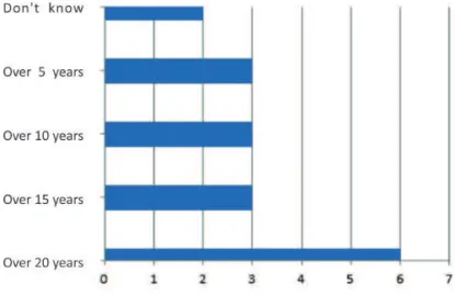
In relation to the time of illness, of the total patients who were conducted to photocoagulation or intravitreal injection of growth factor inhibitors of the vascular endothelium 35% of them had DM for over 20 years (Figure 2).
Regarding the coexistence of SAH, 16 patients (67%) also presented SAH. (Figure 3). Of these, 13 had indication for laser photocoagulation or intravitreal injection of antiangiogenic.
Figure 1: Conduct proposed of the patients assessed
445
Rev Bras Oftalmol. 2016; 75 (6): 443-6
D
ISCUSSIONIt is believed that high blood sugar worsens the clinical condition. Studies have shown that the prevalence of DR increases with the duration of the disease and the age of the patient. After 20 years of disease, almost 99% of insulin-dependent MD patients and 60% of non-insulin-insulin-dependent MD patients have some degree of DR. (15) Prevalence and relation in
accordance with the present study, since in the sample studied 33% of those identified had diabetes for over 20 years.
In turn, patients with type 1 Diabetes will present some degree of D 20 years after the disease duration.(3,16-20). Thus, it
should be reinforced that the duration of DM is strongly associated to the frequency and severity of DR. (21-23)
According to Boelter, the duration of DM is a factor to be taken into consideration in all patients, regardless of glycemic control or the degree of eye involvement.(24) In the study in
question, the duration of the disease was assessed as a risk factor determinant in the rise of retinopathy.
In the population under study, 65% were women who do not match the studies published according to which there is a ratio of 3:4 respectively between males and females.(7)
According to Laakso, SAH is twice as frequent in the population with DM, and seems to play an important role in the pathogenesis of DR.(13) This fact demonstrates compliance with
the study, as the coexistence of systemic arterial hypertension was identified in 16 patients (67%). Of these, 13 (81%) had indication for laser photocoagulation or intravitreal injection of antiangiogenic.
Patients with DM show hyperperfusion of the capillary bed in various tissues.(25) The increased blood pressure increases
the intraluminar pressure worsening the extravasation of the vascular network favoring the filtration of plasma proteins through the endothelium and its replacement in the capillary basement membrane, contributing to the vascular damage and retinal ischemia by increasing the risk of onset and progression of DR. (14) In the sample evaluated it was notorious that the relation of SAH as a risk factor for the development of DR, as 66% presented SAH.
C
ONCLUSIONWe concluded with the present study that there is significant need for assessment of diabetic patients as for the presence of
Figura 3: Proportion of patients who presented SAH
DR. We also concluded that there is strong association between DR and the risk factors assessed in this study: SAH, time from diagnosis of diabetes mellitus, and glycemic control. We can infer that there is a need for awareness of patients monitored by SUS about the importance of glycemic control and blood pressure levels facing the impact of said measures among the diabetic patients.
R
EFERENCES1. American Diabetes Association: clinical practice recommenda-tions 2002. Report of the Expert Committee on the Diagnosis and Classification of Diabetes Mellitus. Diabetes Care. 2002;25( Suppl 1): S5-20.
2. American Academy of Ophthalmology Basic and Clinical Science Course. Section 12: Retina and vitreous. 1999-2000. (Internet). [cited 2003 Mar 12]. Available from : http: //149.142,138.19/ jseiweb/ Education/educ_educact_ basicand clinical.htm
3. Klein R, Klein BE, Moss SE, Davis MD, DeMets DL. The Wiscon-sin epidemiologic study of diabetic retinopathy. II. Prevalence and risk of diabetic retinopathy when age at diagnosis is less than 30 years. Arch Ophthalmol. 1984;102(4):520-6.
4. Silva VB, Temporini ER, Moreira Filho DC, Kara-Jose N . Tratamento da retinopatia diabética: percepções de pacientes em Rio Claro (SP) - Brasil. Arq Bras Oftalmol. 2005;68(3):363-8. 5. Brasil. Ministério da Saúde. Estatísticas [Internet]. [ citado 2003 Mar 3]. Disponível em: http://www.portalweb01.saude.gov.br/ saude/aplicacoes/noticias/noticias_detalhe.cfm?co_seq_noticia HYPERLINK “http://www.portalweb01.saude.gov.br/saude/ a p l i c a c o e s / n o t i c i a s / n o t i c i a s _ d e t a l h e . c f m ? c o _ s e q _ noticia=123 =123
6. Malerbi DA, Franco LJ. Multicenter study of the prevalence of diabetes mellitus and impaired glucose tolerance in the urban Brazilian population aged 30-69 yr. The Brazilian Cooperative Group on the Study of Diabetes Prevalence. Diabetes Care. 1992;15(11):1509-16.
7. Duke-Elder S. Diabetic Retinopathy. In: Duke-Elder S. System of ophthalmology. London: Henry Kimpton; 1967. p.410-48. 8. Hirata CA, Fang T, Casella AMB, Elieser M, Abujamra S.
Prevalência de retinopatia em uma população de diabéticos. Arq Bras Oftalmol. 1986;49(1):31-3
9. Gass JD. Retinal vascular diseases. In: Gass JD. Stereoscopic atlas of macular diseases: diagnosis and treatmen. 4th ed. St. Louis: Mosby; 1997. p.516-27.
10. Engerman RL. Pathogenesis of diabetic retinopathy. Diabetes. 1989 ;38(10):1203-6. Review.
11. Sparrow JM, Mcleod BK, Smith TDW, Birk MK, Rosenthal AR. The prevalence of diabetic retinopathy and maculopathy and their risk factors in the non-insulin-treatde diabetic patients of an English town. Eye (Lond). 1993;7(1): 158-63.
12. Kullberg CE, Arnqvist HJ. Good blood glucose control caracterizes patients without retinopathy after long diabetes duration. Diabet Med. 1994;12(4):314-20
13. Laakso M. Benefits of strict glucose and blood pressure control in type 2 diabetes: lessons from the UK Prospective Diabetes Study. Circulation. 1999; 99(4):461-2.
14. Janka HU, Warram JH, Rand LI, Krolewski AS. Risk factor for progression of background retinopathy in long- standing IDDM. Diabetes. 1989;38(4):460-4.
15. Moreira Jr CA, Ávila M. Retinopatia diabética. In: Moreira Jr CA, Ávila M. Retina e vítreo. Rio de Janeiro: Cultura Médica; 2000.
16. Keen H, Lee ET, Russel D, Miki E, Bennett PH, Lu M, et al. The appearance of retinopathy and progession to proliferative retin-opathy: the WHO multinational study of vascular disease in diabe-tes. Diabetologia. 2001;44(2):22-30.
446
Rev Bras Oftalmol. 2016; 75 (6): 443-6
17. Klein R, Klein BE, Moss SE, Davis MD, DeMets DL. The Wiscon-sin epidemiologic study of diabetic retinopathy. III. Prevalence and risk of diabetic retinopathy when age at diagnosis is less than 30 years. Arch Ophthalmol. 1984;102(4):527-32.
18. American Diabetes Association. Diabetic Retinopathy. Diabetes Care. 2002; 25 ( Suppl 1):S90-3.
19. Cunha- Vaz J. Lowering the risk of visual impairment and blind-ness. Diabet Med 1998;15(4): S47-50.
20. Krolewski AS, Barzila YJ, Warram JH, Martin BC, Pfeifer M, Rand LI. Risk of early-onset proliferative retinopathy in IDDM is closely related to cardiovascular autonomic neuropathy. Dia-betes. 1992;41(4):430-7.
21. Klein R, Klein BEK, Moss SE, Cruickhanks KJ. The Wisconsin Epidemiologic Study of Diabetic Retinopathy ( WESDR). XVII. The 14-year incidence and progression of diabetic retinopathy and associated risk factors in type I diabetes. Ophthalmology. 1998;105(10):1801-15
22. Dyck PJ, Kratz KM, Karnes JR, Litchy WJ, Klein R, Pach JM, et al. The prevalence by staged severity of various types of diabetic neuropathy, retinopathy, and nephropathy in a population-based cohort. The Rochester diabetic neuropathy study. Neurology. 1993;43(4):817-24. Erratum in: Neurology. 1933;43:2345.
Corresponding author:
Denise Borges de Andrade Mendanha Rua 90, nº 304 – Apto. 306 - Setor Marista ZIP Code: 74 – Goiânia (GO), Brazil Contact: (62) 82034779
E-mail: denisemendanha@hotmail.com
23. Chen MS, Kao CS, Chang CJ, Wu TJ, Fu CC, Chen CJ, et al. Prevalence and risk factors of diabetic retinopathy among noninsulin-dependent diabetic subjects. Am J Ophthalmol. 1992;114(6):723-30.
24. Boelter MC, Azevedo MJ, Gross JL, Lavinsky J. Risk factors for diabetic retinopathy. Arq Bras Oftalmol. 2003;66(2):239-47. 25. Forrester JV, Knott RM. Pathogenesis of diabetic retinopathy
and cataract. In: Pickup JC, Williams G. Textbook of diabetes. 2nd ed. Oxford: Blackwell Science; 1997. p.45.