Susceptibility to
Yersinia pseudotuberculosis
Infection
is Linked to the Pattern of Macrophage Activation
A. Tansini & B. M. M. de Medeiros
Introduction
Yersinia pseudotuberculosis is an enteropathogen that causes gastrointestinal disorders, usually self-limiting enteritis and mesenteric lymphadenitis [1]. In the mouse oral infection model, enteropathogenic yersiniae produce sys-temic disease, the bacteria replicating in the small intes-tine, invading the Peyer’s patches and disseminating to the liver and spleen[2, 3].
Several virulence factors have been identified that pro-mote resistance to mouse serum, coordinate gene expres-sion and enable the acquisition of iron by the bacterium [4]. Yersinia contains an extrachromosomal 70 Kb plas-mid, essential for pathogenicity, which encodes the type III protein secretion system (TTSS), the Yop effector pro-teins and non-fimbrial adhesin Yad A[5, 6]. The type III secretion system is used to inject effector proteins from the bacterial cytoplasm into the cytosol of a host cell. Once inside the cell, the Yop effectors interfere with sig-nalling pathways involved in the regulation of the actin
cytoskeleton, phagocytosis, apoptosis and the inflamma-tory response, thus favouring the survival of the bacteria [6].
The early control of infection with Yersinia is medi-ated by mechanisms of innate immunity, involving mac-rophages, NK cells and neutrophils [7–9]. This early response is followed by an adaptive immune response, and the resolution of the infection is mediated by CD4+
Th1 cells that produce cytokines such as IFN-cand IL-2 [8, 9]. An effective way for Yersinia to subvert this immune response would be to inhibit the presentation of its antigens by antigen-presenting cells (APC) and thus impair T-cell stimulation [10].
By producing diverse molecules and presenting anti-gens to T cells, macrophages influence the type of adap-tive immune response, leading to the expansion and differentiation of specific lymphocytes [11]. Microbial antigens, tumour products, effector T cells and their secretory products influence the heterogeneity and the state of activation of macrophage populations [12, 13]. Department of Biological Sciences, School of
Pharmaceutical Sciences, UNESP – Sa˜o Paulo State University, Araraquara, SP, Brazil
Received 18 June 2008; Accepted in revised form 5 December 2008
Correspondence to: B. M. M. de Medeiros, Departamento de Cieˆncias Biolo´gicas, UNESP – Universidade Estadual Paulista, Faculdade de Cieˆncias Farmaceˆuticas, Rodovia Araraquara – Jau´, km 1, 14801-902, Araraquara, SP, Brasil. E-mail: medeiros@fcfar.unesp.br
Abstract
Classical macrophage activation is characterized by: high interleukin-12 (IL-12) production, activating a Th1 cell response, and high production of toxic intermediates, such as nitric oxide (NO) and reactive oxygen intermedi-ates[14]. Many have referred to cells activated in this way as M1[15]. Macrophages exposed to immune complexes, IL-4, IL-13 and IL-10, undergo an alternative form of acti-vation, characterized by an IL-10high and IL-12low pheno-type, and promote a type II response [14, 16]. These alternatively activated macrophages are called M2[15].
L-Arginine metabolism in macrophages has been used as an important parameter to discriminate between classi-cally and alternatively activated macrophages [12]. The activation by the classical pathway results in the produc-tion of NO, synthesized from arginine by inducible NO synthase (iNOS)[13, 17]. The alternative metabolic path-way for arginine is catalysed by arginase, which converts arginine to ornithine and urea [13]. It has been shown that macrophages from Th1 strains of mice (C57BL⁄6, B10D2) preferentially use the iNOS pathway and macro-phages from Th2 strains of mice (BALB⁄c, DBA⁄2) pref-erentially use the arginase pathway[15].
It is possible that susceptibility or resistance to Y. pseudotuberculosisinfection correlates with the activation state of macrophages. We have recently demonstrated that the predominant macrophage population (M1 or M2) at the early period of infection seems to be impor-tant in determiningY. enterocoliticasusceptibility or resis-tance in mice [18]. However, little is known about the responses of M1 and M2 macrophages toY. pseudotubercu-losis infection. Thus, this study was designed to reveal the pattern of macrophage activation inY. pseudotuberculo-sis infection of BALB⁄c (Yersinia-susceptible) and C57BL⁄6 (Yersinia-resistant) mice and the immunostimu-latory capacity of these cells.
Material and methods
Bacterial strain. Yersinia pseudotuberculosis YpIIIpIB1, bearing the virulence plasmid, was generously provided by Dr Hans Wolf-Watz (Umea˚ University, Sweden).
Animals. Six-week-old C57BL⁄6 and BALB⁄c female mice were purchased from CEMIB (Centro Multidiscipli-nar para Investigac¸a˜o Biolo´gica), UNICAMP, SP, Brazil. All mice were maintained in isolators under specific-pathogen-free conditions and provided with sterilized food and water. The study was approved by the School Committee for Ethics in Animal Experimentation, UNESP at Araraquara.
Experimental infection of mice. Groups of 16 mice were infected by gavage with 0.25 ml of a bacterial suspension containing 107CFU⁄ml. Groups of 16 uninfected mice were used as controls. Four mice from each group were sacrificed at intervals of 12 h, 1 day, 3 days and 5 days after infection, with carbon dioxide.
Preparation of heat-killed Yersinia antigen. Viable cells of Y. pseudotuberculosis were killed by incubation for 1 h at 60C. The heat-killed bacteria were collected by cen-trifugation, washed twice and resuspended in PBS. A sus-pension of heat-killed Y. pseudotuberculosis equivalent to 10lg of protein per ml was used in the cultures.
Peritoneal macrophage culture. Macrophages were collected from the peritoneal cavities of infected and con-trol mice in 5.0 ml of sterile PBS, pH 7.4. The cells were washed twice by centrifugation at 200·gfor 5 min
at 4C and 100ll were plated in microplates at 2.5· 106cells⁄ml in RPMI-1640 (Gibco, Carlslad, CA, USA), supplemented with HEPES (12.5 mM), sodium bicarbon-ate (2 g⁄l), L-glutamine (2 mM), penicillin (100 IU⁄ml), streptomycin (100lg⁄ml), 5 ·10)2M 2-mercapto-ethanol (Sigma, Steinheim, Germany) and 10% fetal bovine serum (FBS; Cultilab, Campinas, Brazil). Adher-ent and non-adherAdher-ent cells were separated by plate inver-sion after 1 h incubation at 37C, in an atmosphere of 5% CO2 and 95% relative humidity. Macrophages were
then cultured in supplemented RPMI-1640 in the pres-ence of either 10lg⁄ml of lipopolysaccharide (LPS) (Escherichia coli O111 B4; Sigma) or 10lg⁄ml of heat-killed Yersinia (HKY) or without antigenic stimulation. Supernatants were removed after 48 h and tested for NO and cytokine (IL-10, IL-12, TNF-a, TGF-b) production. Arginase activity was measured in macrophage lysates.
NO production. Nitric oxide production by macrophag-es was determined by measuring nitrite, employing the method of Green et al. [19]. Briefly, 50ll of cell super-natant was removed from each well and incubated with an equal volume of Griess reagent (1% w⁄v sulfanil-amide, 0.1% w⁄v naphthylethylene diamine dihydrochlo-ride, 3.0% H3PO4) at room temperature for 10 min.
Absorbance at 550 nm was determined with an ELISA microplate reader (Model 550; BioRad, Hercules, CA, USA). Nitrite concentration was calculated from an analytical curve for NaNO2.
Determination of arginase activity. Arginase activity was measured in cell lysates [20]. Briefly, cells were lysed with 100ll of 0.1% Triton X-100. After 30 min on a shaker, 100ll of 25 nm Tris–HCl was added to 100 ll of this lysate, 10 ll of 10 mm MnCl2was added and the
enzyme was activated by heating for 10 min at 56C. Arginine hydrolysis was conducted by incubating the lysate with 100 ll of 0.05M L-arginine at 37C for 60 min. The reaction was stopped with 900 ll of H2SO4
(96%)⁄H3PO4 (85%)⁄H2O (1⁄3⁄7). The urea
concentra-tion was measured at 540 nm after addiconcentra-tion of 40ll of a-isonitrosopropiophenone (dissolved in 100% ethanol) followed by heating at 95C for 30 min. One unit of enzyme activity is defined as the amount of enzyme that catalyses the formation of 1 lmol of urea per min.
measured with commercially available kits from BD Biosciences⁄Pharmingen, San Diego, CA, USA (IL-10, IL-12, TNF-a, TGF-b and IL-4) or BioLegend, San Diego, CA, USA (IFN-c).
Immunization of mice. BALB⁄c and C57BL⁄6 mice were immunized with an intraperitoneal injection of 10lg of HKY mixed with 1 mg of aluminium hydrox-ide in 0.15M NaCl. Fourteen days later, the mice received a reinforcement dose of 10lg of the antigen in saline solution. Lymphocytes were collected 7 days later and co-cultured with macrophages from mice infected withY. pseudotuberculosisor uninfected.
T-cell enrichment. Lymphocytes were obtained from the spleens of immunized mice using the Pan T-Cell Isolation Kit (Miltenyi Biotec, Bergisch Galdbach, Ger-many), according to manufacturer’s instructions. The cell enrichment process was accompanied by flow cytometry, using anti-CD3-FITC, anti-CD4-PE and anti-CD8-SPRD (BD⁄PharMingen). The final cell suspension contained approximately 80% CD3, 50% CD4 and 35% CD8.
T-cell antigen-specific activation. T-cell enriched frac-tions were seeded in 96-well plates, at a density of 5· 105cells⁄well, and co-cultured with macrophages (2·104cells⁄well) from mice infected with Y. pseudo-tuberculosisor uninfected, in the presence of HKY, in sup-plemented RPMI-1640, at 37C, in an atmosphere of 5% CO2and 95% relative humidity. After 4 days, 100ll of
supernatant was removed for cytokine analyses and 3-(4,5-dimethyl-2-thiazolyl)-2,5 diphenyl-2H-tetrazolium bro-mide (MTT) was added to the cultures (5 mg⁄ml; 10ll⁄well), which were incubated for an additional 4 h. After dissolving the formazan crystals by incubating with isopropanol, the plates were read at 540 nm.
Statistical analysis. Results are representative of two independent experiments and are presented as mean ± SD of triplicate observations. Data were analysed statistically by Student’s t-test, using the Origin statistical program, version 5.0 (Northampton, MA, USA), with the level of significance set atP< 0.05.
Results
NO production by macrophages from C57BL⁄6 and BALB⁄c
mice
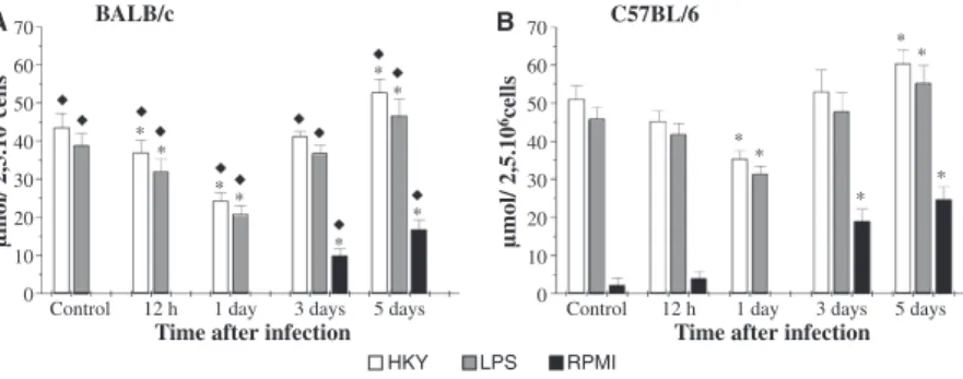
Macrophages from C57BL⁄6 control mice produced 1.2-fold more NO than macrophages from BALB⁄c controls (Fig. 1). Yersinia pseudotuberculosis infection led to a decrease in the production of NO by macrophages from BALB⁄c mice, 12 h post-infection, when these cells were stimulated with LPS or HKY. On the first-day post-infection, there was a significant decrease in the production of NO, in both strains of mice, C57BL⁄6 macrophages producing around 1.5 more NO than those from BALB⁄cmice.
From the third-day post-infection, an increase in NO production was seen; however, the increase became signif-icant only on the fifth-day post-infection, in both C57BL⁄6 and BALB⁄c mouse cells. The amount of NO produced by macrophages from infected C57BL⁄6 mice on the fifth-day post-infection was greater than that for BALB⁄c mice (about 1.2-fold more in the cells stimu-lated with LPS or HKY, and 1.5-fold in macrophages incubated with RPMI medium alone).
Arginase activity
Macrophages from BALB⁄c control mice exhibited higher arginase activity than macrophages from C57BL⁄6 con-trols (Fig. 2). There was no significant difference in argi-nase activity between macrophages from C57BL⁄6 and BALB⁄cmice, 12 h after infection.
Yersinia pseudotuberculosis infection led to a significant increase in the arginase activity on the first-day post-infection, macrophages from BALB⁄c mice showing higher levels than those from C57BL⁄6 mice (around 1.3-fold more in the cells stimulated with LPS or HKY).
The arginase activity of the macrophages collected from BALB⁄c mice on the third- and fifth-day post-infection
Control 0 10
µmol/ 2,5.10
6cells
µmol/ 2,5.10
6cells
20 30 40 50 60
70 BALB/c
A B 70 C57BL/6
60
50
40
30
20
10
0 12 h 1 day 3 days 5 days
HKY LPS RPMI Control 12 h
*
* * *
* * * * *
*
* *
*
*
1 day 3 days 5 days Time after infection Time after infection
Figure 1 Nitrite (NO2)) production by murine peritoneal macrophages collected from BALB⁄c(A) and C57BL⁄6 (B) mice duringYersinia
pseudotu-berculosisinfection. Cells (2.5·106⁄ml) were cultured with HKY, LPS or RPMI-1640 medium, and the supernatants were collected after 48 h of
cul-ture. Nitrite was assayed with Griess reagent. Control refers to average values obtained from uninfected mice throughout the experiment. Results are
representative of two independent experiments and are presented as mean ± SD of triplicate observations. *P< 0.05 versus control and¤P< 0.05
stayed high, although at lower levels than on the first-day post-infection. On the other hand, in macrophages from C57BL⁄6 mice, a significant decrease was observed in the arginase activity on the fifth-day post-infection, in the cells stimulated with LPS or HKY, the levels being 2.5 times lower than those obtained in BALB⁄c mice in the same period.
Cytokine production
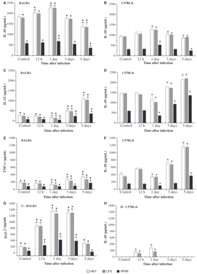
Figure 3 represents the levels of IL-10, IL-12, TNF-a and TGF-b in culture supernatants of macrophages from uninfected orY. pseudotuberculosis-infected mice.
Macrophages from C57BL⁄6 control mice produced higher levels of IL-12 and TNF-athan macrophages from BALB⁄c mice. There was no detectable production of TGF-b by macrophages from uninfected C57BL⁄6 mice. Macrophages from BALB⁄c control mice produced larger amounts of IL-10, and appreciable levels of TGF-b.
In the early period of Y. pseudotuberculosis infection, there was an increase in the IL-10 and TGF-b produc-tion, followed by a decrease in the IL-12 and TNF-a production, in both strains of mice. At 12-h and 1-day post-infection, macrophages from BALB⁄c mice produced significantly higher amounts of IL-10 (around 1.9-fold more at 12 h and 1.8-fold at 1 day post-infection) and TGF-b (about fivefold more when stimulated with HKY and eightfold when incubated with LPS, 12 h post-infec-tion) than macrophages from C57BL⁄6 mice. A small amount of TGF-b was produced by macrophages from C57BL⁄6 mice at 12-h and 1-day post-infection. The lev-els of IL-12 and TNF-a produced by macrophages from BALB⁄c were lower than those by C57BL⁄6 mouse cells: BALB⁄c cells produced levels of IL-12 around seven times lower than C57BL⁄6 in the early period of infection, and levels of TNF-a 3.2 times lower at 12 h post-infection.
On the third- and fifth-day post-infection, an increase in IL-12 and TNF-a levels was observed, followed by a
decrease in IL-10 production, in both strains of mice. At these times, macrophages from C57BL⁄6 mice produced significantly higher levels of IL-12 (about 3.6-fold more on the third-day and twofold on the fifth-day post-infec-tion, in the cells stimulated with LPS or HKY) and TNF-a (approximately threefold more in the macrophag-es stimulated with HKY) than macrophagmacrophag-es from BALB⁄c mice, and lower levels of IL-10. There was no TGF-b production by C57BL⁄6 cells on the third- and fifth-day post-infection, while a non-significant amount was produced by BALB⁄c mouse cells on the fifth-day post-infection.
T-cell antigen-specific activation
The capacity of macrophages from uninfected or infected mice to present the antigen HKY to T lymphocytes was investigated by using the MTT assay to measure cell proliferation. This method is based on reduction of the tet-razolium salt by living cells to produce a blue formazan.
The results show that macrophages obtained from C57BL⁄6 control mice induced higher proliferation of T cells than macrophages obtained from BALB⁄c control mice (Fig. 4). The formazan production was reduced at 12-h and 1-day post-infection for both strains of mice, and the level of proliferation of T cells obtained from BALB⁄c mice remained lower than for the C57BL⁄6 strain. On the third- and fifth-day post-infection, the for-mazan concentration increased to a level exceeding the control, with greater production by cells from C57BL⁄6 mice.
Figure 5 represents the levels of IFN-c and IL-4 in the supernatants of the co-cultures of T cells obtained from HKY-immunized BALB⁄cand C57BL⁄6 mice with macrophages from infected or uninfected mice. Cells from C57BL⁄6 control mice produced higher levels of IFN-c than cells from BALB⁄c control mice. There was no detectable production of IL-4 in supernatants of cells from BALB⁄cand C57BL⁄6 control mice.
Control
µmol/ 2,5.10
6cells
µmol/ 2,5.10
6cells BALB/c
A B C57BL/6
0 200 400 600 800 1000 1200 1400 1600 1800
0 200 400 600 800 1000 1200 1400 1600 1800
12 h 1 day 3 days 5 days
HKY LPS RPMI
Control 12 h 1 day 3 days 5 days Time after infection Time after infection * *
* *
* *
*
* *
* * * * *
*
* *
* * *
* *
Figure 2Arginase activity in murine peritoneal macrophages collected from BALB⁄c(A) and C57BL⁄6 (B) mice duringYersinia pseudotuberculosis
infection. Cells (2.5·106⁄
ml) were cultured with HKY, LPS or RPMI-1640 medium for 48 h and then macrophages were lysed and arginase activ-ity determined. Control refers to average values obtained from uninfected mice throughout the experiment. Results are representative of two
BALB/c C57BL/6
C57BL/6
C57BL/6
H - C57BL/6 BALB/c
BALB/c
G - BALB/c
Time after infection Time after infection
Time after infection
Time after infection
Time after infection Time after infection
Time after infection
Time after infection
IL-10 (pg/mL) IL-10 (pg/mL)
IL-10 (pg/mL)
IL-10 (pg/mL)
IL-10 (pg/mL)
IL-12 (pg/mL)
TNF-α
(pg/ml)
TGF-β
(pg/ml)
2500
A B
C D
E F
G H
2000
1500
1000
500
0
2500
2000
1500
1000
500
0
1400
1200
1000
800
600
400
200
0
1400 1600
1200
1000
800
600
400
200
0 1400
1600
1200
1000
800
600
400
200
0
1400
1200
1000
800
600
400
200
0 2500
2000
1500
1000
500
0 2500
2000
1500
1000
500
0
Control 12 h 1 day 3 days 5 days
Control 12 h 1 day 3 days 5 days
Control 12 h 1 day 3 days 5 days Control 12 h 1 day 3 days 5 days
Control 12 h 1 day 3 days 5 days
Control 12 h 1 day 3 days 5 days
Control 12 h 1 day 3 days 5 days
Control 12 h 1 day 3 days 5 days
HKY LPS RPMI
Figure 3 Cytokine production by murine peritoneal macrophages collected from BALB⁄cand C57BL⁄6 mice duringYersinia pseudotuberculosis
infec-tion. Cells (2.5·106⁄ml) were cultured with HKY, LPS or RPMI-1640 medium, the supernatants were collected after 48 h of culture and the
con-centrations of IL-10 (A,B), IL-12 (C,D), TNF-a(E,F) and TGF-b(G,H) assayed by ELISA. Control refers to average values obtained from uninfected
mice throughout the experiment. Results are representative of two independent experiments and are presented as mean ± SD of triplicate observations.
There was a decrease in the IFN-c production in the co-culture with macrophages obtained in the early period of Y. pseudotuberculosis infection, in both strains of mice. The amount of IFN-cdetected in the supernatants of the co-culture of BALB⁄c-infected mice was lower than that detected in co-culture of C57BL⁄6-infected mice on all days post-infection. On the fifth-day post-infection, a significant increase in the production of IFN-c by cells from C57BL⁄6-infected mice was observed (twofold more in relation to BALB⁄c).
There was no detectable production of IL-4 in the supernatants of the co-culture of C57BL⁄6-infected mice. A small amount of IL-4 was detected in the co-cul-ture of BALB⁄c mice on the first- and third-day post-infection.
Discussion
Our results showed that Yersinia-resistant and -suscepti-ble strains of mice have different patterns of macrophage activation, during Y. pseudotuberculosisinfection.
The NO levels produced by macrophages from C57BL⁄6 control mice were significantly higher than those produced by macrophages from BALB⁄cmice. Con-versely, the arginase activity of cells from C57BL⁄6 con-trol mice was significantly lower than that observed for the BALB⁄c mice. Similarly, macrophages from C57BL⁄6-infected mice, stimulated with HKY or LPS or unstimulated, produced higher levels of NO and lower arginase activity than equivalent macrophages from BALB⁄c mice. These results corroborate reports showing that macrophages from Th1 strains of mice are more eas-ily activated for NO production, and macrophages from Th2 strains of mice use the arginase pathway of arginine metabolism [21, 22].
At 12 h and 1 day after infection, macrophages from C57BL⁄6- and BALB⁄c-infected mice showed decrease in the NO production when compared with macrophages from control mice. These results suggest that Y. pseudotu-berculosis infection can interfere with NO production, as described by Pujol and Bliska [23] in experiments where macrophages were infected in vitrowithY. pseudotuberculo-sisandYersinia pestis.
Control 0
0.1 0.2 0.3 0.4
* *
*
* *
* *
*
0.5 0.6 0.7
OD
540nm
12 h 1 day 3 days 5 days
Time after infection
BALB/c C57BL/6
Figure 4Influence ofYersinia pseudotuberculosisinfection on the capacity of macrophages to stimulate T-cell proliferation. Peritoneal macrophages
obtained from BALB⁄c- and C57BL⁄6-infected mice were co-cultured
with T cells from immunized mice of the same strain, for 4 days in the presence of HKY. T-cell proliferation was measured by the MTT assay. Results are expressed as the OD at 540 nm, representing the production of formazan, and are presented as mean ± SD of quadruplicate
observa-tions. *P< 0.05 versus control and¤P< 0.05 versus C57BL⁄6.
2250
A B
2000
1750
1500
1250
* *
*
*
1000
750
500
250
0
Control 12 h 1 day 3 days 5 days
Time after infection
BALB/c C57BL/6
IFN-g (pg/ml)
0 10 20 30 40 50
Control 12 h 1 day 3 days 5 days
Time after infection
IL-4 (pg/ml)
Figure 5 Cytokine production in supernatants of co-cultures of T cells with macrophages from uninfected or infected mice. Peritoneal macrophages
obtained from infected BALB⁄cand C57BL⁄6 mice were co-cultured with T cells from immunized mice of the same strain, for 4 days in the presence
of HKY, and the concentrations of IL-4 and IFN-cwere assayed in supernatants by ELISA. Control refers to average values obtained from uninfected
mice throughout the experiment. Results are presented as mean ± SD of triplicate observations. *P< 0.05 versus control and ¤P< 0.05 versus
Macrophages activated in different patterns display distinct profiles of cytokine secretion. In general, M1 cells have an IL-12high, IL-10low phenotype, and are effi-cient producers of proinflammatory cytokines (IL-1b, TNF-a, IL-6). M2 macrophages share an IL-12low, IL-10high phenotype, with low production of proinflam-matory cytokines [14]. Consistent with these previously published data, our study showed that macrophages from C57BL⁄6 control mice produced higher levels of IL-12 and TNF-a than macrophages from BALB⁄c mice. On the other hand, macrophages from BALB⁄c control mice produced higher amounts of IL-10 and considerable levels of TGF-b.
During Y. pseudotuberculosis infection, macrophages from C57BL⁄6 mice produced significantly higher amounts of IL-12 and TNF-a, while macrophages from BALB⁄c mice produced significant higher amounts of IL-10 and TGF-b. These observations suggest that the infection does not change the characteristic pattern of activation of macrophages in these mice.
At 12 h and 1 day after infection, there was an increase in the production of IL-10 by macrophages of both strains of mice. IL-10 is a cytokine with broad anti-inflammatory properties that results from its ability to inhibit functions of both macrophages and dendritic cells, including secretion of their proinflammatory cytokines [24] and NO production [25]. Sing et al. [26] reported that low calcium response V (LcrV) or antigen V, a secreted antihost protein with strong immunomodulatory effects that is associated Yersinia virulence, inhibits zymosan-induced production of TNF-a by inducing IL-10. The increase of IL-10 production, in the early period of infection, was consistent with the observed decrease of proinflammatory cytokines and NO production in the same period.
In this study, we verified that infection with Y. pseu-dotuberculosisexerts a suppressor role on TNF-a and IL-12 production by macrophages from BALB⁄c and C57BL⁄6 mice, corroborating in vitro studies that reported the function of LcrV and YopB in the inhibition of the pro-duction of TNF-aand IL-12 in murine peritoneal macro-phages [27] and revealing one survival mechanism used by this bacterium.
In the early period of infection, significant production of TGF-b was seen, with higher levels for BALB⁄c mice. TGF-b is an immunoregulatory cytokine that inhibits the activation of macrophages [28]. Possibly the most important deactivating effect of TGF-b on macrophages is its ability to limit the production of cytotoxic reactive oxygen and nitrogen intermediates [29]. This is coherent with the decrease in NO production seen 12 h and 1 day after infection, mainly in macrophages from BALB⁄c mice.
On the third- and fifth-day post-infection, there was a decrease in the production of TGF-b by macrophages of
BALB⁄c mice and the absence of TGF-b production by macrophages of C57BL⁄6 mice. In the same period, a concomitant increase of NO levels was observed in both strains of mice.
In the course of infection, a decrease of IL-10 produc-tion and increase of TNF-a and IL-12 concentrations were observed in macrophages from BALB⁄c and C57BL⁄6 mice. TNF-a and IL-12 are two major media-tors of inflammatory responses in mammals[30]. IL-12 is a heterodimer cytokine that induces IFN-c production, activates TNF-a and increases NK-cell cytotoxicity, as well as T-cell proliferation[31]. The high TNF-a and IL-12 production and lower IL-10 levels that occurred on the fifth-day post-infection, in macrophages from C57BL⁄6 mice, were coherent with the increase of NO and decrease of arginase activity.
The T cells play a crucial role in the defence of the immune system against Yersinia; an effective way for yersiniae to subvert the host immune response is to inhibit the presentation of their antigens and thus impair T-cell stimulation[10, 32]. In the earlyY. pseudotuberculosis infec-tion, we observed a decrease in the immunostimulatory capacity of macrophages, which led to a lower proliferation of T-cells from BALB⁄cmice. On the third- and fifth-day post-infection, macrophages acquired the capacity to stim-ulate T-cells, higher T-cell proliferation occurring in C57BL⁄6 mice. These results corroborated the published reports [32] and show that macrophages influence the susceptibility of BALB⁄cmice toYersiniainfection.
It is known that T lymphocytes from different strains of mice tend to produce different profiles of cytokines [33]. In this study, we observed that the C57BL⁄6 unin-fected mice produced higher amounts of IFN-c than BALB⁄c-uninfected mice in supernatants of co-cultures. However, neither strain of mice produced IL-4. It is pos-sible that BALB⁄c control mice produced an amount that was below the limit of detection of the test. The results obtained with C57Bl⁄6 mice agree with reports of Hein-zel et al. [34], who verified that T cells from this strain expressed high levels of IFN-c mRNA and that IL-4 mRNA was not detectable.
disease progression [36, 37]. It is known that IFN-c stimulates C57BL⁄6 mice to produce NO[38], justifying the high levels of NO produced by macrophages of this strain of mice.
On the other hand, we observed that BALB⁄c-infected mice produced a typically Th2 response, with IL-4 produc-tion on the first- and third-day post-infecproduc-tion and lower production of IFN-c. IL-4 does not protect againstYersinia infection. The administration of anti-IL-4 antibodies beforeY. enterocoliticainfection transforms BALB⁄c suscep-tible mice into resistant animals, while the same treatment does not affect C57BL⁄6 mice significantly[36].
Macrophages from Yersinia-resistant and Yersinia -sus-ceptible strains not only differ in their ability to be acti-vated in the classical sense, but also respond differently to the same stimuli. It may well be that the susceptibil-ity of BALB⁄cmice to Yersiniais a consequence not only of the weak and delayed T-lymphocyte response[36], but also of the deficient activation of macrophages observed early in the infection.
Our results suggest that the deficient activation of macrophages in BALB⁄c mice (weak levels of NO, high levels of IL-10 and TGF-b1 and a reduced immunostim-ulatory capacity) may contribute to the susceptibility of this strain of mice toYersinia pseudotuberculosis.
Acknowledgment
We thank Vale´ria Aparecida de Arau´jo Mallavolta for technical support. The work reported here was supported by Fundac¸a˜o de Amparo a` Pesquisa do Estado de Sa˜o Paulo-FAPESP⁄Brazil (grant number 06⁄06964–8), Programa de Apoio ao Desenvolvimento Cientı´fico da Faculdade de Cieˆncias Farmaceˆuticas-UNESP⁄Brazil (PADC 2006⁄13-I) and by a fellowship provided by Co-ordenac¸a˜o de Aperfeic¸oamento de Pessoal de Nı´vel Supe-rior-CAPES⁄Brazil (Aline Tansini).
References
1 Barnes PD, Bergman MA, Mecsas J, Isberg RR.Yersinia
pseudotuber-culosisdisseminates directly from a replicating bacterial pool in the
intestine.J Exp Med2006;203:1591–601.
2 Oellerich MF, Jacobi CA, Freund Set al. Yersinia enterocolitica
infec-tion of mice reveals clonal invasion and abscess formainfec-tion. Infect
Immun2007;75:3802–11.
3 Viboud GL, Bliska JB.Yersiniaouter proteins: role in modulation
of host cell signaling responses and pathogenesis.Annu Rev Microbiol
2005;59:69–89.
4 Carniel E. Plasmids and pathogenic islands of Yersinia. Curr Top
Microbiol Immunol2002;264:89–108.
5 Navarro L, Alto NM, Dixon JE. Functions of theYersinia effector
proteins in inhibiting host immune responses.Curr Opin Microbiol
2005;8:21–7.
6 Heesemann J, Sing A, Tru¨lzsch K.Yersinia¢s stratagem: targeting
innate and adaptive immune defense. Curr Opin Microbiol
2006;9:55–61.
7 Hanski C, Naumann M, Grutzkau A et al.Humoral and cellular
defense against intestinal murine infection withYersinia
enterocoliti-ca.Infect Immun1991;59:1106–13.
8 Autenrieth IB, Hantschmann P, Heymer B, Heesemann J. Immuno-histological characterization of the cellular immune response against
Yersinia enterocolitica in mice: evidence for the involvement of T
lymphocytes.Immunobiology1993;187:1–16.
9 Autenrieth IB, Vogel U, Preger S, Heymer B, Heesemann J.
Exper-imentalYersinia enterocoliticainfection in euthymic and Tcell-
defi-cient athymic nude C57BL⁄6 mice: comparison of time course,
histomorphology, and immune response. Infect Immun 1993;61:
2585–95.
10 Kramer U, Wiedig CA. Y. enterocolitica translocated Yops impair
stimulation of T-cells by antigen presenting cells. Immunol Lett
2005;100:130–8.
11 Van Ginderachter JA, Movahedi K, Hassanzadeh Ghassabeh Get al.
Classical and alternative activation of mononuclear phagocytes:
pick-ing the best of both worlds for tumor promotion. Immunobiology
2006;211:487–501.
12 Munder M, Eichmann K, Modolell M. Alternative metabolic states
in murine macrophages reflected by the nitric oxide synthase⁄
argi-nase balance: competitive regulation by CD4+ T cells correlates
with Th1⁄Th2 phenotype.J Immunol1998;160:5347–54.
13 Bronte V, Zanovello P. Regulation of immune responses by
L-argi-nine metabolism.Nature Rev Immunol2005;5:641–54.
14 Mantovani A, Sica A, Sozzani S, Allavena P, Vecchi A, Locati M. The chemokine system in diverses forms of macrophages activation
and polarization.Trends Immunol2004;25:677–86.
15 Mills CD, Kincaid K, Alt JM, Heilman MJ, Hill AM. M-1⁄M-2
macrophages and the Th1⁄Th2 paradigm. J Immunol 2000;
164:6166–73.
16 Gordon S. Alternative activation of macrophages. Nature Rev
Immu-nol2003;3:23–35.
17 Kro¨ncke KD, Fehsel K, Kolb-Bachofen V. Inducible nitric oxide synthase and its product nitric oxide, a small molecule with
com-plex biological activities.Biol Chem Hoppe Seyler1995;376:327–43.
18 Tumitan ARP, Monnazzi LGS, Ghiraldi FR, cilli EM, Medeiros
BMM. Pattern of macrophage activation in Yersinia-resistant and
Yersinia-susceptible strains of mice. Microbiol Immunol
2007;51:1021–8.
19 Green LCD, Wagner DDA, Glogowski J, Skepper PL, Wishnok J,
Tannebaum SR. Analysis of nitrate, nitrite and [15N] nitrate in
bio-logical fluids.Ann Biochem1982;126:131–8.
20 Corraliza IM, Campo ML, Soler G, Modollel M. Determination of
arginase activity in macrophages: a micromethod.J Immunol Methods
1994;174:231–5.
21 Liew FY, Li Y, Moss D, Parkinson M, Rogers V, Moncada S.
Resis-tance toLeishmania major infection correlates with the induction of
nitric oxide synthase in murine macrophages. Eur J Immunol
1991;21:3009–14.
22 Stenger S, Thuring H, Rollinghoff M, Bogdan C. Tissue expression of inducible nitric oxide synthase is closely associated with
resis-tance toLeishmania major.J Exp Med1994;180:783–93.
23 Pujol C, Bliska JB. TurningYersiniapathogenesis outside in:
sub-version of macrophage function by intracellular yersiniae. Clin
Immunol2005;117:216–26.
24 O’Garra A, Vieira P. Th1 cells control themselves by producing
interleukin-10.Nature2007;7:425–8.
25 Ter Steege JC, Van de Ven WC, Forget PP, Buurman WA. Regula-tion of lipopolysaccharide-induced NO synthase expression in the major organs in a mouse model. The roles of endogenous
interferon-gamma, tumor necrosis factor-alpha and interleukin-10.Eur Cytokine
Netw2000;11:39–46.
26 Sing A, Roggenkamp A, Geiger AM, Heesemann J. Yersinia
antigen-induced IL-10 production of macrophages is abrogated in
IL-10-deficient mice.J Immunol2002;168:1315–21.
27 Sharma RK, Sodhi A, Batra HV, Tuteja U. Effect of rLcrV and
rY-opB fromYersinia pestison murine peritoneal macrophagesin vitro.
Immunol Lett2004;93:179–87.
28 Tsunawaki S, Spora M, Ding A, Nathan C. Deactivation of
macro-phages by transforming growth factor beta.Nature1998;334:260–2.
29 Bogdan C, Nathan C. Modulation of immune macrophages function by transforming growth factor beta, 4 and interleukin-10.Ann Acad Sci1993;685:713–39.
30 Ma X. TNF-aand IL-12: a balancing act in macrophage
function-ing.Microbes Infect2001;3:121–9.
31 Del Vecchio M, Bajetta E, Canova Set al.Interleukin-12: biological
properties and clinical application.Clin Cancer Res2007;13:4677–85.
32 Erfuth SE, Grobner S, Kramer Uet al. Yersinia enterocoliticainduces
apoptosis and inhibits surface molecule expression and cytokine
pro-duction in murine dendritic cells.Infect Immun2004;72:7045–54.
33 Hsieh CS, Macatonia SE, O’Garra A, Murphy MK. T cell genetic background determines default T helper phenotype development in
vitro.J Exp Med1995;181:713–21.
34 Heinzel FP, Sadick MD, Mutha SS, Locksley RM. Production of interferon gamma, interleukin 2, interleukin 4, and interleukin
10 by CD4+ lymphocytes in vivo during healing and
progres-sive murine leishmaniasis. Proc Natl Acad Sci USA
1991;88:7011–5.
35 Bohn E, Heesemann J, Ehlers S, Autenrieth IB. Early gamma inter-feron mRNA expression is associated with resistance of mice against
Yersinia enterocolitica.Infect Immun1994;62:3027–32.
36 Autenrieth IB, Beer M, Bohn E, Kaufmann SH, Heesemann J.
Immune responses to Yersinia enterocolitica in susceptible BALB⁄c
and resistant C57BL⁄6 mice: an essential role for gamma interferon.
Infect Immun1994;62:2590–9.
37 Autenrieth IB, Heesemann J. In vivo neutralization of tumor
necro-sis factor-alpha and interferon-gamma abrogates renecro-sistance to
Yer-sinia enterocolitica infection in mice. Med Microbiol Immunol
1992;18:333–8.
38 Dileepan KN, Page JC, Stechchulte JD. Direct activation of mur-ine peritoneal macrophages for nitric oxide production and tumor
cell killing by interferon-c. J Interferon Cytokine Res 1995;15:


