Rev Ass Med Brasil 1999; 45(3): 225-8 2 2 5 LEISHMANIA ANTIGEN IN C57BL/6J MICE
Artigo Original
Persistence of
Leishmania
antigen in C57Bl/6j inbred mice
infected with
Leishmania (Leishmania) amazonensis
C. VASCONCELLOS, M.R. KAUFFMAN, M. N. SOTTO
Department of Dermatology, Hospital do Servidor Público Estadual de São Paulo; Faculdade de Medicina da Universidade de São Paulo, São Paulo, SP.
SUMMARY – PURPOSE. To develop an animal model
for studying mucocutaneous leishmaniasis.
METHODS. The hind footpad of C57Bl/6j inbred
mice was experimentally infected with 107
Leish-mania (LeishLeish-mania) amazonensis promastigote and the skin was studied through light and electron transmission microscopy and immunohistochemis-try (PAP) techniques.
RESULTS. There were morphological evidences of
cellular immune mechanisms and hypersensitivity reaction after eight weeks of infection and
metas-INTRODUCTION
Increasing in tourism and labors migration dis-seminated leishmaniasis throughout the world1. In the New World there is a particular expression of the disease called American Tegumentary Leish-maniasis, comprising three clinical features: Loca-lized Leishmaniasis (LCL), Diffuse Cutaneous Leis-hmaniasis (DCL) and Mucocutaneous Leishma-niasis (MCL)2.
Some authors3 suggested that DCL and MCL could be both originating from Leishmania
(Leish-mania) amazonensis4, but the mechanisms by which Leishmania leads to MCL remains obscure, owing mainly to the lack of an appropriate animal model5-6.
The great majority of papers concerning to animals’ models with different susceptibilities to L.
(L.) amazonensis do so from an immunological point of view. Morphologic studies about the infla-mmatory response of the inoculation site are scarce and brief in follow up periods of infection. The aim of this study is to propose this model to study the human disease, according to the histo-pathologic features of the inoculation site and the clinical aspects, in the course of one-year infection.
MATERIAL AND METHOD
Animals. Female 40-day-old inbred mice C57Bl/ 6j strain from the University of São Paulo Medical School General Colony was used. The animals were
tasis and well shaped parasites at ultrastructural level by fifty-one weeks post infection. Relapse of infection with mucosa lesions occurred around the 50th week after inoculation.
CONCLUSION. The use of this animal model in long
term follow up could be an useful experimental model for human mucocutaneous leishmaniasis.
KEYWORDS: Experimental mucocutaneous leishmaniasis. Inbred mice. Transmission electron microscopy. Immuno-histochemistry.
maintained in plastic cages and received proper food and water ad libitum.
Parasites. Leishmania (Leishmania)
amazo-nensis (WHOM/BR/00LTB0016) - G. Grimaldi, Fio-cruz - RJ. The strain was sustained in the labo-ratory through sequential passages in BALB/c mice, culture in NNN / BHI medium and reino-culation at hind footpad in mice7.
Infection. Experimental groups, each with three animals were inoculated under ether anesthesia with 50 µl of saline solution containing 107 Leishmania
(Leishmania) amazonensis promastigote forms in the stationary phase of growth in the right hind footpad of mice. There was one control animal in each expe-rimental group, inoculated under the same condi-tions with 50 µl of sterile saline solution. Three inoculated animals and one control were killed 2, 6, 8, 10, 20 and 51 weeks post inoculation (WPI).
Techniques. Fragments from right hind footpad skin were fixed in buffered 10% formaldehyde solution, pH 7.2 and processed by usual histology technique and stained with Hematoxilyn and Eo-sin. Fragments from the same tissue were proces-sed for immunohistochemistry (PAP) demonstra-tion of Leishmania antigen in tissues18 and for Transmission Electron Microscopy9.
The experiment was repeated twice.
RESULTS
Rev Ass Med Brasil 1999; 45(3): 225-8
2 2 6
VASCONCELLOS, C et al.
torpid disease with an apparent healing of hind footpads’ cutaneous lesions around 20th WPI. By 50th WPI there was relapse of the lesions at the inoculated footpad and appearance of lesions at the tail and in the nose of all animals.
Histopathology. The C57Bl/6j hind footpad
dis-closed a rather organized tissue response with granuloma formation after the 8th WPI and fibrosis in the dermis, together with hyperplasia of epider-mis, after the 38th WPI. Eosinophils were com-monly seen. The nerve endings of the dermis were surrounded by dense macrophage infiltrate very close to the perineurial sheet.
Late in the course of the infection (51st WPI) there were grouped lymphocytes surrounding the vessels of the dermis that showed fibrinoid dege-neration. Necrosis was extensive and confluent, coming soon after 2nd WPI and followed by granu-loma formation and collagen deposition in the dermis. By the time the dermis of C57Bl/6j mice disclosed granuloma formation and collagen depo-sition, its epidermis displayed exocytosis of mono-nuclear cells, acanthosis and transepidermal eli-mination of parasites (evident by the 20th WPI). The microscopic aspects of control animals were quite normal.
Immunohistochemistry (PAP). This techni-que displayed well-shaped amastigotes forms of
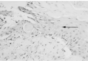
Leishmania stained in golden brown in the inocu-lated hind footpad skin. There was no evidence of amorphous antigenic material inside the cytoplasm of macrophages, as seen in human lesions8. By the 20th WPI there was elimination of cellular debris and parasites through the epidermis (Fig. 1).
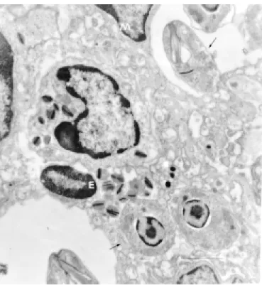
Transmission electron microscopy. The ino-culation site disclosed epidermal keratinocytes mitochondria crystolysis (Fig. 2) throughout the experiment. Some eosinophils showed phagocy-tised parasites (Fig. 3). There were preserved parasites within macrophages (Fig. 4) and in the extracellular space among collagen fibers of the dermis by 51st WPI.
DISCUSSION
The epidermis of C57Bl/6j mice was clearly involved in the inflammatory process since the beginning of the infection and showed areas of ulceration and hyperplasia. In spite of the presence of parasites in the right hind footpad until the 51st WPI, this animal seems to be able in circums-cribing the infection10,11. Necrosis was intense with elimination of parasites through the epidermis and the granuloma surrounded necrosis foci, which were partially replaced by collagen.
C57Bl/6j mice showed a well-developed tissue response (granuloma formation) by the 8th week post infection, in spite of macrophage parasitism. This was coincident with the appearing of morpho-logical evidences of cellular immune mechanisms (granuloma) and of hypersensitivity reaction (fibri-Fig. 1 – Hind footpad – elimination of cellular debris and
parasites (→) through epidermis. Experimental cutaneous leishmaniasis, C57Bl/6j inbred mice, inoculation of 107L.
(L.) amazonensis promastigote forms at right hind
footpad (20th week post infection, PAP, OM 200x).
Fig. 2 – Hind footpad – epidermal keratinocytes mito-chondria crystolysis (→). Esperimental cutaneous leish-maniasis, C57Bl/6j inbred mice, inoculation of 107L. (L.)
amazonensis promastigote forms at right hind footpad
Rev Ass Med Brasil 1999; 45(3): 225-8 2 2 7 LEISHMANIA ANTIGEN IN C57BL/6J MICE
noid degeneration of vessel’s walls and epidermal aggression by the inflammatory infiltrate). This histologic aspect is similar to that of human Muco-cutaneous Leishmaniasis12,13. Besides, by the final period of the experiment, cutaneous lesions reap-peared mucous lesions apreap-peared and well-preser-ved parasites could be seen at the ultrastructural level. Thus, such an animal model presented here may be used for experimental studies of human mucocutaneous leishmaniasis.
Acknowledgments
We are grateful to Ms. Cleusa F. H. Takakura, Carla Pagliari and Debora Aziz Rocha (Department of Pathology FMUSP) who kindly gave us proce-dure assistance.
RESUMO
Persistência do antígeno da Leishmania no camundongo isogênico C57Bl/6j infectado com a Leishmania (Leishmania) amazonensis.
OBJETIVO. Desenvolver um modelo experimental
para o estudo da leishmaniose cutâneo-mucosa.
MÉTODOS. O coxim plantar traseiro de
camun-dongos isogênicos C57Bl/6j foi inoculado com 107
Fig. 4 – Hind footpad – Upriht amastigote forms (→) inside macrophge (M). Experimental cutaneous leish-maniasis, C57Bl/6j inbred mice, inoculation of 107
L.(L.) amazonensis promastigote forms at right hind
footpad (51st week post infection, Transmission Electron Microscopy, Bar = 1 µm).
formas promastigotas da Leishmania(Leishmania) amazonensis e a pele foi estudada através da
microscopia óptica e eletrônica e de técnica imuno-histoquímica (PAP).
RESULTADOS. Ocorreram evidências
morfoló-gicas de mecanismos imunes mediados por célu-las, concomitantemente ao de reação de hipersen-sibilidade, após a oitava semana de infecção e a presença de parasitas com ultraestrutura preser-vada na quinquagésima primeira semana após a infecção. Houve recidiva da infecção com surgi-mento de lesões mucosas por volta da 50a semana
pós inoculação.
CONCLUSÃO. Este modelo animal, com um período
de tempo de seguimento prolongado, poderia ser empregado como modelo para o estudo experi-mental da leishmaniose cutâneo-mucosa. [ Rev Ass Med Brasil 1999; 45(3): 225-8.]
UNITERMOS: Leishmaniose cutânea experimental. Camun-dongos isogênicos. Microscopia eletrônica de transmissão. Imunohistoquímica.
BIBLIOGRAPHIC REFERENCES
1. Schuppli R. Leishmaniasis review. Dermatologica 1982; 165: 1-6.
Fig. 3 – Hind footpad – Leishmania amastigote forms (→) inside eosinophil (E). Experimental cutaneous leisma-niasis, C57BL/6j inbred mice, inoculation of 107L. (L.)
amazonensis promastigote forms at right hind footpad
Rev Ass Med Brasil 1999; 45(3): 225-8
2 2 8
VASCONCELLOS, C et al.
2. Coutinho E, Coelho M. Leishmaniose tegumentar experi-mental. III. Patologia comparada da infecção por amostras de Leishmania braziliensis, Leishmania mexicana e Leish-mania tropica em animais de laboratório.Rev Inst Med Trop
São Paulo, 1972; 14: 12-29.
3. Barral A, Petersen EA, Sacks DL, Neva FA. Late metastatic leishmaniasis in the mouse. A model for mucocutaneous disease. Am J Trop Med Hyg, 1983; 32:277-285.
4. Lainson R, Shaw JJ. Evolution, classification and geogra-phical distribution. In Lainson R, Shaw JJ eds. The
Leishma-niasis in Biology and Medicine, 1st ed. London, W Peters and
Killick-Kendrick Academic Press, 1987; 1. 1-120.
5. Childs GE, Lightner LK, McKinney L, Groves MG, Price EE, Hendricks L. Inbred mice as model host for cutaneous leishmaniasis. I. Resistance and susceptibility to infection with Leishmania brasiliensis, L. mexicana and L.
æthio-pica. Ann Trop Med Parasitol, 198; 478: 25-34.
6. Conceição-Silva F, Dórea RCC, Pirmez C, Schubach A, Coutinho SG. Quantitative studies of Leishmania
brazi-liensis brazibrazi-liensis reactive T cells in peripheral blood and in the lesions of patients with American Mucocu-taneous Leishmaniasis. Clin Exp Immunol, 1990; 79: 221-226.
7. Giorgio S. Estudo da imunidade celular e humoral na imunopatologia e resistência à infecção por L. amazonensis.
Dissertação (Mestrado) - Instituto de Ciências Biomédicas, Universidade de São Paulo, 1989.
8. Sotto MN, Yamashiro-Kanashiro EH, Matta VLR; Brito T. Cutaneous leishmaniasis of the New World: diagnostic immunopathology and antigen pathways in skin and mu-cosa.Acta Trop. (Basel), 1989; 46: 121-130.
9. Duarte MIS, Mariano ON, Takakura CFH, Uip DE, Corbett CEP. A fast method for processing biologic material for electron microscopic diagnosis in infectious disease.
Ultras-truct Pathol, 1992; 16: 475-482.
10. Hill JO. Pathophysiology of experimental leishmaniasis: the role of parasite physiology in the development of metastatic disease. Am J Trop Med Hyg, 1988; 39: 256-260.
11. Vasconcellos, C. & Sotto, M. N. – Experimental cutaneous leishmaniasis: transmission electron microscopy of the in-oculation site. Int J Exp Path, 1997; 78: 81-89.
12. Magalhães A.V., Moraes M.A.P., Raick A.N., Llanos-Cuen-tas A., Costa J.M.L., Cuba C.C. & Marsden P.D. - Histopa-tologia da leishmaniose tegumentar por Leishmania
brazi-liensis brazibrazi-liensis. 1. Padrões histopatológicos e estudo evolutivo das lesões. Rev Inst Med Trop São Paulo, 1986;28: 253-262.