Faculdade de Ciências Médicas da Santa Casa de Misericórdia de São Paulo Mailing address: Roberto Franken – Rua Franco da Rocha, 163/52 – 05015040 -São Paulo, SP, Brazil
Objective – To analyze the immune response in peri-pheral blood of patients with infective endocarditis.
Methods – We studied 10 patients with infective en-docarditis, age range from 20 to 50 years-old, males and females, and 20 healthy subjects in the same age range. The diagnosis of the disease was based on the clinical pic-ture, echocardiogram, and hemoculture based upon sam-ples drawn and tested before the treatment started. The were no history of atopy or malnutrition, no autoimmune disease, and they were not using any immunosuppressant or antibiotic medication.
Results – The patients with endocarditis had signifi-cantly higher T and B lymphocyte, CD4+ and CD8+ cell counts, IgM and IgG serum levels, and C4 component of the complement than the control group; no significant diffe-rence concerning serum IgA and neutrophil oxidative me-tabolism; a significant decrease in C3, chemotaxis, and monocyte phagocytosis;cryoglobulins were detected in 66.6% of patients and they were formed by IgG, IgM, IgA, C3, and C4.
Conclusion – The patients with infective endocarditis were immunocompetent in most sectors of immune respon-se and, at a certain moment, an autoimmune component may be present.
Key words: cryoglobulin, endocarditis, immune respon-se, autoimmunity
Arq Bras Cardiol, volume 76 (nº 1), 48-52, 2001
W ilma C. N eves Forte, Aline C. Mario, Adilson da Costa, Luciana S. Henriques, Carla L. Gonzales, Roberto A. Franken
São Paulo, SP - Brazil
Immunologic Evaluation in Infective Endocarditis
Endocarditis starts with the formation of a sterile ve-getation composed by fibrin and platelets due to an endo-cardial lesion. This process is called non-bacterial thrombo-tic endocarditis 1-3. In that site, a favorable environment exists for the adherence of microorganisms that are involved by new layers of fibrin and platelets. This picture is called infective endocarditis and usually develops in places whe-re blood goes from a high-pwhe-ressuwhe-re source through an ori-fice to a low-pressure site, because the effects of blood pres-sure and turbulence are favorable for the deposition of bac-teria from the fast stream. It is therefore predominantly loca-ted on the left side of the heart, where the mitral and aortic valves are the valves most affected.
All this makes it clear that, for endocarditis to develop, several pathogenetic mechanisms – hemodynamic, immu-nologic, and microbiologic – are required 2.
A variety of microbial agents cause infective endocar-ditis 2, but the most outstanding are bacteria, first found to be present by Virchow in 1869 and by Winge and Heiberg in 1872. Among these microorganisms, the most important are the following: Streptococcus viridans 4-6, Streptococcus
bovis 4, Streptococcus agalactiae 4, Streptococcus
gordo-nii 7, Haemophilus sp 4, Actinobacillus
actinomycetemco-mitans 4,8, Cardiobacterium hominis, Eikenella sp, and
Kingella Kingae 4. Staphylococcus aureus and
Entero-coccus faecalis also belong to this list, mainly when
asso-ciated with community infection and absence of an alterna-tive focus of infection 4. Less frequently, other microorga-nisms such as Staphylococcus epidermidis 4, Coxiella
bur-netii 4,9-11, Pseudomonas aeruginosa 12, and others may also cause endocarditis,
Cicatrization of this process starts even without treat-ment, but it only becomes complete after antibiotic therapy, when vegetations decrease and become endothelial. The healed valve becomes fibrous and calcified, remaining sus-ceptible to infection for life.
li-ning occurs, and it is produced in great quantities during the repair of such lesions 1, being capable of activating the complement system. Certain microorganisms were shown to be able to bind to fibronectin by specific receptors 14. The number of fibronectin receptors is directly correlated with the virulence of the microorganism 15. Staphylococcus
aureus strains isolated from patients with infective
endo-carditis were shown to have a great number of such recep-tors 15. A great number of links between fibronectin and this bacterium (56.2%) were also found 16.
Certain authors admit that, at a certain point of in the di-sease, the clinical picture can be maintained by immune pro-cesses, regardless of the presence of the microorganism 17. In this regard, other authors found immune complexes to be present in 97% of the studied cases and a rheumatic factor in 50% of cases, depending on their etiology 18.
Some papers 5,17,19-23 suggest that extracardiac pro-blems, such as articular, renal, vascular, and skin propro-blems, may be the result of the deposit of circulating immune com-plexes in the different affected organs. A major proof of the participation of the immune system in infective endocarditis is the renal aggression that coexists with the disease 18.
Based on the above, what could be the reason for some patients developing only pictures of tonsillitis, whereas others – exposed to the same microorganisms – develop pictures of bacterial endocarditis? Could some a transient immunodeficiency lead to the development of endocardi-tis? It is known that endocarditis is the result of certain fac-tors predisposing the endothelium to infections (several cardiac lesions) and of situations leading to transient bac-teremia.
We decided to perform the present study starting from the existing studies on an immune component in infective endocarditis and from the lack of studies encompassing all sectors of immune response in patients with active infective endocarditis, in addition to the importance of this disease.
Methods
Peripheral blood drawn from 11 patients with infective endocarditis was analyzed. The disease was diagnosed based on the clinical picture (unspecific signs and symp-toms of insidious onset suggesting a picture of infection), on an echocardiogram to view vegetations, and on blood culture for the detection of bacteria (Duke’s criteria). Pa-tients ranged between 20 and 50 years of age, were males and females, had no atopy or malnourishment, autoimmune disease or any history of autoimmunity, and other infective diseases, were not using immunosuppressive drugs or anti-biotics upon sample collection, which was done immedia-tely before the beginning of treatment (group II). The data found were compared with that from the control group (group I), made up of 20 healthy subjects aged 20 to 50 years. Material collection was performed upon hospital ad-mittance of the patients at the Lung and Heart Unit (Unida-de (Unida-de Pulmão e Coração) - UPCOR - of Hospital Central da Santa Casa de São Paulo, at the same time as samples for
other tests were collected, whereby patients gave their consent for the use of part of these tests for research purposes.
radial immunodiffusion plates, where the diameter of the halo was measured after 48 and 72 hours; parallel heating tests of the cryoprecipitate were made, to exclude fibrinogen.
The monoclonal antibody methodology with radial immunodiffusion dosage has been widely employed. The antibodies used were from Dako Corporation, and the dilu-tions were 1:10 for CD4, CD8, CD3, CD19, and 1:20 for the conjugate. The nitro-blue tetrazolium test is useful for the evaluation of the oxidative metabolism of polymorphonu-clear neutrophils because, if the phagocytosis digestion step of these cells is functionally viable, a liberation of elec-trons occurs, which are captured by the yellow stain nitro-blue tetrazolium, that is then reduced causing formazan, which forms a deposit of blue grains in the cell cytoplasm. The tests on chemotaxis and phagocytosis by mononu-clear phagocytes show the directed migration and the in-gestion step of these cells, evaluating their functionality, for chemotaxis and phagocytosis depend on the integrity of the monocyte receptors for C3a-C5a and C3b-C5b, respectively. The statistical method used was Student’s t test, with values <0.05 being considered significant.
Results
The results are shown in table I (control group) and table II (group of patients with infective endocarditis).
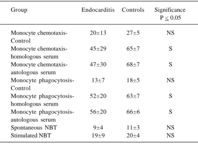
Table I shows a significant increase in T and B lym-phocytes, CD4 and CD8 cells, serum IgM and IgG, and the C4 complement component, a significant decrease in the C3 complement component without any significant IgA altera-tion in the patients with endocarditis. Table II shows a signi-ficant decrease in the monocyte phagocytosis and chemo-taxis values in the endocarditis cases. No significant diffe-rences were found between the values of the nitro-blue te-trazolium test.
In addition to that, in eight of the eleven patients with in-fective endocarditis, the presence of cryoprecipitates was observed, all of them constituted by IgM, IgG, IgA, C3, and C4.
Discussion
In an attempt to have infective endocarditis as the only variable, a selection was made immediately before on-set of treatment of patients with this disease, but without any atopy, malnutrition, or other systemic diseases and wi-thout any autoimmunity antecedents.
Experiments were performed in duplicate and well-established techniques were used 24,25.
The significantly increased results found for bursa-equivalent lymphocytes (CD19 positive cells), thymus-de-pendent lymphocytes (CD3 positive cells) and subpopula-tions of auxiliary (CD4 positive) and cytotoxic (CD8 positi-ve) cells, as well as the increase of serum levels of IgM, IgG, and the C4 complement component in the endocarditis pa-tients are consistent with values usually observed in the presence of infections.
Papers 26,27 published in the literature show high IgM, IgG, and IgA titles in patients with infective endocarditis. No studies were found with an evaluation of T and B lym-phocytes and positive CD4 and CD8 cells in active endo-carditis, except for a rare and atypical form of endocarditis caused by Coxiella burnetii that develops with rare vegeta-tions and a negative blood culture 11. In this case, in addi-tion to cryoglobulinemia and polyclonal gammopathy 11, a decrease of B (CD19 positive) and T (CD3 positive) lympho-cytes was shown, the decrease being greater in the CD4 po-sitive cells than in the CD8 popo-sitive cells 28, suggesting that this type of endocarditis might be associated with a sup-pression of lymphocyte response to specific antigens 10. A significant increase in of alpha-TNF and beta-IL-1, proin-flammatory cytokines produced by activated monocytes during the inflammation process, besides an increase of IgG and IgA and of immune complexes were also found in this type of endocarditis 10.
Table I – Results in absolute numbers of B and T lymphocytes, positive CD4 and CD8 cells, serum levels of immunoglobulins, and C3 and C4 complement components in patients with infective
endocarditis and in normal subjects
Mean Standard deviation
Group Endocarditis Controls Significance P < 0.05 T Lymphocytes 2914± 1562 1601 ± 568 S B Lymphocytes 647± 282 343 ± 147 S CD4 + cells 2317± 791 1213 ± 467 S CD8 + cells 1426± 1589 533 ± 175 S IgM (mg/dl) 243± 101 239 ± 116 S IgG (mg/dl) 1971± 307 1496 ± 507 S IgA (mg/dl) 353± 141 398 ± 156 NS
C3 (mg/dl) 86± 87 156 ± 58 S
C4 (mg/dl) 42± 15 29 ± 29 S
S- significant; NS- nonsignificant.
Table II – Chemotaxis results (distances in micra), monocyte phagocytosis (percentage of monocytes, out of a fixed number of 200, which phagocyte 3 or more Zymosan particles) and nitro-blue
tetrazolium (NBT) test for neutrophils in patients with infective endocarditis (group II) and normal subjects (group I)
Group Endocarditis Controls Significance P < 0.05
Monocyte chemotaxis- 20±13 27±5 NS Control
Monocyte chemotaxis- 45±29 65±7 S homologous serum
Monocyte chemotaxis- 47±30 68±7 S autologous serum
Monocyte phagocytosis- 13±7 18±5 NS Control
Monocyte phagocytosis- 52±20 63±7 S homologous serum
Monocyte phagocytosis- 56±20 66±6 S autologous serum
Spontaneous NBT 9±4 11±3 NS
Stimulated NBT 19±9 20±4 NS
Cryoglobulinemia was also observed in another study on subacute bacterial endocarditis 29.
The presence of a cryoprecipitate formed by IgG, IgM, IgA, C3, and C4, observed in most of the patients in this study suggests that the disease may have an autoimmune character. The data found on the decrease of the C3 comple-ment component and the monocyte activity contribute fur-ther to this hypothesis. For instance, complement is fre-quently consumed in active autoimmune diseases. Likewi-se, mononuclear phagocytes need to be perfectly functio-nal to be able to break down circulating immune complexes. Factors involved in the adhesion of monocytes or bacteria, or both to the surface of vegetations can affect phagocy-tosis of bacteria by mononuclear phagocytes 30. It was ob-served that fibronectin, a component of the extracellular ma-trix, may be involved in the adhesion of the bacterium to the surface of the endocardial vegetations by causing a strong adhesion and thus leading to a decreased monocyte phago-cytosis 30.
The mononuclear phagocytes of the studied patients with endocarditis showed decreased chemotaxis and pha-gocytosis, which might contribute to a smaller breakdown of circulating immune complexes and the consequent pre-sence of cryoglobulins. Reduction of monocyte chemotaxis and phagocytosis does not usually occur in infections.
When phagocytosis is evaluated in the presence of homologous serum, the C3 component is supplied, and it will link to the C3 receptor present in monocytes, allo-wing phagocytosis to occur. If this is decreased, it implies that an intrinsic cellular problem exists in the monocyte, a
References
fact that is confirmed by supplying autologous serum and there being no difference between phagocytosis with homologous and with autologous serum (both equally decreased).
Earlier studies found a decrease in the C3 and C4 complement components and the presence of circulating immune complexes in patients with endocarditis 5,17-21,31. Some authors describe an increase in circulating immune complexes as the disease progresses and a decrease with treatment 5. Other investigators correlate the immune complex findings with the endocardial infection rather than with the endocardial defect 17,20. Some papers show a genetic predisposition in patients with infective endocardi-tis, the most frequent haplotype being HLA-B35 32, sugges-ting a greater susceptibility of certain individuals to the disease.
Our results, showing a significant increase of T and B lymphocytes, CD4+ and CD8+ cells, IgM, IgG, and C4 complement component, are consistent with those obser-ved in immunocompetent individuals during infections. On the other hand, the significant decrease observed with regard to the C3 complement component, chemotaxis, and phagocytosis by monocytes, and the presence of cryopre-cipitates in the serum of patients with endocarditis lead to the conclusion that the disease has also an autoimmune component.
It is possible that infective endocarditis is determined by an aggressor microorganism associated with an autoim-mune component, as suggested by the presence of cryoglo-bulin in the serum of these patients.
1. João SR, Afonso MRB, Júnior LM, Júnior BNA, França HH. Aspectos patogê– nicos e imunitários da endocardite infecciosa. Arq Bras Cardiol 1990; 54: 69-72. 2. Anguita M, Torres F, Castillo JC, Valles F. Etiopathogenesis of infective endocar-ditis: predisposing heart diseases and casual microorganisms. Rev Esp Cardiol 1998; 2: 11-5.
3. Assef MAS, Wüstbuf AR, Cleiter LY, et al. Análise da febre em 58 casos de endo-cardite infecciosa. Arq Bras Cardiol 1992; 58: 107-12.
4. Durack DT, Lukes AS, Bright DK. New criteria for diagnosis of infective endocar-ditis: utilization of specific echocardiographic findings. Am J Med 1994; 96: 200-9. 5. Deck CR, Guarda ES, Bianchi CC, et al. Complejos immunes circulantes en
endo-carditis infecciosa. Rev Mèd Chile 1988; 116: 1101-4.
6. Jiang Y, Magli L, Russo M. Bacterium-dependent induction of cytokines in mo-nonuclear cells and their pathologic consequences in vivo. Infec Immun 1999; 67: 2125-30.
7. Sommer P, Gleyzal C, Guerret S, Etienne J, Grimaud J. Induction of a putative lami-nin-binding protein of Streptococcus gordonii in human infective endocarditis. Infective Immun 1992; 60: 360-5.
8. Wilson ME, Genco RJ. The role of antibody, complement and neutrophils in host defense against Actinobacillus actinomycetemcomitans. Immunol Invest 1989; 18: 187-209.
9. Atzpodien E, Baumgartner W, Artelt A, Thiele D. Valvular endocarditis occurs as a part of a disseminated Coxiella burnetii infection in immunocompromised BALB/cJ (H-2d) mice infected with the nine mile isolate of C. burnetii. J Infect Dis 1994; 170: 223-6.
10. Capo C, Zugun F, Stein A, et al. Upregulation of tumor necrosis factor alpha and interleukin-1 beta in Q fever endocarditis. Infec Immun 1996; 64: 1638-42. 11. Glasseim M, Agger WA, Vanscoy RE, Howe GB. Chronic sternal wound
infec-tion and endocarditis with Coxiella burnetii. Clin Infect Dis 1999; 28: 1249-51.
12. Oshima T, Nakaya T, Saito K, Maeda H, Nagano T. Child neglect followed by mar-ked thymic involution and fatal systemic Pseudomonas infection. Int J Legal Med 1991; 104: 167-71.
13. Assef MAS, Gandra SMA, Franken RA. Endocardite infecciosa. Estudo de 83 ca-sos no Hospital da Santa Casa de São Paulo. Arq Bras Cardiol 1991; 56: 195-9. 14. Mosher DF, Proctor RA. Binding and factor XIII a-mediated cross-linking of a 27-kilodalton fragment of fibronectin to Staphylococcus aureus. Science 1980; 209: 927-9.
15. Proctor RA, Cristman G, Mosher DF. Fibronectin induced agglutination of Staphy-lococcus aureus correlates with invasiveness. J Lab Clin Med 1984; 104: 455-69. 16. Scheld WM, Strunk RW, Balian G, Calderone RA. Microbial adhesion to fibronectin
in vitro correlates of endocarditis in rabbits. Proc Soc Exp Biol 1985; 180: 474-82. 17. Kaufmann RH, Thompson J, Valentijn RM, Daha MR, Vanes LA. The clinical im-plications and the pathogenetic significance of circulating immune complexes in infective endocarditis. Am J Med 1981; 71: 17-25.
18. Sabbagh AZ, Cohen RV, Tsunematsu EK, Bicego VC, Saber S, Júnior EBA. Endo-cardite infecciosa – a utilidade do enfoque imunológico. An Paul Med Cir 1985; 112: 23-9.
19. Roblot P, Barrier J, Roblot F, Muller A, Marechaud R. Is the immunologic evalua-tion in endocarditis of value? Rev Med Interne 1993; 14: 1031.
20. Bayer AS, Theofilopovios NA, Eisenberg R, Dixon FJ, Guze LB. Circulating im-mune complexes in infective endocarditis. N Engl J Med 1976; 295: 1500-5. 21. Cabane J, Godeau H, Acar M, Bach JF. Fate of circulating immune complexes in
in-fective endocarditis. Am J Med 1979; 66: 277-81.
23. Harris PS, Cobbs CG. Cardiac cerebral and vascular complications of infective endocarditis. Cardiol Clin 1996; 14: 437-50.
24. Forte WCN, Gonzales CCL, Carignam S, Mimica I. Avaliação de neutrófilos na desnutrição moderada. Rev Ass Med Bras 1999; 45: 147-51.
25. Forte WCN, Campos JVM, Leão RC. Non specific immunological response in mo-derate malnutrition. Allerg Immunopathol 1984; 12: 489-95.
26. Planes AM, Bernejo B, Tornos MP, Fernandez F, Arcalis L. Serologic study in pati-ents with streptococcal infective endocarditis. Med Clin 1996; 107(18):693-7. 27. Agarwal R, Bahl VK, Malaviya NA, Krishnan S, Chopra P. Immunologic
pa-rameters in infective endocarditis: a prospective study. Indian Heart J 1991; 43: 179-83.
28. Sabatier F, Dignat-George F, Mege JL, Brunet C, Raoult D, Sampol J. CD4+ T-cell lymphopenia in Q fever endocarditis. Clin Diagn Lab Immunol 1997; 4: 89-92.
29. Agarwal A, Clements J, Sedmak DD, et al. Subacute bacterial endocarditis mas-querading as type III essential mixed cryoglobulinemia. J Am Soc Nephrol 1997; 8: 1971-6.
30. Bancisi MJ, Veltrop MH, Bertina RM, Thompson J. Role of phagocytosis in acti-vation of the coagulation system in Streptococcus sanguis endocarditis. Infect immun 1996; 64: 5166-70.
31. Barrier J, Roblot P, Ramassamy A, Becq-Geraudon B. Immunologic sutdies in the differential diagnosis of infectious endocarditis and septicemia without endo-cardiac lesion. Rev Med Interne 1996; 17: 21-4.