Transcriptome Profile of the Response of
Paracoccidioides spp. to a Camphene
Thiosemicarbazide Derivative
Lívia do Carmo Silva1, Diana Patrícia Tamayo Ossa2, Symone Vitoriano da Conceição Castro1, Ludmila Bringel Pires3, Cecília Maria Alves de Oliveira3,
Cleuza Conceição da Silva4, Narcimário Pereira Coelho4, Alexandre Melo Bailão1, Juliana
Alves Parente-Rocha1, Célia Maria de Almeida Soares1, Orville Hernández Ruiz2, Juan G. McEwen Ochoa2, Maristela Pereira1*
1Laboratório de Biologia Molecular, Instituto de Patologia Tropical e Saúde Pública Universidade Federal de Goiás, Goiânia, Brazil,2Unidad de Biología Celular y Molecular, Corporación para Investigaciones Biológicas (CIB) and Facultad de Medicina Universidad de Antioquia, Medellín, Colombia,3Laboratório de Produtos Naturais, Instituto de Química, Universidade Federal de Goiás, Goiânia, Brazil,4Laboratório de Fitoquímica e Síntese Orgânica, Departamento de Química, Universidade Estadual de Maringá, Paraná, Brazil
Abstract
Paracoccidioidomycosis (PCM) is a systemic granulomatous human mycosis caused by fungi of the genusParacoccidioides, which is geographically restricted to Latin America. Inha-lation of spores, the infectious particles of the fungus, is a common route of infection. The PCM treatment of choice is azoles such as itraconazole, but sulfonamides and amphotericin B are used in some cases despite their toxicity to mammalian cells. The current availability of treatments highlights the need to identify and characterize novel targets for antifungal treat-ment of PCM as well as the need to search for new antifungal compounds obtained from natu-ral sources or by chemical synthesis. To this end, we evaluated the antifungal activity of a camphene thiosemicarbazide derivative (TSC-C) compound onParacoccidioidesyeast. To determine the response ofParacoccidioides spp. to TSC-C, we analyzed the transcriptional profile of the fungus after 8 h of contact with the compound. The results demonstrate that Paracoccidioides lutziiinduced the expression of genes related to metabolism; cell cycle and DNA processing; biogenesis of cellular components; cell transduction/signal; cell rescue, defense and virulence; cellular transport, transport facilities and transport routes; energy; pro-tein synthesis; propro-tein fate; transcription; and other propro-teins without classification. Addition-ally, we observed intensely inhibited genes related to protein synthesis. Analysis by fluorescence microscopy and flow cytometry revealed that the compound induced the pro-duction of reactive oxygen species. Using an isolate with down-regulatedSOD1gene expres-sion (SOD1-aRNA), we sought to determine the function of this gene in the defense of Paracoccidioidesyeast cells against the compound. Mutant cells were more susceptible to TSC-C, demonstrating the importance of this gene in response to the compound. The results presented herein suggest that TSC-C is a promising candidate for PCM treatment.
a11111
OPEN ACCESS
Citation:do Carmo Silva L, Tamayo Ossa DP, Castro SVdC, Bringel Pires L, Alves de Oliveira CM, Conceição da Silva C, et al. (2015) Transcriptome Profile of the Response of Paracoccidioides spp. to a Camphene Thiosemicarbazide Derivative. PLoS ONE 10(6): e0130703. doi:10.1371/journal.pone.0130703
Editor:Oscar Zaragoza, Instituto de Salud Carlos III, SPAIN
Received:February 3, 2015
Accepted:May 23, 2015
Published:June 26, 2015
Copyright:© 2015 Silva et al. This is an open access article distributed under the terms of the
Creative Commons Attribution License, which permits unrestricted use, distribution, and reproduction in any medium, provided the original author and source are credited.
Data Availability Statement:The ESTs obtained were submitted to the National Center for Biotechnology Information (NCBI) under accession numbers: LIBEST_028508 Paracoccidioides thiosemicarbazide Library.
Introduction
Paracoccidioidomycosis (PCM) is a systemic mycosis geographically restricted to Latin America caused by thermodimorphic fungi of the genusParacoccidioides. The fungi usually infect the host through the respiratory tract by inhalation of conidia, which are the infectious propagules found in the environment. In the lungs, these propagules differentiate into the pathogenic form in a temperature-dependent manner, corresponding to the yeast phase of the fungus, and spreads to other organs through lymphohematogenous dissemination. Because this mycosis affects mainly rural males of working age between the ages of 30 and 50 years, the disease has socioeconomic repercussions due to its potential to debilitate. In addi-tion to the lungs, PCM frequently compromises the mucous membranes, lymph nodes, liver, spleen and bone marrow [1,2].
Treating PCM remains a challenge due to the toxicity of the antifungals commonly used to treat this mycosis—sulfonamides, azoles and polyenes [3,4]. Additionally, despite the use of antifungals, individuals with PCM have persistent latent foci, which slow down treatment and may extend it over months or years depending on the severity of the disease and the site of injury [5,6,7]. Thus, the need to research and develop new therapeutic approaches is increasingly evident. With this aim, our group has invested effort into identifying and charac-terizing novel targets for antifungal drugs againstParacoccidioides spp. [8–16] and searching for new antifungal compounds obtained from natural sources or their synthetic derivatives [17,18,19].
The monoterpenoids are the components of essential oils, which are produced in large quantities by plants. These molecules are significant due to their therapeutic potential, low cost as well as the commercial availability, being used as starting material for synthesis of bio-active compounds [20,21]. Following this approach a series of thiosemicarbazides and thiose-micarbazones deriving from bisabolol, kaurenoic acid, limonene and camphene were
synthetized by our research group [22,23,24]. Among them, the tiosemicarbazide camphene derivative (TSC-C) showed remarkable antifungal activity. The previous study showed that TSC-C inhibited the growth ofTrichophyton mentagrophytesby damaging the cell wall struc-ture or interfering with its formation during the process of cell division, growth or morpho-genesis [24]. Based on these results, we elected TSC-C to study its activity and mode of action onParacoccidioides brasiliensis.
We constructed a cDNA library to obtain expressed sequences tags (ESTs) fromP.lutziiin response to TSC-C with the ultimate aim to identify the likely mode of action of the compound in the fungus. We performed assays to confirm the transcriptome data toP.lutziiand Paracoc-cidioides brasiliensis, such as quantitative real-time PCR (qRT-PCR), fluorescence microscopy, DNA fragmentation, cell cycle analysis by flow cytometry and enzymatic assays.
Materials and Methods
General procedure for the preparation of compounds
The TSC-C was prepared as described by Yamaguchi [24].Microorganism and cell culture
TheP.lutziiATCC MYA 826 andP.brasiliensisATCC 60855 strains were used in the assays. Yeast cells were maintained in Fava-Netto liquid medium [25] for 3 days. The cells were then transferred and grown overnight in McVeigh Morton (MMcM) liquid medium overnight [26] and subsequently used in experiments.
Goiás), CAPES (Coordenação de Aperfeiçoamento de Pessoal de Nível Superior), FINEP (Financiadora de Estudos e Projetos), and INCT-IF (Instituto Nacional de Ciência e Tecnologia para Inovação Farmacêutica). Additionally, LCS was supported by fellowship from CNPq and SVCC, LBP, NPC from CAPES. The funders had no role in study design, data collection and analysis, decision to publish, or preparation of the manuscript.
Determination of inhibitory concentration (IC
50)
Preparation of resazurin. Resazurin powder (Sigma Aldrich, St. Louis, MO, USA) was dissolved in sterile distilled water at a final concentration of 0.02%, sterilized by filtration and stored at 4°C until use.
Preparation of the camphene thiosemicarbazide derivative. The stock solution of TSC-C was prepared in dimethyl sulfoxide (10% DMSO) and diluted to obtain the evaluated concentrations (316μM, 158μM, 79μM, 39.5μM and 19.5μM).
The determination of IC50was performed according to the micro-dilution method described in the Clinical and Laboratory Standards Institute (CLSI) [27] and De Paula et al. [28]. Were inoculated 1x10⁶cells/mL ofP.lutziiyeast cells per microplate well in MMcM liq-uid medium supplemented with 316μM, 158μM, 79μM or 39.5μM TSC-C. To determine the maximum growth rate (positive control), some wells received culture medium in place of the 100 mL of test compound dilution. The plates were incubated at 36°C with shaking at 150 rpm for 48 h. Each well then received 15μL of the resazurin solution, and the plate was re-incubated for 24 h. The IC50was defined as the concentration of compound capable of inhibiting 50% of cell growth of the fungus according to the absorbance at 600 nm.
Determination of the susceptibility of
P
.
lutzii
to the camphene
thiosemicarbazide derivative
The TSC-C sensitivity test was carried out on plates containing Fava-Netto semi-solid medium supplemented with TSC-C. The concentrations tested were 316μM, 158μM, 79μM and 39.5μM. Negative control plates were prepared in the absence of TSC-C. A total of 105, 106 and 107yeast cells were inoculated on each plate. The plates were incubated for 7 days at 36°C and photographed.
Viability curve
Cell viability was determined using trypan blue staining and standard cell count techniques in a Neubauer chamber. We inoculated 1x10⁶cells/mL ofP.lutziiyeast cells in MMcM liquid medium supplemented with TSC-C at 79μM—the IC50concentration—for 0, 1, 2, 3, 4, 8 and 24 h of incubation. The negative control was performed in the absence of TSC-C. For counting, samples were collected at specific time points, and 10μL of the cell solution was added to 190μL trypan blue solution and diluted to a final volume of 1 mL. Yeast cells were observed under light microscopy with a 40X lens.
RNA extraction and purification of mRNA
Total RNA was extracted after the incubation ofParacoccidiodies spp. yeast with TSC-C at 79μM for 8 h of cultivation. The RNA was extracted with Trizol reagent (Invitrogen), precipi-tated with isopropanol, and resuspended with diethyl pyrocarbonate- (DEPC-) treated water. The mRNA was purified using the GenElute mRNA kit (Sigma Aldrich).
cDNA library construction and DNA sequencing
cDNA inserts were sequenced from the 5’end by employing standard fluorescence labeling with the DYEnamic ET dye terminator kit with an M13 flanking vector primer. Automated sequence analysis was performed in a MegaBACE 1000 DNA sequencer (GE Healthcare, Upp-sala, Sweden).
Pipeline processing and annotation of ESTs
PHRED [29], Crossmatch (http://www.macvector.com/Assembler/trimmingwithcrossmatch. html) and CAP3 [30] tools were integrated into a pipeline (http://www.lbm.icb.ufg.br/ pipelineUFG/). Only sequences with at least 50 nucleotides and a PHRED quality greater or equal to 20 were considered for assembly and cluster formation. ESTs were screened for vector sequences against the UniVec data. All of the clustered sequences were queried for similarity using BLASTX (http://www.ncbi.nlm.nih.gov/BLAST) sequence comparison software against the nucleotide database generated from theP.lutzii Pb01 structural genome (http://www. broad.mit.edu/annotation/genome/paracoccidioides_brasiliensis/MulHome.html). Sequences were grouped into functional categories with the PEDANT3 database (http://pedant.
helmholtz-muenchen.de/index.jsp). Similarities with E-values10−5were considered
signifi-cant. The Munich Information Center for Protein Sequences (MIPS) (http://mips.gsf.de/) data-base was used to assign functional categories. EC numbers were obtained by the Enzyme Database-Brenda (http://www.brenda-enzymes.info)
In silico
determination of up-regulated genes
To assign a differential expression character, ESTs from contigs formed from yeast cells treated with TSC-C were statistically evaluated using the method by Audic and Claverie [31]. Overex-pressed genes, determined by comparison to theP.lutziitranscriptome database (https://dna. biomol.unb.br/Pb/), were determined with a 95% confidence rate.
Generation of
P
.
brasiliensis SOD1
-aRNA isolate
DNA from theP.brasiliensiswild-type strain ATCC 60855 (WT) was extracted from yeast cultures during exponential growth. We employed a high-fidelity Platinum Taq DNA poly-merase (Invitrogen, Carlsbad, CA, USA) to amplify aRNA oligonucleotides designed on the PABG_03954 (www.broadinstitute.org) sequence of theSOD1gene.P.brasiliensisplasmid construction for aRNA andAgrobacterium tumefaciens-mediated transformation were per-formed as previously described [32]. Briefly, the amplifiedSOD1-aRNA oligonucleotides were inserted into the pCR35 plasmid under the control of the Calcium Binding Protein 1 (CBP-1) promoter region fromHistoplasma capsulatum[33]. The pUR5750 plasmid was used as a parental binary vector to harbor the aRNA cassette within the transfer DNA (T-DNA). The constructed binary vectors were introduced intoA.tumefaciensLBA1100 ultracompetent cells by electroporation as described previously [34] and isolated by kanamy-cin selection (100 mg/mL).
P.brasiliensisandA.tumefacienswere combined in a 1:10 ratio and incubated for 3 days of co-culture at 28°C. Selection ofP.brasiliensistransformants was performed in BHI solid media containing hygromycin B (Hyg; 200 mg/mL) over a 15 day incubation period at 36°C. Ran-domly selected Hyg resistant transformants were tested for mitotic stability.P.brasiliensis
Determination of the susceptibility of
P
.
brasiliensis
and the
SOD1
-aRNA
isolate to TSC-C
To evaluate the susceptibility ofP.brasiliensisto TSC-C, the WT, EV andSOD1-aRNA isolate strains were grown in Fava-Netto liquid medium for 72 h under constant shaking at 150 rpm and 36°C. Yeast were then transferred into MMcM liquid medium and cultured overnight. Yeast cells were then washed with 1X PBS, and the assays were performed with 1x10⁶cells. The different isolates were distributed in solid BHI medium supplemented with 316μM, 158μM, 79μM and 39.5μM TSC-C. The controls were carried out in the same medium without the addition of TSC-C. TheSOD1-aRNA isolated was growth in the presence of TSC-C added of ascorbic acid aiming to validate the influence of TSC-C as indutor agente of ROS. Initially, the concentrations from 0.08 to 100 mM ascorbic acid were used to determinate IC50(data not shown). So, 0.2 mM ascorbic acid was added at 316μM, 158μM, 79μM and 39.5μM TSC-C. All plates were incubated for 6 days at 36°C before being photographed.
Gene expression analysis by qRT-PCR
Total RNA was obtained fromParacoccidioides spp. yeast cells grown in the presence or absence of TSC-C for 8 h. After treatment with DNase, the cDNA was synthesized from total RNA using Superscript II reverse transcriptase (Invitrogen) according to the manufacturer's instruc-tions. The primers for, ATP synthase, Superoxide dismutase (SOD1) [PABG_03954 (www. broadinstitute.org)], Heat shock protein 30 kDa (HSP30), alcohol dehydrogenase (ADH), alde-hyde dehydrogenase (ALDH) andα-tubulin genes were designed using the Primer Express
soft-ware (Applied Biosystems, Foster City, CA, USA). The sequences of the oligonucleotide primers are shown inTable 1. The qRT-PCR analyses were performed in triplicate with the Ste-pOnePlus real-time PCR system (Applied Biosystems). The expression values were calculated using the alpha tubulin transcript (XM_002796593) as the endogenous control as reported pre-viously [35]. For transcripts of interest, relative expression levels were calculated using the stan-dard curve method for relative quantification [36]. The relative stanstan-dard curve was generated by pooling cDNAs from all conditions and serially diluting them from 1:5 to 1:625.
Preparation of protein extracts from
P
.
lutzii
Protein extracts were obtained after 8 h incubation in MMcM in the presence of 79μM TSC-C or in its absence. Yeast cells were centrifuged at 10,000 xgfor 10 min at 4°C, and the proteins were extracted using extraction buffer (20 mM Tris-HCl pH 8.8; 2 mM CaCl2) with a mixture of protease inhibitors (GE Healthcare). After the addition of glass beads (0.45 mm), the cells were lysed in a bead-beater, followed by centrifugation at 10,000 xgfor 15 min at 4°C. The supernatant was collected and used in enzyme activity assays. The protein concentrations were determined using the Bradford reagent (Sigma-Aldrich), as previously described [37].
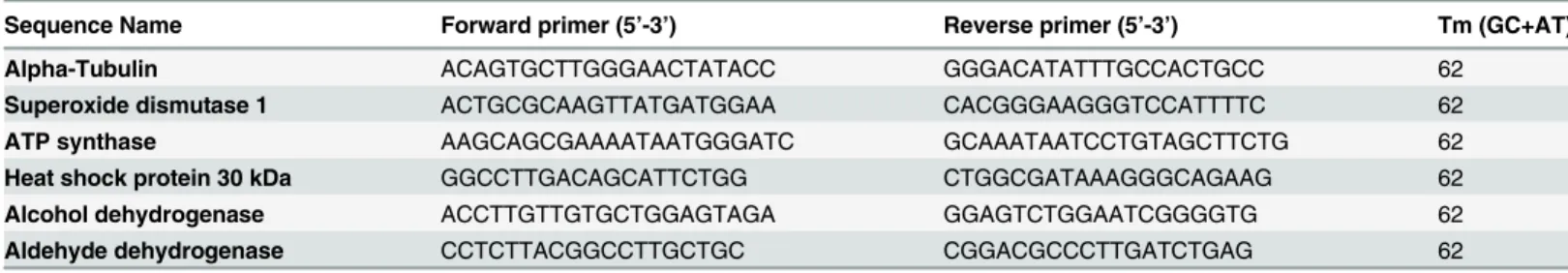
Table 1. Oligonucleotide primers used in qRT-PCR.
Sequence Name Forward primer (5’-3’) Reverse primer (5’-3’) Tm (GC+AT)
Alpha-Tubulin ACAGTGCTTGGGAACTATACC GGGACATATTTGCCACTGCC 62
Superoxide dismutase 1 ACTGCGCAAGTTATGATGGAA CACGGGAAGGGTCCATTTTC 62 ATP synthase AAGCAGCGAAAATAATGGGATC GCAAATAATCCTGTAGCTTCTG 62 Heat shock protein 30 kDa GGCCTTGACAGCATTCTGG CTGGCGATAAAGGGCAGAAG 62 Alcohol dehydrogenase ACCTTGTTGTGCTGGAGTAGA GGAGTCTGGAATCGGGGTG 62 Aldehyde dehydrogenase CCTCTTACGGCCTTGCTGC CGGACGCCCTTGATCTGAG 62
Determination of enzymatic activity
SOD activity was measured using a commercially available kit (SOD assay Kit Sigma-Aldrich) following the manufacturer's instructions. The SOD assay kit utilizes the water-soluble tetrazo-lium salt-WST-1 (2-[4- Iodophenyl]-3-[4-nitrophenyl]-5-[2,4-disulfophenyl]-2H-tetrazotetrazo-lium, monosodium salt), which produces a water-soluble formazan dye upon reduction with a super-oxide anion, and the product can be detected by a colorimetric method at 440 nm. 1μg/mL of proteins was used in assay, and the levels of SOD activity were quantified by measuring the decrease in absorbance.
Reactive oxygen species (ROS) detection
Intracellular H2O2was measured by detecting the fluorescence intensity of 2`,7`-dichloro-fluorescein, the oxidation product of 2`,7`-dichlorofluorescein diacetate. After treatment with 79μM TSC-C for 4, 8 and 12 h, yeast cells were centrifuged and incubated with 20μM 2`,7`- dichlorofluorescein diacetate for 30 min at 37°C. After washing with PBS, yeast cells were resuspended in 1 mL PBS and analyzed with a BD Accuri C6 flow cytometer (Accuri Cytometers, Ann Arbor, MI, USA). A total of 10,000 cells per sample were acquired with the FL1-H channel.
Fluorescence microscopy
Yeast cells were inoculated in 100 mL MMcM medium at 1x10⁶cells/mL. The cultures were incubated overnight at 36°C with gentle shaking. Cells were then centrifuged at 5,000 xgfor 5 min and transferred into MMcM media containing 79μM TSC-C for 4, 8 and 12 h. Control cells were incubated in MMcM without TSC-C. To detect ROS, cells were centrifuged and incubated with 20μM 2`,7`- dichlorofluorescein diacetate for 30 min at 37°C. The specimens were analyzed with an Axio Scope A1 microscope and Axio Vison LE software (Carl Zeiss AG, Germany).
DNA fragmentation assay
Yeast cells were treated with 79μM TSC-C for 4, 8 and 12 h. Samples were centrifuged, the cell pellet was resuspended in 300 mL of cell lysis buffer (10 mM Tris, 0.5% Triton X-100, pH 7.5), and the sample was incubated on ice for 30 min. The lysates were centrifuged at 12,000 xgfor 10 min at 4°C, and the supernatants were extracted once with buffered phenol and once with chloroform. DNA was precipitated with 3 M sodium acetate and butanol. DNA samples were resuspended in 50μL Tris-EDTA buffer (10 mM Tris, 1 mM EDTA, pH 7.5) treated with RNa-seA. Extracted DNA was electrophoresed through a 2% agarose gel and stained with ethidium bromide.
Cell cycle analysis
Mitochondrial membrane potential measurement
The mitochondrial membrane potential was measured using rhodamine 123 (Rho123). Yeast cells were treated with 79μM TSC-C for 4, 8 and 12 h. After treatment, the cells were collected by centrifugation and incubated with 20μM Rho123 for 20 min at room temperature. After a PBS wash, the cells were resuspended in 1 mL PBS and analyzed using a BD Accuri C6 flow cytometer (Accuri Cytometers) with excitation and emission wavelengths of 488 and 530 nm, respectively.
Statistical analysis
Descriptive statistics were calculated from the results, and charts were created in Microsoft Office Excel 2003 (Microsoft, Redmond, WA, USA). In this study, all of the values were expressed as arithmetic means with S.D. of triplicates. The significant differences between the groups were analyzed by Student’s t-test andp-values0.05 were considered statistically significant.
Results and Discussion
The camphene thiosemicarbazide derivative affects
Paracoccidioides
spp
. growth and viability
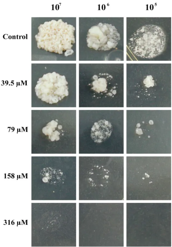
Here, we aimed to evaluate the effect of TSC-C onP.lutzii. The cells were incubated in the presence of TSC-C.Fig 1Ademonstrates that TSC-C inhibited yeast growth in a dose-depen-dent manner. TSC-C at a concentration of 79μM inhibited the cellular growth by 50% and became the IC50value of TSC-C forParacoccidioidesyeast. Additionally, the cellular viability of the fungus was monitored in the presence of 79μM TSC-C for 24 h.Fig 1Breveals that the yeast cell viability drops to 85% after 8 h of exposure to TSC-C, time used for the transcrip-tomic analysis. The dose-dependent inhibition was also observed in yeast cells grown on the solid medium supplemented with different concentrations of TSC-C (Fig 2). TSC-C (79μM) was not toxic to Balb 3T3 cells (data not shown), These results confirmed the antifungal activity of this compound.
cDNA library construction and overview of ESTs from
P
.
lutzii
exposed to
TSC-C
A cDNA library was constructed to determine the expression profile ofParacoccidioides spp. exposed to TSC-C. The dosage and duration of antifungal treatment are known to be critical steps in adaptive gene expression [38]; thus, the choice of these parameters was necessary for the construction of a cDNA library. The concentration used in the experiments was 79μM cor-responding to IC50of TSC-C forP.lutzii. The fungus was exposed to TSC-C for 8 h, since exhibited 85% viability.
We obtained a total of 2,012 clones, and 1,844 of these were successfully sequenced. All sequences were arranged into 68 contigs and 686 singlets representing different transcripts. Of these, 33 genes were down-regulated and 84 genes were up-regulated when compared to the transcriptomeP.lutziiyeast cells grownin vitro. A total of 64 genes were unique to TSC-C-treatedP.lutziiyeast cells. The ESTs obtained were submitted to the National Center for Bio-technology Information (NCBI) under accession numbers: LIBEST_028508Paracoccidioides
Functional annotation and analysis of sequences
All up- and down-regulated ESTs were compared toParacoccidioides Pb01 genes in the Broad Institute database with the Blast X program. Only ESTs with e-value<10−5were considered in
this analysis. All contigs and singlets were annotated with Blas2GO. The ESTs were grouped according to the MIPS functional annotation scheme (Munich Information Center for Protein Sequences) into general functional categories affected by TSC-C. The ESTs were related to
Fig 1. Effect of TSC-C onP.lutziiyeast cell growth. (A)Inhibition ofParacoccidioides cell growth after treatment with TSC-C. The inhibition was visualized by addition of resazurin reagent to culture and measuring the absorbance at 600 nm. To calculate the IC50value, two absorbance readings were performed; ‘1° day’refers to reading at the beginning of the experiment,‘3° days’refers to reading after 3 days of incubation with 316μM, 158μM, 79μM and 39.5μM
TSC-C. The positive control was performed in the absence of the compound.(B) Cell viability after 1, 2, 3, 4, 8 and 24 h exposure to TSC-C. The data are presented as percentage of cell viability. The Student’st-test was used for statistical comparisons, and the observed differences were statistically significant (p0.05). The error bars represent the standard deviation of three biological replicates.
Fig 2. Susceptibility ofP.lutziiyeast cells exposed to TSC-C.Samples containing 1x10⁷, 1x10⁶and 1x10⁵yeast cells were spotted on Fava-Netto plates supplemented with TSC-C at the concentrations indicated above. The plates were incubated for 7 days at 36°C before photo documentation.
metabolism; cell cycle and DNA processing; biogenesis of cellular components; cellular com-munication/signal transduction mechanism; cell rescue, defense and virulence; energy; protein synthesis; protein fate; translation; and unclassified proteins (Table 2).
Graphs were plotted to demonstrate the statistically enriched MIPS functions with up- or down-regulated genes after exposure to the compound. A total of 51% (161 ESTs) were associ-ated with proteins of unknown function (Fig 3A). Transcriptome analysis revealed that ESTs associated with metabolism (9%) and protein synthesis (9%) were the most highly represented after 8 h of TSC-C exposure (Fig 3B). The TSC-C treatment resulted in the up- and down-regu-lation of genes involved in different biological processes (Table 2; Fig3Cand3D). The groups with the highest percentage of up-regulated genes were unclassified proteins (50%); metabo-lism (12%); cell cycle and DNA processing (8%); energy (6%); transcription (5%); protein fate (5%); cellular transport, transport facilities and transport routes (5%); biogenesis of cellular components (2%); protein synthesis (1%); and cell rescue, defense and virulence (1%) (Fig 3C). The highest percentage of down-regulated genes were grouped within protein synthesis (43%); unclassified proteins (27%); cellular transport, transport facilities and transport routes (9%); energy (6%); and cell rescue, defense and virulence (3%) (Fig 3D).
We analyzed transcript occurrence by determining the number of ESTs found for each tran-script. The transcripts with the highest occurrence of up-regulated ESTs were as follows: hypo-thetical protein PAAG_02996 (18 ESTs), histone H 4.1 (8 ESTs), hypohypo-thetical protein
PAAG_03567 (6 ESTs), hypothetical protein PAAG_07875 (5 ESTs), histone H2a (5 ESTs), membrane-associated progesterone receptor component 1 (5 ESTs), 3-demethylquinone-9 3-methyltransferase (5 ESTs), hydroxymethylglutaryl-CoA lyase (5 ESTs) and superoxide dis-mutase (5 ESTs). For down-regulated ESTs, the highest abundance were as follows: hypotheti-cal protein PAAG_04431 (7 ESTs), hypothetihypotheti-cal protein PAAG_03385 (5 ESTs), nucleoside diphosphate kinase (5 ESTs) and ribosomal protein 60S–L31 (5 ESTs).
Description of transcripts changed during exposure to TSC-C
ABC transporterCDR4was induced in TSC-C-treatedP.lutziiyeast cells. These are transmem-brane proteins that utilize energy generated by the hydrolysis of adenosine triphosphate (ATP) to carry out biological processes including the translocation of various substrates across mem-branes [39]. In addition, they are involved in multidrug resistance in other human pathogens such asCandida albicans[40,41]Aspergillus fumigatus[42,43] andCryptococcus neoformans
[44]. Notably, its induction has been correlated with the protection ofAspergillus nidulans
against cytotoxic agents [45].
Similarly, most genes related to protein fate were induced in the presence of TSC-C. Con-versely, genes related to protein synthesis, mainly ribosomal proteins, were inhibited. It is well established that ribonuclease inhibitors such as the vanadyl ribonucleoside complex (VRC) can inhibit RNases involved in ribosomal subunit formation, resulting in a decreased rate of ribo-somal subunit synthesis [46].
Another gene strongly repressed in the presence of TSC-C was the endoplasmic reticulum and nuclear membrane proteinNPL4. InSaccharomyces cerevisiae, the Npl4p protein is part of a highly conserved protein complex required for the proteasome-mediated processing and acti-vation of ER-membrane-bound transcription factors, resulting in proper membrane fluidity and organelle function. Furthermore, the perturbation of membrane composition in mutant npl4 cells leads to the loss of ER/nuclear envelope integrity, which in turn causes the observed defects in nuclear transport [47].
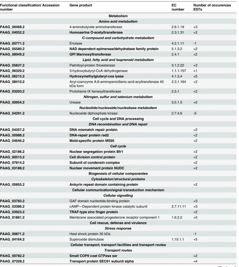
Table 2. Functional classification of up and down-regulated genes fromP.lutziiyeast cells in the presence TSC-C.
Functional classification/ Accession number
Gene product EC
number
Number of occurences ESTs
Metabolism
Amino acid metabolism
PAAG_00468.2 4-aminobutyrate aminotransferase 2.6.1.19 +3 PAAG_04052.2 Homoserine O-acetyltransferase 2.3.1.31 +2
C-compound and carbohydrate metabolism
PAAG_00771.2 Enolase 4.2.1.11 -1
PAAG_05580.2 NAD dependent epimerase/dehydratase family protein 5.1.3.2 +2
PAAG_08949.2 GPI Mannosyltransferase 2.4.1 +2
Lipid,fatty acid and isoprenoid metabolism
PAAG_05837.2 Palmitoyl-protein thioesterase 3.1.2.22 +2 PAAG_06329.2 3-hydroxybutyryl-CoA dehydrogenase 1.1.1.157 +1 PAAG_06215.2 Hydroxymethylglutaryl-coa lyase 4.1.3.4 +5 PAAG_08410.2 Acyl-coenzyme A:6-aminopenicillanic-acid-acyltransferase 40
kDa form 2.3.1.164 +2
PAAG_03203.2 Protoheme IX farnesyltransferase 2.5.1 +2
Nitrogen,sulfur and selenium metabolism
PAAG_00954.2 Urease 3.5.1.5 +2
Nucleotide/nucleoside/nucleobase metabolism
PAAG_04291.2 Nucleoside diphosphate kinase 2.7.4.6 -5
Cell cycle and DNA processing
DNA recombination and DNA repair
PAAG_04357.2 DNA mismatch repair protein +2
PAAG_05988.2 DNA-repair protein rad2 +2
PAAG_04646.2 Mold-specific protein MS95 +2
Cell cycle
PAAG_02186.2 Nuclear segregation protein Bfr1 +2
PAAG_00513.2 Cell division control protein +2
PAAG_07814.2 Subunit of condensin complex +2
PAAG_03188.2 Nuclear movement protein NUDC +2
Biogenesis of cellular componentes
Cytoskeleton/structural proteins
PAAG_05855.2 Ankyrin repeat domain containing protein +2 Cellular communication/signal transduction mechanism
Cellular signalling
PAAG_03783.2 GAF domain nucleotide-binding protein +3
PAAG_03386.2 cAMP—Dependent protein kinase catalytic subunit 2.7.11.11 +3
PAAG_03923.2 TRAF-type zincfinger protein +2
PAAG_01861.2 Membrane associated progesterone receptor component 1 1.6.2.2 +5 Cell rescue, defense and virulence
Stress response
PAAG_00871.2 Heat shock protein 30 kDa -1
PAAG_04164.2 Superoxide dismutase 1.15.1.1 +5
Cellular transport, transport facilities and transport routes
Transport routes
PAAG_00782.2 Small COPII coat GTPase sar +2
PAAG_07328.2 Transport protein SEC61 subunit alpha +4
Table 2. (Continued)
Functional classification/ Accession number
Gene product EC
number
Number of occurences ESTs
PAAG_09049.2 ENTH domain-containing protein +2
PAAG_05643.2 Endoplasmic reticulum and nuclear membrane proteinc Npl4 -3
PAAG_08587.2 GPR1/FUN34/yaaH family protein -1
PAAG_05251.2 High affinity copper transporter -1
Transported compounds (substrates)
PAAG_00635.2 ABC transporter CDR4 3.6.3.44 +3
Energy
Glycolysis and gluconeogenesis
PAAG_00403.2 Alcohol dehydrogenase 1.1.1.1 -4
Energy conversion and regeneration
PAAG_04570.2 ATP synthase D chain, mitochondrial 3.6.3.14 -2
Respiration
PAAG_08901.2 Glyoxylate reductase 1.1.1.26 +2
PAAG_05249.2 Aldehyde dehydrogenase 1.2.1.3 -2
Electron transport and membrane-associated energy conservation
PAAG_06595.2 3-demethylubiquinone-9 3-methyltransferase +5 PAAG_05031.2 NADH-ubiquinone oxidoreductase 40 kDa subunit 1.6.5.3 +3 PAAG_01307.2 NADH dehydrogenase iron-sulfur protein 1.6.99.3 +2 PAAG_07593.2 Cytochrome-c oxidase chain VIIc 1.9.3.1 +2
Protein synthesis
Ribosome biogenesis
PAAG_09043.2 Ribossomal protein 40S—S2 -1
PAAG_01785.2 Ribossomal protein 40S—S3 -1
PAAG_05017.2 Ribossomal protein 40S—S10-A -3
PAAG_01433.2 Ribossomal protein 40S—S14 -1
PAAG_00088.2 Ribossomal protein 60S—L3 -2
PAAG_07955.2 Ribossomal protein 60S—L18 -1
PAAG_00205.2 Ribossomal protein 60S—L24 -1
PAAG_04965.2 Ribossomal protein 60S—L31 -5
PAAG_07841.2 Ribossomal protein 60s—P1 -2
PAAG_03664.2 Ribossomal protein L28e -2
PAAG_00206.2 Ribossomal protein S30 -2
PAAG_07649.2 Ribossomal protein S36 -2
PAAG_03828.2 Ribossomal protein 40 S-S9 -1
Translation
PAAG_02024.2 Elongation factor 1-alpha -2
PAAG_07105.2 Isoleucyl-tRNA synthetase 6.1.1.5 +2
Protein fate (folding, modification, destination)
Assembly of protein complexes
PAAG_05879.2 Complex I intermediate-associated protein -2 PAAG_02594.2 Phosphoprotein phosphatase 2C 3.1.3.16 +2
Protein folding and stabilization
PAAG_05788.2 Peptidyl-prolyl cis-trans isomerase A2 5.2.1.8 +2
Protein/peptide degradation
PAAG_05052.2 AAA Family ATPase +2
Protein modification
Table 2. (Continued)
Functional classification/ Accession number
Gene product EC
number
Number of occurences ESTs
PAAG_05777.2 Dual specificity phosphatase catalytic domain containing protein
+2
Transcription
RNA synthesis
PAAG_07098.2 Histone H4.1 +8
PAAG_08917.2 Histone H2a +5
PAAG_08532.2 Ribonuclease Z 3.1.26.11 +2
PAAG_00891.2 AT hook motif Family protein +2
Unclassified Proteins
PAAG_04823.2 Hypothetical protein +3
PAAG_03567.2 Hypothetical protein +6
PAAG_05112.2 Hypothetical protein +2
PAAG_04156.2 Hypothetical protein +2
PAAG_01567.2 Hypothetical protein +3
PAAG_03475.2 Hypothetical protein +2
PAAG_03684.2 Hypothetical protein +4
PAAG_03129.2 Hypothetical protein +3
PAAG_06864.2 Hypothetical protein +2
PAAG_01497.2 Hypothetical protein +2
PAAG_07875.2 Hypothetical protein +5
PAAG_04268.2 Hypothetical protein +3
PAAG_02546.2 Hypothetical protein +4
PAAG_08699.2 Hypothetical protein +3
PAAG_04869.2 Hypothetical protein +4
PAAG_02037.2 Hypothetical protein +2
PAAG_02676.2 Hypothetical protein +3
PAAG_07947.2 Hypothetical protein +3
PAAG_08808.2 Hypothetical protein +2
PAAG_00149.2 Hypothetical protein +2
PAAG_01940.2 Hypothetical protein +3
PAAG_07462.2 Hypothetical protein +3
PAAG_01216.2 Hypothetical protein +2
PAAG_02607.2 Hypothetical protein +3
PAAG_05412.2 Hypothetical protein +2
PAAG_05607.2 Hypothetical protein +4
PAAG_05415.2 Hypothetical protein +2
PAAG_07420.2 Hypothetical protein +2
PAAG_07885.2 Hypothetical protein +3
PAAG_00947.2 Hypothetical protein +2
PAAG_02407.2 Hypothetical protein +2
PAAG_08549.2 Hypothetical protein +3
PAAG_08355.2 Hypothetical protein +2
PAAG_02237.2 Hypothetical protein +2
PAAG_02996.2 Hypothetical protein +18
PAAG_07710.2 Hypothetical protein +3
PAAG_04455.2 Hypothetical protein +4
an essential cofactor for a wide variety of enzymes in crucial biological processes critical for cell growth, differentiation and survival [48,49]. Some studies suggest that copper acquisition plays an important role in the virulence ofC.neoformans[50]. However, at high intracellular concen-trations, copper can be toxic due to the perturbation of the cellular redox potential, which increases production of reactive free radicals and indirectly increases oxidative stress [51]. InS.
cerevisiae, the high-affinity copper transport proteins, which play an important role in regulating copper homeostasis, are induced by copper deprivation and repressed by copper excess [52,53].
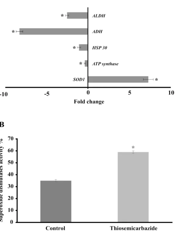
The mitochondrial electron transport chain performs the transfer of electrons from glycoly-sis and the Krebs cycle and thereby creates an electrochemical gradient and energy, which is used for a variety of vital processes that include, ATP synthesis [54], ion homeostasis [55], pro-tein import [56] and programmed cell death [57]. Because energy metabolism and redox state are potential targets, the development of drugs that specifically compromise the structural and functional integrity of mitochondria may provide novel opportunities to combat fungal infec-tions [58]. Studiesin vitrohave demonstrated the interaction between drugs and mitochondria that may prove useful in several therapies [59,60,61]. In this sense, mitochondrial insult or fail-ure can rapidly lead to the inhibition of cell survival and proliferation [62]. Furthermore, here we uncovered that genes related to electron transport, such as NADH-ubiquinone oxidoreduc-tase 40 kDa subunit, NADH dehydrogenase iron-sulfur protein and cytochrome-c oxidase chain VII c, were induced in the presence of TSC-C; however, the expression of the ATP synthase D chain was inhibited (Fig 4A), suggesting that TSC-C could destabilize the electron transport chain and, as a consequence, decrease the production of ATP inP.lutzii.
TSC-C induces
SOD1
up-regulation as a consequence of
TSC-C-induced ROS
Superoxide dismutases (SODs) constitute the primary antioxidant defense against ROS, pro-moting dismutation of the superoxide radical (O2−) into molecular oxygen and H2O2, which
Table 2. (Continued)
Functional classification/ Accession number
Gene product EC
number
Number of occurences ESTs
PAAG_06704.2 Hypothetical protein +2
PAAG_08976.2 Hypothetical protein +2
PAAG_04152.2 Hypothetical protein +2
PAAG_05097.2 Hypothetical protein +2
PAAG_06142.2 Hypothetical protein +2
PAAG_01456.2 Hypothetical protein +2
PAAG_04691.2 Hypothetical protein -2
PAAG_04913.2 Hypothetical protein -1
PAAG_04707.2 Hypothetical protein -2
PAAG_03385.2 Hypothetical protein -5
PAAG_07334.2 Hypothetical protein -4
PAAG_04431.2 Hypothetical protein -7
PAAG_08722.2 Hypothetical protein -2
PAAG_00415.2 Hypothetical protein -3
PAAG_03232.2 Hypothetical protein -2
Genes in bold correspond to single genes in condition TSC-C. The signs + and - represent induced and repressed genes, respectively.
are less toxic for the cell. ROS-generating agents induce fungal SODs, among other proteins [63].SOD3protectsH.capsulatumyeast cells from host-derived oxidative stress by detoxifying ROS produced by macrophages and neutrophils, thereby enabling the survival of the fungus [64]. In the case ofParacoccidioides spp., the induction ofSOD1protein is similarly observed in cells exposed to H2O2[65]. InC.albicans, another fungal pathogen, the inactivation of ROS detoxifying enzymes has been shown to attenuate its virulence [66].
Here, we confirmed the up-regulation ofSOD1inP.lutziiandP.brasiliensisby qRT-PCR (Figs4Aand6D) and by enzymatic activity assay inP.lutzii(Fig 4B), suggesting that TSC-C could induce the formation of ROS leading to oxidative stress inParacoccidioides spp. yeast cells. Thus, to confirm our hypothesis, we evaluated the production of ROS by means of
Fig 3. Statistically enriched MIPS functions. (A)Total ESTs represented by classified and unclassified categories.(B)Genes expressed differentially in the presence of camphene thiosemicarbazide derivate. Up-(C)or down-(D)regulatedP.lutziigenes after exposure of yeast cells to TSC-C. The functional classification was based on the MIPS functional annotation scheme. Each functional class is represented as a color-coded segment and expressed as a percentage of the total number of ESTs.
Fig 4. Effect of TSC-C on the genes andSOD1activity ofP.lutzii(A)Gene expression profile of yeast cells exposed to TSC-C for 8 h. Changes in the gene expression levels were calculated by the relative standard curve method using the non-treated control samples to calibrate. Each error bar represents the standard error of the mean (±SD), and significant fold changes are denoted by asterisks in the figure (*p0.05). Data were normalized with the transcript encoding theα-tubulin protein.(B)SOD1 activity. Yeast cells were grown in the presence of TSC-C for 8 h, and total protein was extracted to measure SOD1 activity. The Student’st-test was used for statistical comparisons, and the observed differences were statistically significant (p0.05). Error bars represent the standard deviation of three biological replicates.
Fig 5. Formation of ROS by TSC-C.(A) Fluorescence microscopy ofP.lutziiyeast cells stained with 2`,7`-dichlorofluorescein diacetate. Yeast cells were grown in the absence of TSC-C fori)4 h,ii)8 h andiii)12 h and in the presence of TSC-C foriv)4 h,v)8 h andvi)12 h.(B)Flow cytometry analysis of yeast cells grown in the absence or in the presence of TSC-C. The cells were monitored fori)4 h,ii)8 h andiii)12 h stained with 2`,7`-dichlorofluorescein diacetate. Black histograms represent control yeast cells, and green histograms represent yeast cells treated with TSC-C.
fluorescence microscopy and flow cytometry. In yeast cells treated with TSC-C, we observed an increase in fluorescence in a time-dependent manner, indicating that this compound could be inducing the formation of ROS inP.lutzii(Fig5Aand5B). The main classes of antifungal drugs used in the treatment of invasive fungal infections, such as azoles, polyenes and echino-candins, are also capable of inducing ROS production [67,68].
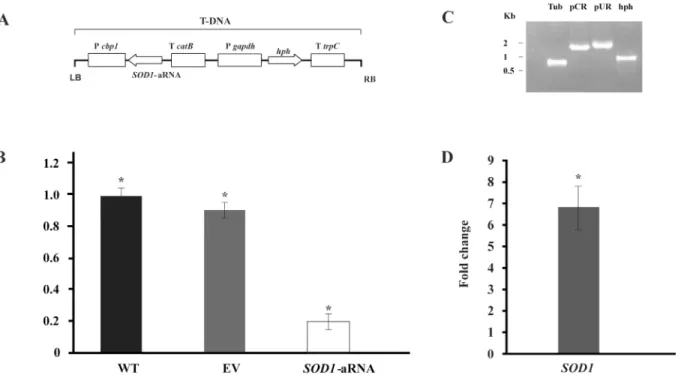
Taking into account the above results, we inquired about the importance ofSOD1during TSC-C treatment. To validate our assumption, we created aP.brasiliensisisolate (SOD1 -aRNA) with down-regulatedSOD1gene expression (Fig6Aand6B). The PCR analysis was performed to confirm the integration of the a-RNA cassette in theP.brasiliensisgenome (Fig 6C).SOD1-aRNA was obtained to ATCC 60855 isolate sinceSOD1was also induced in this isolate (Fig 6D) and the protocol to obtainment of mutant toP.brasiliensishas not yet stan-dardized. The susceptibility of ATCC60855 andSOD1-aRNA to TSC-C was evaluated (Fig 7). WhenSOD1-aRNA isolate cells were exposed to TSC-C, we observed a reduced growth rate relative to WT and EV yeast cells. The growth was restored in the presence of antioxidant, ascorbic acid. This result corroborates our transcriptional data that indicate an up-regulation ofSOD1during TSC-C treatment and suggests that the up-regulation of this gene is important forParacoccidioidessurvival in the presence of the compound.
Fig 6. Gene silencing ofSOD1inP.brasiliensis. (A)Transfer DNA (T-DNA) inserted into the genome ofP.brasiliensisyeast cells via ATMT in order to silence theSOD1gene. The antisense oligonucleotide was directed to exon 3 (black box) that amplify a length of 85 bp. This AS oligonucleotide was placed under the control of the calcium binding protein (CBP-1) with a terminator (CAT-B); the plasmid contained hygromycin B phosphotransferase (HPH) under the control of glyceraldehyde 3-phosphate ofAspergillus nidulans(PGPDA) with a terminator (TTRCP).(B)PbSOD1gene expression levels obtained by RT-qPCR. The measurement was normalized with the housekeeping gene alpha-tubulin in WT, EV andSOD1-aRNA yeast cells growing in the exponential phase. Mitotic stability was confirmed by sub-culturingP.brasiliensis SOD1-aRNA yeast cells and testing for low expression levels in this isolate after successive sub-cultures. (C) Validation by PCR of the presence and integration of the Transfer DNA (T-DNA) into the genome ofP.brasiliensistransformant. The genomic DNA from theSOD1-aRNA isolate was tested by PCR using specific primers for the alpha-tubulin geneTUB(Tub, lane 3), for the
transformation constructs pCR35 (pCR, lane 4) and pUR5750 (pUR, lane 5) and for the hygromycin resistance gene (hph, lane 6). (D)SOD1expression profile inP.brasiliensisafter exposure to TSC-C. RNA was extracted after 8 h of exposure of yeast cells to TSC-C. Changes in gene expression levels were calculated by the relative standard curve method using the non-treated control samples to calibrate.
TSC-C-induced ROS leads to the collapse of the
P
.
lutzii
mitochondrial
membrane
Mitochondrial ROS production can lead to the oxidative damage of mitochondrial proteins, membranes and DNA, thereby impairing the ability of this organelle to synthesize ATP and carry out its wide range of metabolic functions; such functions include the tricarboxylic acid cycle, fatty acid oxidation, the urea cycle and amino acid metabolism, which are pivotal for the normal function of most cells [69]. Furthermore, ROS also would cause a transition in mito-chondrial permeability. This transition consists of the loss of the mitomito-chondrial membrane
Fig 7. Susceptibility ofP.brasiliensis SOD1-aRNA to TSC-C.1x106yeast cells ofP.brasiliensisWT60855, EV60855 andSOD1-aRNA were spotted on solid BHI supplemented with 39.5, 79 and 158μM TSC-C. Control cells were spotted on BHI without TSC-C or with 39.5, 79 and 158μM TSC-C and ascorbic acid. The plates were incubated for 7 days at 36°C before photo documentation.
potential, resulting from the formation of pores, and subsequent cell death [70]. Because TSC-C induces the production of ROS, we evaluated the mitochondrial membrane integrity by estimating the electric potential (ΔCm) with fluorescence in yeast grown in the presence of
TSC-C for 4, 8 and 12 h. Flow cytometry analysis revealed aΔCm decrease in the yeast cells
exposed to the compound relative to control cells (Fig 8), suggesting that the TSC-C-induced ROS lead to the collapse of the mitochondrial membrane.
TSC-C inhibits cell proliferation by changing the expression profile of
genes related to the cell cycle
Cell cycle and DNA processing were among the major classes of overexpressed genes inP. lut-ziicells exposed to TSC-C. From these, 8 were unique to yeast cells grown in the presence of TSC-C, and these included DNA mismatch repair protein and DNA repair proteinRAD2. Therefore, we explored the possibility that the induction of these genes was associated with DNA damage through a DNA fragmentation assay. In fact, we did not observe DNA fragmen-tation in the samples cultured in the presence of TSC-C for any of the times tested (S1 Fig).
Considering the identity of the genes differentially regulated by the presence of the com-pound, we evaluated theP.lutziicell cycle by analyzing DNA content by flow cytometry. The phase of the cell cycle was determined by the difference in DNA content between cells in the pre-replicative (G0 and G1) phases, the replicative (S) phase (DNA synthesis) and the post-replicative plus mitotic (G2+M) phases [71]. The results showed that the percentage of yeast cells in the G1 phase increased in a time-dependent manner after exposure to TSC-C; further-more, the number of cells in the S and G2 phases decreased (Fig 9). Altogether, these results indicate that TSC-C inhibits cell proliferation by changing the expression profile of genes related to the cell cycle.
Conclusion
TSC-C seems to induce the formation of ROS inParacoccidioides spp., leading to the collapse of the mitochondrial membrane, and also to inhibit cell proliferation by changing the expres-sion of genes related to the cell cycle. Relevant genes related to protein synthesis, copper homeostasis and cellular response induced by drugs or stress conditions were also observed in
Fig 8. Effect of TSC-C on the mitochondrial membrane potential ofP.lutzii.The mitochondrial membrane potential (ΔΨm) was determined by flow cytometry analysis of yeast cells treated with TSC-C forA)4 h,B)8 h andC)12 h and stained with rodhamine123. Histograms in black represent the controls, and red histograms represent cells treated with TSC-C.
Fig 9. Effect of TSC-C on theP.lutziicell cycle.The DNA content of yeast in each cell cycle phase was analyzed by flow cytometry in the absence of TSC-C forA)4 h,B)8 h andC)12 h or in the presence of TSC-C forD)4 h,E)8 h andF)12 h and subsequently stained with ethidium iodide as represented by histograms.
Paracoccidioidesyeast cells exposed to TSC-C. The high percentage of unclassified proteins found here indicates that further studies are needed in order to better understand how TSC-C affectsParacoccidioides spp.
Supporting Information
S1 Fig. DNA fragmentation assay.DNA fragmentation was carried out in P. lutzii yeast cells exposed to TSC-C at 79μM for 4, 8 and 12 h. The controls were performed with yeast cells incubated in the absence of TSC-C.
(TIF)
Author Contributions
Conceived and designed the experiments: LCS MP OHR JGMO CMAO CCS. Performed the experiments: LCS DPTO SVCC LBP NPC. Analyzed the data: LCS CMAO AMB JAPR CMAS MP. Contributed reagents/materials/analysis tools: CCS CMAS OHR MP. Wrote the paper: LCS DPTO CMAO AMB JAPR OHR JGMO MP.
References
1. Bagagli E, Theodoro RC, Bosco SM, McEwen JG.Paracoccidioides brasiliensis: phylogenetic and eco-logical aspects. Mycopathologia 2008; 165: 197–2007. PMID:18777629
2. Bocca AL, Amaral AC, Teixeira MM, Sato PK, Shikanai-Yasuda MA, Soares Felipe MS. Paracoccidioi-domycosis: eco-epidemiology, taxonomy and clinical and therapeutic issues. Future Microbiol. 2013; 8: 1177–1179. doi:10.2217/fmb.13.68PMID:24020744
3. Marques SA. Paracoccidioidomycosis: epidemiological, clinical, diagnostic and treatment up-dating. An Bras Dermatol. 2013; 88: 700–711. doi:10.1590/abd1806-4841.20132463PMID:24173174
4. Travassos LR, Taborda CP, Colombo AL. Treatment options for paracoccidioidomycosis and new strat-egies investigated. Expert Rev Anti Infect Ther. 2008; 6: 251–262. doi:10.1586/14787210.6.2.251
PMID:18380607
5. Borges SR, Silva GM, Chambela Mda C, Oliveira Rde V, Costa RL, Wanke B, et al. Itraconazole vs. tri-methoprim-sulfamethoxazole: A comparative cohort study of 200 patients with paracoccidioidomyco-sis. Med Mycol. 2014; 52: 303–310. doi:10.1093/mmy/myt012PMID:24577007
6. Bocca AL, Amaral AC, Teixeira MM, Sato PK, Shikanai-Yasuda MA, Soares Felipe MS. Paracoccidioi-domycosis: eco-epidemiology, taxonomy and clinical and therapeutic issues. Future Microbiol. 2013; 8: 1177–1191. doi:10.2217/fmb.13.68PMID:24020744
7. Yasuda MA. Pharmacological management of paracoccidioidomycosis. Expert Opin Pharmacother. 2005; 6: 385–397. PMID:15794730
8. De Oliveira KM, da Silva Neto BR, Parente JA, da Silva RA, Quintino GO, Voltan AR, et al. Intermolecu-lar interactions of the malate synthase ofParacoccidioides spp. BMC Microbiol. 2013; 13: 107. doi:10. 1186/1471-2180-13-107PMID:23672539
9. Santana LA, Vainstein MH, Tomazett PK, Santos-Silva LK, Góes AM, Schrank A, et al. Distinct chiti-nases are expressed during various growth phases of the human pathogenParacoccidioides brasilien-sis. Mem Inst Oswaldo Cruz. 2012; 107: 310–316. PMID:22510825
10. Cruz AH, Brock M, Zambuzzi-Carvalho PF, Santos-Silva LK, Troian RF, Góes AM et al. Phosphoryla-tion is the major mechanism regulating isocitrate lyase activity inParacoccidioides brasiliensisyeast cells. FEBS J. 2011; 278: 2318–2332. doi:10.1111/j.1742-4658.2011.08150.xPMID:21535474
11. Tomazett PK, Castro Nda S, Lenzi HL, de Almeida Soares CM, Pereira M. Response of Paracocci-dioides brasiliensisPb01 to stressor agents and cell wall osmoregulators. Fungal Biol. 2011; 115: 62–
69. doi:10.1016/j.funbio.2010.10.005PMID:21215956
12. Tomazett PK, Félix CR, Lenzi HL, de Paula Faria F, de Almeida Soares CM, Pereira M. 1,3-β-d-Glucan synthase ofParacoccidioides brasiliensis: recombinant protein, expression and cytolocalization in the yeast and mycelium phases. Fungal Biol. 2010; 114: 809–816. doi:10.1016/j.funbio.2010.07.007
PMID:20943190
14. Zambuzzi-Carvalho PF, Cruz AH, Santos-Silva LK, Goes AM, Soares CM, Pereira M. The malate synthase ofParacoccidioides brasiliensisPb01 is required in the glyoxylate cycle and in the allantoin degradation pathway. Med Mycol. 2009; 47: 734–744. doi:10.3109/13693780802609620PMID: 19888806
15. da Silva Neto BR, de Fátima da Silva J, Mendes-Giannini MJ, Lenzi HL, de Almeida Soares CM, Per-eira M.The malate synthase ofParacoccidioides brasiliensisis a linked surface protein that behaves as an anchorless adhesin. BMC Microbiol. 2009; 9: 272. doi:10.1186/1471-2180-9-272PMID:20034376
16. Bonfim SM, Cruz AH, Jesuino RS, Ulhoa CJ, Molinari-Madlum EE, Soares CM, et al. Chitinase from Paracoccidioides brasiliensis: molecular cloning, structural, phylogenetic, expression and activity anal-ysis. FEMS Immunol Med Microbiol. 2006; 46: 269–283. PMID:16487309
17. Zambuzzi-Carvalho PF, Tomazett PK, Santos SC, Ferri PH, Borges CL, Martins WS, et al. Transcrip-tional profile ofParacoccidioidesinduced by oenothein B, a potential antifungal agent from the Brazilian Cerrado plantEugenia uniflora. BMC Microbiol. 2013; 13: 227. doi:10.1186/1471-2180-13-227PMID:
24119145
18. Santos GD, Ferri PH, Santos SC, Bao SN, Soares CM, Pereira M. Oenothein B inhibits the expression of PbFKS1 transcript and induces morphological changes inParacoccidioides brasiliensis. Med Mycol. 2007; 45: 609–618. PMID:18033615
19. Prado RS, Alves RJ, Oliveira CMA, Kato L, Silva RA, Quintino GO, et al. Inhibition ofParacoccidioides lutzii Pb01 isocitrate lyase by the natural compound argentilactone and its semisynthetic derivatives. PLoS One. 2014; 21; 9(4):e94832 doi:10.1371/journal.pone.0094832PMID:24752170
20. Figueiredo IM, Santos LV, Costa WF, Carvalho JE, Silva CC, Sacoman JL, Kohn LK, Sarragiotto MH. Synthesis and Antiproliferative Activity of Novel Limonene Derivates with a Substituted Thiourea Moi-ety. Braz. J. Chem. 2006; 5: 954–960.
21. Piochon M, Legault J, Gauthier C, Pichette A. Synthesis and cytotoxicity evaluation of natural alpha-bisabolol beta-D-fucopyranoside and analogues. Phytochem. 2009; 70: 228–236. doi:10.1016/j. phytochem.2008.11.013PMID:19136127
22. Da Silva AP, Martini MV, de Oliveira CM, Cunha S, de Carvalho JE, Ruiz AL, da Silva CC. Antitumor activity of (-)-alpha-bisabolol- based thiosemicarbazones agains human tumor cell lines. Eur J Med Chem. 2010; 45: 2987–2993. doi:10.1016/j.ejmech.2010.03.026PMID:20413188
23. Haraguchi SK, Silva AA, Vidotti GJ, dos Santos PV, Garcia FP, Pedroso RB, Nakamura CV, de Oliveira CM, da Silva CC. Antitrypanasomal activity of novel benzaldehyde-thiosemicarbazone derivates from kaurenoic acid. Molecules. 2011; 16: 1166–1180. doi:10.3390/molecules16021166PMID:21270733
24. Yamaguchi UM, Silva APB, Nakamura TU, Filho BPD, Silva CC, Nakamura CV. Effects of a thiosem-carbazide camphene derivative onTrichophyton mentagrophytes. Molecules. 2009; 14: 1796–1807. doi:10.3390/molecules14051796PMID:19471200
25. Fava-Netto C. Estudos quantitativos sobre a fixação de complemento na blastomicose sul-americana, com antigeno polissacarídico. Arq Cir Clin Exp.1955; 18: 197–254. PMID:13363721
26. Restrepo A, Jiménez BE. Growth ofParacoccidioides brasiliensisyeast phase in a chemically defined culture medium. J Clin Microbiol. 1980; 12: 279–281. PMID:7229010
27. Clinical Laboratory Standards Institute. Reference method for broth dilution antifungal susceptibility testing of filamentous fungi: Approved Standards 2nd Edition M27-A2. 2002.
28. De Paula e Silva ACA, Oliveira HC, Silva JF, Sangalli-Leite F, Scorzoni L, Fusco-Almeida AM, et al. Microplate alamar Blue Assay forParacoccidioidesSusceptibility Testing. J Clin Microbiol. 2013; 51: 1250–1252. doi:10.1128/JCM.02914-12PMID:23345296
29. Ewing B, Hillier L, Wendl MC, Green P. Base-calling of automated sequencer traces using phred. I. Accuracy assessment. Genome Res. 1998; 8: 175–185. PMID:9521921
30. Huang X, Madan A. CAP3: a DNA sequence assembly program. Genome Res. 1999; 9: 868–877.
PMID:10508846
31. Audic S, Claverie JM. The significance of digital gene expression profiles. Genome Res. 1997; 7: 986–
995. PMID:9331369
32. Almeida AJ, Carmona JA, Cunha C, Carvalho A, Rappleye CA, Goldman WE, et al. Towards a molecu-lar genetic system for the pathogenic fungusParacoccidioides brasiliensis. Fungal Genet Biol. 2007; 44: 1387–1398. PMID:17512227
33. Rappleye CA, Engle JT, Goldman WE. RNA interference in Histoplasma capsulatum demonstrates a role for alpha-(1,3)-glucan in virulence. Mol microbiol. 2004; 53: 153–165. PMID:15225311
34. Den Dulk-Ras A, Hooykaas PJ. Electroporation of Agrobacterium tumefaciens. Methods Mol Biol. 1995; 55: 63–72. PMID:8528423
adhesion to type I collagen and fibronectin: identification of potential adhesins. Res Microbiol. 2012; 163: 182–191. doi:10.1016/j.resmic.2012.01.004PMID:22306611
36. Bookout AL, Cummins CL, Mangelsdorf DJ, Pesola JM, Kramer MF. High-throughput real-time quanti-tative reverse transcription PCR. Curr Protoc Mol Biol. 2006; 15: 8. doi:10.1002/0471142727. mb1508s73PMID:18265376
37. Bradford MM. A rapid and sensitive method for the quantitation of microgram quantities of protein utiliz-ing the principle of protein-dye bindutiliz-ing. Anal Biochem. 1976; 7: 248–254.
38. Freiberg C, Brotz-Oesterhelt H. Functional genomics in antibacterial drug discovery. Drug Discov Today. 2005; 10: 927–935. PMID:15993812
39. Goffeau A, de Hertogh B, Baret PV. ABC transportadores. In: Enciclopédia of Biol. Chemistry. 2004; 1: 1–5.
40. Franz R, Michel S, Morschhauser J. A fourth gene from theCandida albicansCDR family of ABC trans-porters. Gene. 1998; 220: 91–98. PMID:9767132
41. Prasad R, De WP, Goffeau A, Balzi E. Molecular cloning and characterization of a novel gene of Can-dida albicans, CDR1, conferring multiple resistance to drugs and antifungals. Curr Genet. 1995; 27: 320–329. PMID:7614555
42. Slaven JW, Anderson MJ, Sanglard D, Dixon GK, Bille J, Roberts IS, et al. Increased expression of a novelAspergillus fumigatusABC transporter gene, atrF, in the presence of itraconazole in an itracona-zole resistant clinical isolate. Fungal Genet Biol. 2002; 36: 199–206. PMID:12135575
43. Tobin MB, Peery RB, Skatrud PL. Genes encoding multiple drug resistance-like proteins inAspergillus fumigatusandAspergillus flavus. Gene. 1997; 200: 11–23. PMID:9373135
44. Posteraro B, Sanguinetti M, Sanglard D, La SM, Boccia S, Romano L, et al. Identification and character-ization of aCryptococcus neoformansATP binding cassette (ABC) transporter-encoding gene, CnAFR1, involved in the resistance to fluconazole. Mol Microbiol. 2003; 47: 357–371. PMID:
12519188
45. Andrade AC, Van Nistelrooy JG, Peery RB, Skatrud PL, de Waard MA. The role of ABC transporters fromAspergillus nidulansin protection against cytotoxic agents and in antibiotic production. Mol Gen Genet. 2000; 263: 966–977. PMID:10954082
46. Frazier AD, Champney WS. Inhibition of ribosomal subunit synthesis inEscherichia coliby the vanadyl ribonucleoside complex. Curr Microbiol. 2013; 67: 226–223. doi:10.1007/s00284-013-0350-5PMID:
23512123
47. Hitchcock AL, Krebber H, Frietze S, Lin A, Latterich M, Silver PA.The conserved npl4 protein complex mediates proteasome-dependent membrane-bound transcription factor activation. Mol Biol Cell. 2001; 12: 3226–3241. PMID:11598205
48. Kim BE, Nevitt T, Thiele DJ. Mechanisms for copper acquisition, distribution and regulation. Nat Chem Biol. 2008; 4: 176–185. doi:10.1038/nchembio.72PMID:18277979
49. Samanovic MI, Ding C, Thiele DJ, Darwin KH. Copper in microbial pathogenesis: meddling with the metal. Cell Host Microbe. 2012; 11: 106–115. doi:10.1016/j.chom.2012.01.009PMID:22341460
50. Waterman SR, Hacham M, Hu G, Zhu X, Park YD, Shin S, et al. Role of a CUF1/CTR4 copper regula-tory axis in the virulence ofCryptococcus neoformans. J Clin Invest. 2007; 117: 794–802. PMID: 17290306
51. Park HJ, Nguyen TT, Yoon J, Lee C. Role of reactive oxygen species inEscherichia coliinactivation by cupricion. Environ Sci Technol. 2012; 46: 11299–11304. doi:10.1021/es302379qPMID:22998466
52. Dancis A, Haile D, Yuan DS, Klausner RD. The Saccharomyces cerevisiae copper transport protein (Ctr1p). Biochemical characterization, regulation bycopper, and physiologic role in copper uptake. J Biol Chem. 1994; 269: 25660–25667. PMID:7929270
53. Pena MM, Puig S, Thiele DJ. Characterization of theSaccharomyces cerevisiaehigh affinity copper transporter Ctr3. J Biol Chem. 2000; 275: 33244–33251. PMID:10924521
54. Dimroth P, Kaim G, Matthey U. Crucial role of the membrane potential for ATP synthesis by F(1)F(o) ATP synthases. J Exp Biol J. 2000; 203: 51–59. PMID:10600673
55. Nicholls DG. Mitochondria and calcium signaling. Cell Calcium. 2005; 38: 311–317. PMID:16087232
56. Martin J, Mahlke K, Pfanner N. Role of an energized inner membrane in mitochondrial protein import. Delta psi drives the movement of presequences. J Biol Chem.1991; 266: 18051–18057. PMID:
1833391
58. Ghannoum MA, Rice LB. Antifungal agents: mode of action, mechanisms of resistance, and correlation of these mechanisms with bacterial resistance. Clin Microbiol Rev. 1999; 12: 501–517. PMID: 10515900
59. Dias N, Bailly C. Drugs targeting mitochondrial functions to control tumor cell growth. Biochem Pharma-col. 2005; 70: 1–12. PMID:15907809
60. Grivicich I, Regner A, da Rocha AB, Grass LB, Alves PA, Kayser GB, et al. Irinotecan/5-fluorouracil combination induces alterations in mitochondrial membrane potential and caspases on colon cancer cell lines. Oncol Res. 2005; 5: 385–392.
61. Moreno-Sanchez R, Bravo C, Vasquez C, Ayala G, Silveira LH, Martínez-Lavín M. Inhibition and uncoupling of oxidative phosphorylation by nonsteroidal anti-inflammatory drugs: study in mitochondria, submitochondrial particles, cells, and whole heart. Biochem Pharmacol. 1999; 57: 743–752. PMID:
10075080
62. Ruy F, Vercesi AE, Kowaltowski AJ. Inhibition of specific electron transport pathways leads to oxidative stress and decreasedCandida albicansproliferation. J Bioenerg Biomembr. 2006; 38:129–135.63. PMID:17053999
63. Angelova MB, Pashova SB, Spasova BK, Vassilev SV, Slokoska LS. Oxidative stress response of fila-mentous fungi induced by hydrogen peroxide and paraquat. Mycol Res. 2005; 109: 150–158. PMID:
15839099
64. Youseff BH, Holbrook ED, Smolnycki KA, Rappleye CA. Extracellular superoxide dismutase protects Histoplasma yeast cells from host-derived oxidative stress. PLoS Pathog. 2012; 8: e1002713. doi:10. 1371/journal.ppat.1002713PMID:22615571
65. De Arruda Grossklaus D, Bailão AM, Vieira Rezende TC, Borges CL, de Oliveira MA, Parente JÁ, et al. Response to oxidative stress in Paracoccidioides yeast cells as determined by proteomic analysis. Microbes Infect. 2013; 15: 347–364. doi:10.1016/j.micinf.2012.12.002PMID:23421979
66. Hwang CS, Rhie GE, Oh JH, Huh WK, Yim HS, Kang SO. Copper- and zinc-containing superoxide dis-mutase (Cu/ZnSOD) is required for the protection ofCandida albicansagainst oxidative stresses and the expression of its full virulence. Microbiol. 2002; 148: 3705–3713. PMID:12427960
67. Delattin N, Cammue BP, Thevissen K. Reactive oxygen species-inducing antifungal agents and their activity against fungal biofilms. Future Med Chem. 2014; 6: 77–90. doi:10.4155/fmc.13.189PMID: 24358949
68. Vandenbosch D, Braeckmans K, Nelis HJ, Coenye T. Fungicidal activity of miconazole against Can-didaspp. Biofilms. J Antimicrob Chemother. 2010; 65: 694–700. doi:10.1093/jac/dkq019PMID:
20130024
69. Murphy MP. How mitochondria produce reactive oxygen species. Biochem J. 2009; 417: 1–13. doi:10. 1042/BJ20081386PMID:19061483
70. Vercesi AE, Kowaltowski AJ, Grijalba MT, Meinicke AR, Castilho RF. The role of reactive oxygen spe-cies in mitochondrial permeability transition. Biosci Rep. 1997; 17: 43–52. PMID:9171920