Neuroendocrine Cells from Mesoderm Is Regulated by
Notch Signaling
Sangbin Park1, Erika L. Bustamante1, Julie Antonova1, Graeme W. McLean1,2, Seung K. Kim1,2,3*
1Department of Developmental Biology, Stanford University School of Medicine, Stanford, California, United States of America,2Howard Hughes Medical Institute, Stanford, California, United States of America,3Department of Medicine (Oncology), Stanford University School of Medicine, Stanford, California, United States of America
Abstract
Drosophila neuroendocrine cells comprising the corpora cardiaca (CC) are essential for systemic glucose regulation and represent functional orthologues of vertebrate pancreatica-cells. AlthoughDrosophila CC cells have been regarded as developmental orthologues of pituitary gland, the genetic regulation of CC development is poorly understood. From a genetic screen, we identified multiple novel regulators of CC development, including Notch signaling factors. Our studies demonstrate that the disruption of Notch signaling can lead to the expansion of CC cells. Live imaging demonstrates localized emergence of extra precursor cells as the basis of CC expansion inNotchmutants. Contrary to a recent report, we unexpectedly found that CC cells originate from head mesoderm. We show that Tinman expression in head mesoderm is regulated by Notch signaling and that the combination of Daughterless and Tinman is sufficient for ectopic CC specification in mesoderm. Understanding the cellular, genetic, signaling, and transcriptional basis of CC cell specification and expansion should accelerate discovery of molecular mechanisms regulating ontogeny of organs that control metabolism.
Citation:Park S, Bustamante EL, Antonova J, McLean GW, Kim SK (2011) Specification ofDrosophilaCorpora Cardiaca Neuroendocrine Cells from Mesoderm Is Regulated by Notch Signaling. PLoS Genet 7(8): e1002241. doi:10.1371/journal.pgen.1002241
Editor:Norbert Perrimon, Harvard Medical School, Howard Hughes Medical Institute, United States of America
ReceivedApril 21, 2011;AcceptedJune 28, 2011;PublishedAugust 25, 2011
Copyright:ß2011 Park et al. This is an open-access article distributed under the terms of the Creative Commons Attribution License, which permits unrestricted use, distribution, and reproduction in any medium, provided the original author and source are credited.
Funding:The work was supported by the Howard Hughes Medical Institute. ELB was supported by NSRA pre-doctoral fellowship (5F31GM079930-03). The funders had no role in study design, data collection and analysis, decision to publish, or preparation of the manuscript.
Competing Interests:The authors have declared that no competing interests exist. * E-mail: seungkim@stanford.edu
Introduction
Recent work has revealed multiple features of evolutionary conservation in endocrine regulation of glucose metabolism. For example, in the fruit fly Drosophila melanogaster, insulin-producing cells (IPCs) in the brain and adipokinetic hormone-producing corpora cardiaca (CC) cells in the neuroendocrine ring gland are the respective functional orthologues of mammalian pancreaticb -cells and a-cells [1–4]. Insect CC cells resemble neurons in multiple ways; CC cells are peptidergic secretory cells [5] that harbor dense core vesicles [6], and have axon-like projections to vascular, gut and brain targets [3,4,7]. Similar to pancreatic islet cells and neuronal cell subsets, CC cells also use KATPchannels to regulate AKH secretion [3]. Targeted CC ablation results in marked hypoglycemia [3,4], demonstrating their role in glucose homeostasis. Thus, the molecular and physiological mechanisms governing CC endocrine function are strikingly similar to those of vertebrate pancreatic islets and neuroendocrine cells.
Despite their crucial role in regulating systemic glucose balance, the embryonic origin of CC cells remains unclear. Based, in part, on their emergence near embryonic foregut, CC cells were initially proposed to originate from a placode in the foregut that produces the stomatogastric nervous system [8]. The CC cell anlage was later inferred to be the most anterior part of mesoderm, based on studies of gene expression in the embryonic head region [9,10]. Most recently, it was proposed that the CC cells originate from neuroectoderm-derived neuroblasts [11]. This latest study concluded that CC precursors originate from the same placode in which insulin
producing neurons are born, and suggested that the developmental relationship between IPC and CC cells may be similar to that of hypothalamus and neuronal pituitary gland. Likewise, while a survey of candidate mutations revealed several genes required for CC development based on ontogenic similarities to pituitary development [9], a systematic, unbiased mutant screen to identify genetic regulators of CC development has not been previously reported.
Here we used genetic screens and gain-of-function studies to investigate specification of CC cell lineage. From a genetic deficiency screen, we discovered that Notch signaling factors are essential regulators of CC development. Our studies demonstrate that Notch signaling controls the number of emerging CC precursor cells. We unexpectedly found that CC cells develop from head mesoderm. Expression oftinmanin head mesoderm is regulated by Notch signaling and the combination oftinmanand
daughterless is sufficient to specify programs leading to ectopic development of CC cell precursors and their AKH+ progeny.
Thus our studies reveal genetic and cellular mechanisms underlying precursor specification and expansion of neuroendo-crine cells crucial for metabolic homeostasis inDrosophila.
Results
A deficiency screen identifies novel regulators of corpora cardiaca development
corresponding to approximately 50% of the genome. We generated strains harboring the akh-RedHStinger (akh-RHS) reporter gene which marks the nuclei of CC cells at embryonic stage 17 (see Materials and Methods). We observed thatakh-RHS+
cells were undetectable in 39 deficiency lines, and successfully identified mutations in 18 lines that mapped to 14 genes using publicly available mutant alleles (Table S1). In agreement with the previous study [9], we found that mutations in giant (gt), short
gastrulation (sog),sine oculis (so), andglass (gl) prevented embryonic development of akh-RHS+
cells. These findings validated our strategy to screen the DrosDel deficiency collection. In addition, we discovered that mutations incrooked neck (crn),spitz (spi),dimmed (dimm),phyllopod (phyl),double parked (dup),three rows (thr),Polycomblike (Pcl), ETS-domain lacking (edl), andheartless (htl) also result in the complete loss of AKH-expressing cells (Table S1). Thus, our deficiency line screen has revealed new regulators required for CC development.
Corpora cardiaca cell expansion from Notch signaling disruption
In contrast to loss of akh-RHS+
cells in 39 deficiency lines, analysis revealed expansion of akh-RHS+
cells in the
Df(3R)ED5942line. The deficiency in this line included theDelta
gene, which encodes an essential conserved activator of Notch signaling. We subsequently confirmed thatDeltamutations resulted in the CC cell expansion phenotype observed inDf(3R)ED5942. We detected an average of 14.060.8akh-RHS+
cells in stage 17 control embryos (n= 16; Figure 1A and 1C), while inDeltamutants we detected an average of 110.2623.7 akh-RHS+
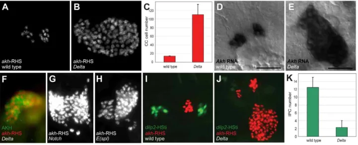
cells (n= 16; Figure 1B and 1C). In situ hybridization and immunostaining revealed expansion of cells expressingakhmRNA (Figure 1E) and AKH protein (Figure 1F) in Delta mutants, demonstrating expanded CC cells in these mutants. Thus,Deltais required for regulating CC cell number.
To identify additional conserved Notch signaling factors required for CC development, we examined akh-RHS reporter expression in Notch, Enhancer of split (E(spl)), Serrate (Ser), and
Suppressor of Hairless(Su(H)) mutant embryos.Notch(Figure 1G) and
E(spl) (Figure 1H) mutant embryos had CC cell expansion indistinguishable from that inDeltamutants, whileSer andSu(H)
mutants had no detectable change in CC cell number (data not shown). Together, these findings suggest that Notch signaling restrains development ofDrosophilaCC cells.
Author Summary
The requirement for glucose regulation is conserved in metazoans and crucial for metabolism, growth, and survival. In fruit flies and other insects, neurons secrete insulin-like hormones and neuroendocrine corpora cardi-aca cells secrete adipokinetic hormone, a peptide with functional similarities to glucagon. Both hormones are essential for systemic glucose control in Drosophila. To understand the mechanisms governing formation and function of corpora cardiaca cells, we sought to identify their embryonic origin and investigate their developmen-tal genetic regulation. Based on prior reports suggesting a neuroectodermal origin, we were surprised to discover— using genetic lineage tracing methods—that embryonic corpora cardiac progenitors derive from anterior head mesoderm. To our knowledge, this is the first demonstra-tion of neuroendocrine differentiademonstra-tion from mesoderm in Drosophila. Genetic studies reveal that Notch signaling restricts the number of corpora cardiaca progenitors, and we show that Notch signaling inactivation results in significant expansion of corpora cardiac cells. Loss- and gain-of-function studies identified transcription factors both necessary and sufficient for corpora cardiaca development. These and other findings reveal similarities in the development of fly corpora cardiaca cells and mammalian neuroendocrine cells that develop in the pancreas, pituitary, and from neural crest.
Figure 1. Disruption of Notch signaling results in the expansion of CC cells.(A) Late stage 17 wild type embryo showing 14 CC cells marked byakh-RHS. (B) Late stage 17Deltaembryo with 93 CC cells marked byakh-RHS. (C) Quantification of CC cells in wild type andDeltamutants. Average CC cells in wild type embryos is 14.060.8 (n= 16) whileDeltamutants show 110.2623.7 (n= 16). (D)AkhmRNAin situhybridization in stage 17 wild type embryo. Scale bar is equal to 10mm. (E)AkhmRNAin situin stage 17Deltamutant embryo shows expanded CC cells. (F) Stage 17Deltamutant
showing expanded CC cells marked by AKH antibody staining (green) andakh-RHS reporter (red). (G–H) BothNotch(G) andE(spl)(H) mutants at stage 17 show CC cell expansion. (I) Stage 17 wild type embryo show 12 IPCs marked bydilp2-HSti reporter (green). (J) Stage 17Deltamutant embryo exhibits reduced IPCs to 4 cells (pale green). (K) Quantification of IPCs in wild type andDeltamutants. Average IPCs in wild type embryos is 12.562.5 (n= 16) whileDeltamutants show 2.361.7 (n= 16). Where indicated, data represent the mean6standard deviation. See also Figure S1.
A prior study suggested that precursors of CC cells and
Drosophila insulin producing cells (IPCs) are adjacent in anterior neuroectoderm [11]. To assess the effect of mutations disrupting Notch signaling on IPC development, we generated a dilp2 -HStinger reporter (dilp2-HSti; see Materials and Methods) to mark IPC nuclei and facilitate IPC counting in stage 17 embryos. We detected an average of 12.562.5 IPCs (n= 16; Figure 1I and 1K) in control embryos, which was significantly different from the average of 2.361.7 IPCs inDeltamutants (n= 16; Figure 1J and 1K). Thus,Deltamutants have CC cell expansion accompanied by IPC hypoplasia, and these distinct outcomes suggest that Notch signaling has distinct roles in regulating developmental programs of CC cells and IPCs.
Deltais required before embryonic stage 11 to restrain corpora cardiaca development
To determine whenDeltafunction is required to restrict CC cell number, we inactivatedDeltafunction at specific embryonic stages using the temperature sensitiveDeltaRFallele. During continuous development at 18uC, CC cell number was normal in DeltaRF
mutants (13.363.1, n= 5; Figure 2B and 2H). However, during development at 29uC, CC cell number quadrupled in DeltaRF
embryos (67.2627.2, n= 13; Figure 2B and 2C), indicating that
Deltafunction was efficiently inactivated at 29uC. Based on these findings, we next used temperature shift from 18uC to 29uC at specific developmental stages inDeltaRF
embryos (summarized in Figure 2A).Deltainactivation at 7 or 8 hours after egg lay (hAEL) resulted in CC cell expansion (79.3620.9,n= 11 and 74.3619,
n= 30, respectively; Figure 2D). By contrast, lesser CC cell expansion resulted from temperature shift to 29uC at 9 hAEL (59.2617.6, n= 18; Figure 2E) or 10 hAEL (21.3611.1, n= 7; Figure 2F). Shift from 18uC to 29uC at 11 hAEL (corresponding to embryonic stage 11) or thereafter produced CC cell numbers indistinguishable from those observed during continuous develop-ment at 18uC (Figure 2B and 2G). These results suggest thatDelta
function is essential for restricting CC cell number before stage 11. To better define better the period when Delta restricts CC cell number, we also performed temperature ‘down-shift’ studies at specific stages duringDeltaRFembryonic development. When the temperature was shifted down from 29uC to 18uC at stage 10, CC cells were not expanded, although their position appeared to be more anterior (Figure S1E). However, temperature shift to 18uC at early stage 11 or thereafter led to CC cell expansion (Figure S1F– S1H). Together, our up- and down-shift experiments suggest that
Deltais required in a brief period from the end of embryonic stage 10 to the beginning of stage 11 to regulate CC cell number, but may be dispensable before or after.
The emergence of multiple Glass+CC precursors inNotch
mutants
The earliest known CC cell lineage markerglassis detected at embryonic stage 11 [9]; thus, we postulated that the requirement forDeltaprior to this stage indicated that Notch signaling specifies the number of Glass+
CC precursors. Glass protein is first detected in AKHnegCC cell precursors, a pair of single cells emerging near the dorsal head midline at early stage 11 (red arrowheads in Figure 3B) [11]. Between stages 11 and 13, the number of Glass+
CC precursors increases to 14–16 cells (comprised of two clusters of 7–8 cells; Figure 5A) that migrate posteriorly to become AKH+
CC cells [11]. To investigate the basis of CC cell expansion in
Delta,Notch, andE(spl)mutants, we first examined the emergence of Glass+
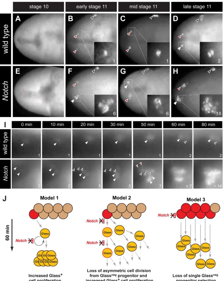
CC precursors near the head midline. At stage 10, in both wild type and Notch mutant embryos, no Glass+
CC
precursors were detectable (Figure 3A and 3E). Thus, the CC cell lineage did not develop precociously inNotchmutants. In early stage 11 (see Materials and Methods), the first pair of midline Glass+
CC precursors emerged in wild type embryos (red arrowheads in Figure 3B). In contrast, up to 6 Glass+
CC precursors were detectable inNotchmutants at early stage 11 (white arrowhead and insert in Figure 3F). We observed variant increases in left and right groups of Glass+
CC precursors at this stage (2 cells indicated by red arrowhead in Figure 3F). In mid stage 11, a pair of Glass+CC precursors remained as single cells in wild type
embryo (red arrowheads in Figure 3C) while clusters of 4–6 Glass+
CC precursors were detectable inNotch mutants (white and red arrowheads in Figure 3G). By late stage 11 in wild type embryo, CC precursors commenced division to increase the number of Glass+
cells from 1 to 2 (white arrowhead and insert in Figure 3D). Likewise, the number of Glass+
CC precursors increased from 6–7 to an average of 14 cells in late stage 11Notchmutant embryos (18 cells indicated by white arrowhead and insert in Figure 3H). These findings suggest that the increase of AKH-expressing CC cells found inDelta,NotchandE(spl)mutants reflects emergence of extra Glass+CC precursors at early stage 11.
Our analysis of static images did not preclude that a single Glassneg progenitor or Glass+ CC precursor might continuously
proliferate in early stage 11 to produce an expanded number of Glass+
CC precursors (Figure 3J, models 1 and 2). To evaluate this possibility, we continuously imaged live embryos expressing a
glass5.2-RedHStinger reporter (glass5.2-RHS) with fluorescence microscopy (Video S1). Nuclear localized fluorescent protein produced from this reporter permitted detection and counting of emerging glass-expressing CC precursors in early stage 11 wild type embryos (Figure 3I ‘wild type’, white arrowhead at t = 0 minutes). The signal intensity of the glass5.2-RHS reporter continuously increased until late stage 11 when the CC precursor divided to produce two adjacent progeny cells with equivalent reporter emission intensity (Figure 3I, t = 60 and 80 minutes). In
Notchmutants, we observed a different sequence of cell appearance and reporter labeling (Video S2). Twoglass5.2-RHS+
cells initially emerged (Figure 3I ‘Notch’, white arrowheads at t = 0 minutes). 20 minutes later, three additional glass5.2-RHS+
cells appeared (Figure 3I, black arrowheads at t = 20 minutes in panels labeled ‘Notch’). The threeglass5.2-RHS+
cells appearing at this later time are not adjacent to the first twoglass5.2-RHS+
cells. The emission intensity of these ‘new’ cells is fainter than that of the initial two cells. Thus, it is unlikely these new cells which appeared within 10 minutes represent progeny of the first twoglass5.2-RHS+
cells. At 50 minutes, two additional glass5.2-RHS+ cells appeared
(Figure 3I ‘Notch’, red arrowheads at t = 50 minutes), resulting in seven CC precursors. As in wild type embryos, CC precursor division begins at 60 minutes, and by 80 minutes the number of
glass5.2-RHS+cells in theNotchmutant was doubled. The number
and density ofglass5.2-RHS+
cells in theNotchmutant precluded further imaging and analysis. Thus, we did not detect accelerated proliferation by the first CC precursors appearing in Notch
mutants. Rather, these data suggest that emergence of excess Glass+
CC precursors from Glassnegprogenitors is the basis for CC cell expansion following disruption of Notch signaling (Figure 3J, model 3).
Corpora cardiaca precursors originate from head mesoderm
A recent study suggested CC cells develop from neuroectoderm [11] (site marked ‘2’ in Figure 5G), based on immunohistochem-ical detection of Glass in a subset of ectodermal cells labeled by a
Figure 2.Deltaregulates CC cell number before embryonic stage 11.(A) Temperature shift conditions applied toDeltaRFmutants at different
time points following 1-hour egg lay. hAEL is hours after egg lay. (B) CC cell number quantification inDeltaRFmutants resulting from the different temperature shift conditions shown in (A). (C)DeltaRFmutant grown at 29
uC exhibitsakh-RHS+
CC cell expansion. (D)DeltaRFmutant grown at 18
uC for 8 hours followed by a shift to 29uC until stage 17 shows a similar CC cell expansion. (E)DeltaRFmutant shifted from 18 to 29uC at 9 hAEL showing moderate CC cell expansion. (F)DeltaRFmutant with a temperature shift at 10 hAEL shows a slight increase in CC cell number. (G)DeltaRFmutant with
a temperature shift at 11 hAEL exhibits normal CC cell number. (H)DeltaRFmutant grown at 18uC shows normal CC cell number. Error bars are6the standard deviation of the mean.
Figure 3. The emergence of multiple Glass+CC precursors inNotchmutants.(A–D) CC precursor development in wild type embryo during
embryonic stages 10 and 11. Early, mid, and late embryonic stages 11 are determined by Bolwig’s organ precursor number (black arrowheads, Material and Methods). (A) Glass expression is not detected in stage 10 wild type embryo. Glass+CC precursors increase from single cell in early-mid
stage 11 embryo (red arrowheads and inserts in B and C) to 2 cells in late stage 11 embryo (white arrowhead and insert in D). (E–H) Glass+
this developmental origin, and controlling Notch signaling in progenitors of the CC cell lineage, we generated a gt1-GAL4 transgenic line (with an enhancer identical to the reportedgt1-lacZ construct; see Materials and Methods). Anterior head expression of b-galactosidase (b-gal) in our gt1-GAL4; UAS-lacZ.NLS embryos (Figure S2D) was identical to the expression ofgt1-lacZ expression reported previously (Figure S2A) [11]. However, the cytoplasmicb-gal signal fromgt1-lacZ appeared diffuse, and was difficult to discern at single cell resolution (Figure S2A). Nuclearb -gal expression ingt1-GAL4; UAS-lacZ.NLS marked several cells near the Glass+CC precursors (Figure 4A), but to our surprise we
did not detect nuclearb-gal in Glass+CC cell precursors in stage
11 embryos (Figure 4D). gt1-GAL4 cell lineage marking using FLP-recombinase (see Materials and Methods) traced gt1-GAL4 expression to third instar larval IPCs marked by the dilp2-HSti reporter (arrowheads in Figure 4H). This result is consistent with the reported origin of IPCs fromgt1-lacZ expressing cells [11], and validates use ofgt1-GAL4 for lineage tracing. However,gt1-GAL4 cell lineage marking did not trace to larval CC cells expressing the
akh-RHS reporter (red in Figure 4H), showing that CC cells do not originate fromgt1-expressing head neuroectoderm.
Based on expression and mutant phenotype analysis of genes that expressed in embryonic head, De Velasco et al [9,10] suggested that CC cells originate from cells adjacent to the anterior ventral furrow (site marked ‘1’ in Figure 5G). To test if CC cells derive from twist-expressing mesoderm cells at this anterior junction between embryonic endoderm and mesoderm, we used the twi.26PE-GAL4 line to label progeny of 12–14 ventral most mesodermal cells, as previously described [14]. Nuclei of the mesodermal cells and their progeny were labeled withb-gal through stage 11 in twi.26PE-GAL4; UAS-lacZ.NLS embryos (Figure 4J). A subset of theseb-gal+
mesodermal progeny co-expressed Glass (Figure 4I). Thus, ventral twist-expressing mesodermal cells invaginate and migrate toward the dorsal midline where Glass+CC precursors are specified (blue domain
in Figure 5G). In third instar larvae, lineage tracing oftwi.26
PE-GAL4+cells using FLP-recombinase revealed nuclear localization
of b-gal in the majority of akh-RHS+ CC cells (arrowheads in
Figure 4N). By contrast, IPCs were alwaysb-galneg(green nuclei in Figure 4N). We also usedMef2-GAL4 line to trace embryonic and larval lineages derived from all muscle lineages beginning at stage 7 embryos [15] (purple domain in Figure 5G). Similar to our findings withtwi.26PE-GAL4, we observed labeling of Glass+
CC precursors with Mef2-GAL4; UAS-lacZ.NLS at stage 11 (Figure S3A–S3C), and labeling of mature larval AKH+
CC cells with
Mef2 lineage tracing (arrowheads in Figure S3D). These results demonstrate that Glass+
CC precursors originate from head
mesoderm, and that IPC and CC cells derive from distinct germ layers inDrosophila.
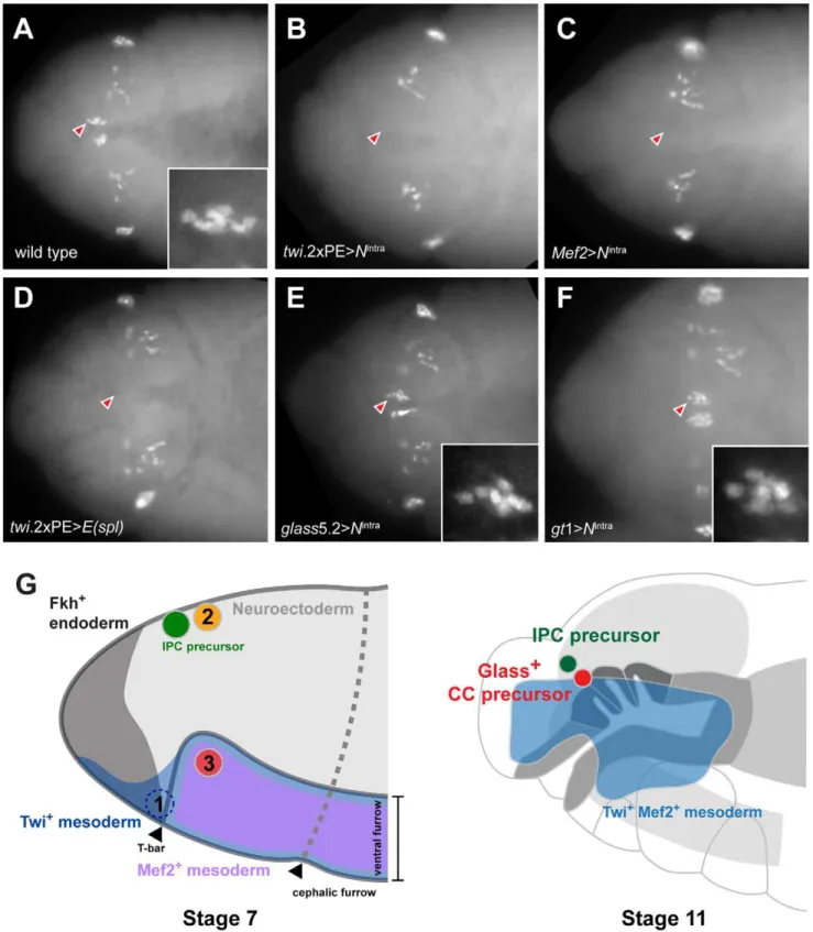
To test this conclusion further, we asked if impaired CC development resulted from Notch signaling disruption in head mesoderm that expressedtwistorMef2. Based on our disruption of Notch signaling using loss-of-function or conditional mutations, we postulated that head mesodermal expression of theNintra allele, which encodes a constitutively activate form ofNotch[16], orE(spl)
prior to stage 11 should reduce or eliminate development of embryonic stage 12 Glass+
CC precursors. By contrast, Notch signaling activation after formation of Glass+
CC precursors should not impair subsequent CC development. In twi.26 PE-GAL4; UAS-Nintra embryos and in Mef2-GAL4; UAS- Nintra
embryos, we failed to detect Glass+CC precursors (Figure 5B and
5C), confirming that CC cells originate from mesoderm that expresses twist and Mef2. Likewise, we observed elimination of Glass+
CC precursors in stage 12 twi.26PE-GAL4, UAS-E(spl)
embryos (Figure 5D). In contrast to these results, the number of Glass+ CC precursors at stage 12 was not detectably altered in
glass5.2-GAL4; UAS-Nintraembryos compared to control embryos (Figure 5E). Thus, consistent with our studies of the conditional
DeltaRFmutants, these results indicate that Notch signaling may be dispensable afterglass-expressing CC precursors are established at the early stage 11. To test if activation of Notch signaling in neuroectoderm affects CC development in the adjacent meso-derm,Nintrawas expressed in head neuroectoderm bygt1-GAL4. The number of Glass+
CC precursors at stage 12 was not altered in gt1-GAL4; UAS-Nintra embryos (Figure 5F), suggesting that Glass+
CC precursors develop independently of Notch signaling in neuroectoderm. Taken together, these results argue that CC cells originate from head mesoderm.
daughterlessandtinmanare required for CC cell development
During trunk mesoderm development, bHLH transcription factors, encoded bydaughterless(da) andtwist(twi), are necessary for the allocation of mesodermal cells to specific fates [17–19]. Prior study showed Twist is required for CC development [9], but it was not known ifdaughterlessor specific Twist targets were required for CC development. Thus, we assessed requirements for Daughter-less and Twist targets in CC cell development from head mesoderm. In late stage 12 wild type embryos, two groups of 6– 7 Glass+
CC precursors are detectable near the dorsal midline (red arrowhead in Figure 6A). In stage 12 mutants lackingdaughterlessor
twist, these Glass+
precursors are absent (red arrowheads in Figure 6B, 6C). Twist regulates expression of several transcription factors required for trunk mesoderm differentiation, includingZn
precursors are expanded inNotchmutant embryos during embryonic stage 11. (E) Glass expression is not detected in stage 10Notchmutant embryo. (F)Notchmutant at early stage 11 showing 2 (red arrowhead) or 6 (white arrowhead and insert) Glass+CC precursors. (G)Notchmutant at mid stage
11 showing 4 (red arrowhead) or 6 (white arrowhead and insert) Glass+
CC precursors. (H)Notchmutant at late stage 11 showing 18 (white arrowhead and insert) Glass+
CC precursors. All embryos are dorsal views with anterior to the left. (I) Glass+
CC precursors in live wild type andNotchmutant embryos are identified byglass5.2-RHS expression. The time at which the first precursor is detected is set to 0 minutes. The number of cells counted is labeled in the lower right corner of each image. In wild type, the first CC precursor (white arrowhead) divides at 60 minutes as shown by the emergence of the second precursor (black arrowhead). InNotchmutants, two CC precursors first emerge (white arrowheads) at 0 minutes. After 20 minutes, three precursors (back arrowheads) appear without apparent cell division. Two additional precursors (red arrowheads) arise at 50 minutes. The older precursors begin to divide at 60 minutes, indicating that in both the wild type andNotchmutants the rate of CC precursor cell division is 60 minutes/division (See also Videos S1 and S2). (J) Models for Glass+
CC precursor expansion inNotchmutants. Model 1 depicts the possibility that Notch signaling regulates proliferation of Glass+CC precursors. InNotchmutants, the speed of this proliferation is increased, resulting
in appearance of multiple Glass+
CC precursors. Model 2 depicts that Notch signaling normally regulates asymmetric cell division of a GlassnegCC progenitor (red), resulting in one Glassnegprogenitor and one Glass+
CC precursor followed by subsequent proliferation of the Glass+
daughter. In Notchmutants, both daughter cells become Glass+CC precursors followed by additional rapid symmetric divisions. Model 3 depicts the possibility
that Notch signaling restricts the development of GlassnegCC progenitors. InNotchmutants, increased numbers of GlassnegCC progenitors (red)
produce multiple Glass+cells that are CC precursors.
finger homeodomain 1 (zfh1),Myocyte enhancer factor 2 (Mef2),held out wings(how), andtinman(tin) [20–25]. To test if these transcription factors are also required for CC development, we assessed CC precursor development in mutant embryos. Normal numbers of CC precursors were detected in embryos harboring mutations in
zfh1, Mef2, and how (data not shown). By contrast, Glass+
CC precursors were not detected in stage 12 embryos with mutations intinman(red arrowheads, Figure 6D). Since proneural genes are required for stomatogastric nerve cell precursor formation [8] and specification of muscle progenitors [26], we also tested mutant embryos deficient for the genes achaete, scute, lethal of scute, and
asense, which encode proneural bHLH factors. However, the number of CC precursors was not altered in mutant embryos (Figure 6E), suggesting these proneural genes are not required for CC development. Thus, our mutant analysis revealed a specific requirement fortwist,daughterless, and tinmantranscription factors during CC precursor specification from head mesoderm.
Tinman expression in head mesoderm is regulated by Notch signaling
We first postulated that regulation oftwistexpression by Notch signaling in head mesoderm, like in trunk mesoderm [27], might underlie CC cell expansion in Notch mutant flies. In wild type embryos, Twist expression by immunostaining was restricted to head mesoderm, and Glass+
CC precursors co-expressed Twist (inserts and arrowheads in Figure 6F, 6G). In stage 11 Notch
mutants, we observed that two clusters of multiple Glass+
CC precursors co-localized with these Twist+
cells (one cluster enlarged in Figure 6H, 6I), indicating that the multiple Glass+
CC precursors inNotchmutants originate also from Twist+head
mesoderm. To test whether head mesodermal Twist expression may be regulated by Notch signaling, we asked if the ectopic activation of Notch signaling in head mesoderm in stage 11
twi.26PE-GAL4; UAS-Nintraembryos results in loss or reduction of Twist expression. A normal pattern of Twist expression in head mesoderm was observed in these embryos but Glass+ CC
precursors were absent (arrowhead in Figure 6J), providing additional evidence that the level of Twist expression in head mesoderm may not be regulated by Notch signaling.
Tinman expression is restricted to the anterior dorsal region of head mesoderm in stage 11 wild type embryos (Figure 6L for dorsal view and Figure S4C for lateral view), and Tinman+
head mesodermal cells at stage 11 include Glass+
CC precursors (arrowheads and enlarged in Figure 6K, 6L, and Figure S4A). In stage 12 embryos, Tinman expression in Glass+
CC precursors was extinguished, while adjacent Glassneg cells - which include the procephalic vascular rudiment [10] - maintained Tinman expression (Figure S4E–S4H). In Notch mutants, the number of Glass+
Tinman+
CC precursors in head mesoderm increased (outlined in Figure 6M and 6N). In addition, Glassneg Tinman+
cells adjacent to Glass+
CC precursors also appear expanded (brackets in Figure S4J and S4L), suggesting that Tinman expression in head mesoderm may be regulated by Notch signaling, To test this possibility, we asked if ectopic Notch signaling activation in head mesoderm resulted in loss of Tinman expression. Expression of both Glass and Tinman was abolished in the head mesoderm of twi.26PE-GAL4; UAS-Nintra
embryos (arrowhead in Figure 6O). Together, these results show that Tinman expression is regulated by Notch signaling in head mesoderm, and suggest the possibility thattinmanmis-expression in this context underlies CC lineage expansion in Notch signaling disruption.
Co-expression of Tinman and Daughterless in mesoderm is sufficient for CC cell lineage specification
Since expanded Tinman expression in Notch mutant head mesoderm accompanied CC lineage expansion, we investigated if ectopic expression oftinmanmight be sufficient to expand CC cells. However, in twi.26PE-GAL4 UAS-tinman embryos, the popula-tion of Glass+
CC precursors was not expanded (Figure 7A), suggesting that additional factors may be required to specify the CC cell lineage in head mesoderm. During trunk mesoderm differentiation, Twist activity is inhibited by its dimerization partner Daughterless to allocate mesodermal cells to various tissue fates [19]. Therefore, we next investigated effects of mis-expressing
daughterless or twist in mesoderm. In twi.26PE-GAL4
UAS-daughterless embryos, Glass+
CC precursors do not increase in head mesoderm (Figure 7B), although we reproducibly observed appearance of ectopic Glass+
cells in the trunk of these embryos (black arrowhead in Figure 7B). In contrast, Glass+CC precursors
were absent intwi.26PE-GAL4 UAS-twistembryos (Figure 7C),
suggesting that CC precursor specification is inhibited by excessive Twist activity. Ectopic CC cell development from mis-expression ofdaughterlessortinmanin head mesoderm was also eliminated by co-expression of twist (Figure 7D, 7E), supporting the view that excess Twist activity can suppress CC development.
Daughterless protein contains a repression domain, and can heterodimerize with Twist to regulate Twist activity [28]. Thus, we postulated that CC cell lineage specification may be regulated bytinmanin mesodermal cells with increased Daughterless activity. To test this possibility, we co-expressedtinmanwithdaughterlessin mesoderm. In twi.26PE-GAL4 UAS-tinman UAS-daughterless em-bryos, the number of Glass+
CC precursors in head mesoderm was markedly expanded (red arrowheads in Figure 7F). In addition to extra Glass+
cells in head mesoderm, we also detected ectopic Glass+cells in the trunk (black arrowheads in Figure 7F). To test if
these Glass+cells developed further toward a fate resembling CC
cells, we assessed akh-RHS marker expression at stage 17. Compared to normal akh-RHS+
cell numbers in twi.26 PE-GAL4 control embryos at stage 17 (Figure 7G), we detected
Figure 4. Copora cardiaca precursors originate from head mesoderm.(A–B) Locations of Glass+
CC precursor in stage 11 embryos in the dorsal view (A) and the lateral view (B). (C–E)gt1-GAL4; UAS-lacZ.NZ embryo expressesb-gal in anterior head neuroectoderm.gt1-GAL4 is expressed in the anterior head and several cells posterior to Glass+
CC precursors.gt1-GAL4 does not express in CC precursors as shown by the arrowheads. (F, G) Single confocal plane image ofgt1-GAL4; UAS-lacZ.NZ embryonic head region stained forb-gal. Glass+
CC precursors (F, arrowheads) are not co-localized withgt1-GAL4 expression. (H) Lineage tracing ofgt1-GAL4+cells in third instar larval IPC and CC cells. IPC and CC cells are marked by
dilp2-HSti (green) andakh-RHS (red) respectively.b-gal expression (blue) fromgt1-GAL4; UAS-FLP; Act5C(FRT.polyA)lacZ.nls1 is co-localized with a subset of IPCs (green), resulting in IPCs with cyan (arrowheads). CC cells (red) are not labeled bygt1-GAL4 lineage tracing, and therefore do not show any cells in magenta. (I–K) Glass+
CC precursors in stage 11 embryo (arrowheads) are a part of the dorsal mesoderm marked bytwi.26PE-GAL4; UAS-lacZ.NZ. (I)
Merged image shows that Glass+cells are located in outer part of the head mesoderm. (J)b-gal expression marks the dorsal head mesoderm. (L, M)
Single confocal plane image oftwi.26PE-GAL4 (M,b-gal) expression in a stage 11 embryo showing Glass+CC precursor (L, arrowhead) originates from
head mesoderm. (N) Lineage tracing oftwi.26PE-GAL4 expressing cells in third instar larval CC cells. Lineage was traced byb-gal expression (blue) in twi.26PE-GAL4; UAS-FLP; Act5C(FRT.polyA)lacZ.nls1 larvae.twi.26PE-GAL4 lineage-traced in several CC cells (magenta with arrowhead), but not in
Figure 5. Ectopic activation of Notch signaling prior to Glass expression in head mesoderm disrupts CC precursor development.(A) Wild type embryo shows two clusters of Glass+CC precursors, each one with 6 precursors (arrowhead and insert) in stage 12 embryo. Other Glass+
cells are larval eye precursors and brain primordia. (B) Activation of Notch signaling in mesoderm bytwi.26PE-GAL4; UAS-Nintraremoves the CC
precursors (arrowhead) in stage 12 embryo. (C) Activation of Notch signaling in muscle lineage byMef2-GAL4; UAS-Nintraremoves the CC precursors
(arrowhead) in stage 12 embryo. (D) Ectopic expression of bHLH repressorE(spl)in mesoderm bytwi.26PE-GAL4; UAS-E(spl)also removes Glass+CC
precursors (arrowhead) in stage 12 embryo. (E) Activation of Notch signaling after Glass+expression initiates in CC cell lineage does not perturb their
development.glass5.2-GAL4; UAS-Nintraembryo in stage 12 maintains normal number of Glass+
CC precursors (arrowhead and insert show 6 cells). (F) Activation of Notch signaling in neuroectoderm bygt1-GAL4; UAS-Nintradoes not disrupt the development of CC precursors (arrowhead and insert
show 6 cells) in stage 12 embryo. All embryo images are dorsal views with anterior to the left. (G) Relative locations of CC and IPC precursors at stage 7 and 11 embryos. Embryos are drawn in the lateral view with anterior to the left. (1)twi+gt+
increased numbers of akh-RHS+
cells in twi.26PE-GAL4
UAS-daughterless UAS-tinman embryos at this stage (red arrowheads in Figure 7H). Unexpectedly, we also detected ectopicakh-RHS+
cells in embryonic trunk of these embryos at stage 17 (black arrowheads in Figure 7H). Taken together, these results show that co-expression of Daughterless and Tinman is sufficient to activate CC cell developmental programs and to promote CC cell lineage expansion both in head mesoderm and ectopic sites. Collectively, the results strongly suggest that the CC cell lineage is specified by a combinatorial transcription code in embryonic mesoderm.
Discussion
Identification of novel genes required for CC cell development
Although DrosDel deficiency lines used in this study cover only
,50% of Drosophila genome, we successfully identified several
genes previously not implicated in CC cell development. Mutations incrooked neck (crn), spitz (spi), dimmed (dimm), phyllopod (phyl), double parked (dup), three rows (thr), Polycomblike (Pcl), ETS-domain lacking (edl), andheartless (htl) result in the complete loss of
Akhexpression. Expression ofdimmin CC cells has been previously reported [29], and dimm is required for the differentiation of central and peripheral neuroendocrine cells. Thus, dimmmay be required for CC cell maturation.spi,edl, andphylare components of the Epidermal Growth Factor signaling pathway andhtlencodes a Drosophila Fibroblast Growth Factor Receptor. Thus, these results suggest that MAPK signaling pathways regulate CC cell development. thr, dup, and crn are required for the cell cycle control, suggesting that the regulation of cell cycle control is also important for proper CC cell development.
Disruption of Notch signaling leads to the expansion of neuroendocrine precursor cells
Prior studies suggest that development of stomatogastric endocrine cells from endoderm, and IPCs from neuroectoderm is regulated by Notch and MAPK signaling [30–32]. Here, we found that Notch signaling disruption from mutations inNotch,Delta or E(spl)led to expansion of CC cells, reminiscent of the expansion of endocrine islet
a-cells during mammalian pancreas development of Dll1or Hes1
mutant mice [33,34]. Notch signaling is required to maintain undifferentiated mammalian pituitary progenitors (reviewed in [35]), and mutations disrupting Notch signaling also result in the expansion of specific pituitary cell types [36]. Thus, signaling pathways controlling CC cell development may reflect ancient conserved genetic programs for endocrine cell specification. Using time-lapsein vivo imaging, we detected the emergence of multiple Glass+
CC precursors in stage 11Notchmutants. The most rapid mitotic divisions in Drosophilaoccur prior to embryonic cellularization, and require approximately 10 minutes [37]. Thus, we calculate that the emergence of 7 Glass+
CC precursors within 20 minutes in Notch
mutant embryos is unlikely to result from accelerated division of a single Glass+CC precursor or loss of asymmetric cell division from
Glassneg CC progenitor. Rather, our data suggest that Notch signaling restricts head mesodermal fate specification possibly by a lateral inhibition mechanism (model 3 in Figure 3J). After the initial Glass+
CC precursors are formed, maturation process from a single Glass+
CC precursor to a cluster of 7–8 AKH+
CC cells appears to be
Notch signaling independent. Conditional mutant studies using a temperature sensitive allele of Delta, or using Notch signaling activation in Glass+CC precursor cells further support this possibility.
Mesodermal origin of neuroendocrine cells
Prior studies suggested that corpora cardiaca neuroendocrine cells inDrosophila may derive from the most anterior region of head mesoderm expressing twist and gt [9,10]. Recently, an alternate neuroectodermal origin for CC cells was proposed [11]. CC cells manifest neuron-like features, lending plausibility to the suggestion that CC cells derived from neuroectoderm expressing gt1-lacZ. However our study identified that the corpora cardiaca originates from head mesoderm expressingtwist,Mef2andtinman. The absence of CC precursors intwistandtinmanmutants also strongly support this view. Lineage tracing studies by cell marking withgt1-GAL4 here confirmed a neuroectodermal origin for insulin-producing neurons in the protocerebrum; however, we did not detect tracing of CC precursors or mature CC cells fromgt1-expressing cells. Thus, CC cells and IPCs have distinct embryonic origins and our data provide conclusive evidence from lineage tracing that neuroendo-crine CC cells derive from mesoderm. The origins of IPCs and CC cells from different germ layers is consistent with the observation that mutations preventing CC cell development do not detectably impair IPC formation [38]. Thus, cell interactions between IPCs and CC cells may not be essential for development of these two cell types. A prior study speculated that corpus allatum cells in the larval ring gland, which produce juvenile hormone, derive from gnathal mesoderm [9], but this origin has not been demonstrated with methods like lineage tracing. Thus, to our knowledge, CC cells may represent the sole example, thus far, of neuroendocrine cell development from mesoderm inDrosophila.
Vascular access and dispersion of hormones is a defining feature of endocrine organs. In mammals, signaling between vascular and endocrine progenitors is an important mechanism for regulating development of organs like the pancreas [39]. Tinman+
cells in
Drosophila head mesoderm also form the procephalic vascular rudiment [10], whose progeny establish the contractile dorsal vessel (Drosophilaheart), and prior studies have demonstrated that axon-like projections from larval CC cells terminate on the dorsal vessel [3]. In addition, similar to the posterior migration of head-mesodermal rudimentary vascular cells, Glass+
AKHneg CC progenitors migrate posteriorly during their maturation into AKH+
cells. De Velasco and colleagues have previously speculated that developing CC precursors might interact with other head mesoderm cells [9] during CC development. Our demonstration that CC cells originate from Tinman+ Glassneg
head mesoderm further supports this possibility. The proximity of embryonic CC cell progenitors to dorsal vessel progenitors may enhance cell-cell interactions that govern hallmark CC cell properties, including AKH expression and physical connections to their vascular targets. Together, these observations suggest that key morphoge-netic and developmental signaling relationships between endocrine and vascular precursors may be conserved from flies to mammals.
Encoding neuroendocrine lineage specification by transcription factor combinations
Many human diseases result from excessive or inadequate endocrine cell mass or function. Thus, there is intense interest in an origin of CC cells by De Velasco et al [9,10]. (2) Neighboring cells from the anterior neuroectoderm were proposed as origins for IPC and CC cells by Wang et al [11]. (3) The origin of CC cells identified by lineage tracing from Twist and Mef2 expressing head mesoderm in this study. By stage 11, Twi+
Mef2+mesoderm has generated Glass+CC precursors that are located near IPC precursors (green) and the endodermal foregut invagination (outlined
identifying evolutionarily-conserved transcriptional codes for neuroendocrine cell development and expansion. Our study identified a unique cell signaling context in mesoderm where neuroendocrine precursor cells can be specified by the two transcription factors Tinman and Daughterless. Allocation of trunk mesodermal fates is regulated by Twist and Daughterless activity [19,28], and here we showed that CC cell specification in head mesoderm is also regulated by a combination of transcription factors.tinman expression in a small subset of head mesoderm is regulated by Notch signaling, reminiscent oftinmanregulation in trunk cardiogenic mesoderm by Notch signaling [40]. However, only two cells within Tinman+
domain in head mesoderm develop into Glass+
CC progenitors. These observations suggest that other factors, in addition to Tinman, are required to specify the CC cell lineage. Consistent with this possibility, we show that Tinman mis-expression is not sufficient to expand CC development. By contrast, co-expression of Tinman and Daughterless led to increased development of head mesoderm into CC cells; thus, Tinman and Daughterless collaborate to specify the CC lineage. The combination of Tinman and Daughterless also induced ectopic AKH+
cells in the embryonic trunk, suggesting that trunk
mesodermal cells may also be competent to develop into CC cells. We speculate that over-expression of Daughterless in mesoderm suppresses Twist activity, and the mesodermal cells in this context are competent to become CC lineage upon Tinman expression, but further studies are required to test this possibility. Our study identified a transcription factor combination whose reconstitution is sufficient for differentiation by a subset of mesodermal cells toward a neuroendocrine fate. However, most embryonic mesodermal cells failed to express Glass or Akh upon mis-expression of Tinman and Daughterless, suggesting additional factors are likely required to re-specify mesoderm into CC cells. Moreover, additional studies are needed to determine how Daughterless, which is ubiquitously expressed, might regulate Twist activity in differentiating mesoderm to give rise to distinct cell fates.
In summary, work here reveals embryonic and molecular mechanisms regulating development of Drosophila CC cells. We demonstrated that Notch signaling restricts CC precursor cell fate in head mesoderm and regulates Tinman expression. We used cell lineage tracing and genetic analysis to demonstrate that CC cells originate from embryonic mesoderm. We also showed that a
Figure 6. Tinman expression in head mesoderm is regulated by Notch signaling.(A) Glass expression in stage 12 wild type embryonic head region. CC precursors are located in the dorsal midline (red arrowheads). Black arrowheads indicate Glass+ larval eye precursors while white
arrowheads indicate Glass+
brain primordia. (B) Indamutant at stage 12, both CC (red arrowheads) and eye precursors (black arrowheads) are missing, but Glass expression in the brain primordia (white arrowheads) is maintained. (C) Intwimutant at stage 12, CC precursors are missing in the dorsal midline (red arrowheads) while brain (white arrowheads) and eye precursors (black arrowheads) are intact. (D) Intinmutant at stage 12, CC precursors are missing in the dorsal midline (red arrowheads). (E) In stage12Df(1)BSC530mutant in which proneural genes are removed, CC precursors (red arrowheads) are intact. (F, G) Dorsal view of stage 11 wild type embryonic head shows Glass+
CC precursors (arrowhead and green in insert of F) co-localized with Twist+cells (red). (H, I) Dorsal view of stage 11Notchmutant shows multiple Glass+CC precursors (outlined and green in
insert) co-localized with Twist+
cells (red). (J) Twi expression in head mesoderm is maintained in stage 11 embryo when Notch signaling is activated in mesoderm bytwi.26PE-GAL4; UAS-Nintra. (K, L) Dorsal view of stage 11 wild type embryonic head shows Glass+CC precursors (arrowhead and green
in K) co-localized with Tinman+cells (red). (M, N) Dorsal view of stage 11Notchmutant shows multiple Glass+CC precursors (outlined and green in
insert) co-localized with Tinman+
cells (red). (O) Both Glass and Tinman expression in head mesoderm is abolished at stage 11 embryo when Notch signaling is activated in mesoderm bytwi.26PE-GAL4; UAS-Nintra. All embryo images are dorsal views with anterior to the left.
combination of the transcription factors Tinman and Daughterless is necessary and sufficient to specify CC cell lineage in mesoderm. Findings from this study should accelerate advances in our understanding of the conserved molecular mechanisms controlling differentiation and expansion of endocrine organs essential for metabolic regulation.
Materials and Methods
Drosophilastrains
y1 w1118 strain was used as the wild type stock. DrosDel deficiency lines were obtained from Bloomington Stock Center. The following mutant alleles and transgenic lines were used in this
Figure 7. Co-expression of Daughterless and Tinman in mesoderm is sufficient for ectopic CC cell lineage specification in mesoderm.(A) Glass+CC precursors are not expanded (red arrowheads) in stage 12twi.2
6PE-GAL4 UAS-tinmanembryo. (B) CC precursors (red
arrowheads) are developed normally in stage 12twi.26PE-GAL4 UAS-daughterlessembryos. Ectopic Glass expressing cells in trunk region is marked
by black arrowhead. (C–E) CC precursors are absent (read arrowheads) whentwist is over-expressed alone (C) or twist is co-expressed with daughterless(D) ortinman(E) in head mesoderm. (F) Glass+
CC precursors in head mesoderm are expanded in stage 12twi.26PE-GAL4 UAS-tinman
UAS-daughterlessembryo (red arrowheads). Ectopic Glass expressing cells are also detected in trunk region (black arrowheads). (G) CC cells are marked byakh-RHS expression in stage 17 control embryo. (H) Expansion of CC cells (red arrowheads) and ectopic CC cells in trunk region (black arrowheads) are detected in stage 17twi.26PE-GAL4 UAS-tinmanUAS-daughterlessembryo. (I) Magnified view of a box marked in (H) to show the
study:Dl9P,DlRF,N264-39,E(spl)rv1,SerRX82,Su(H)IB115,da10,twi1,
zfh100865, Mef2X1, howstru-3R-3, Df(1)BSC530, twi.26PE-GAL4, GAL4-Mef2.R and UAS-lacZ.NZ (Bloomington Stock Center).
gt1-lacZ was provided by Dr. Stephen Small (New York University) [13]. tin346, tinEC40 and UAS-tin were provided by Dr. Rolf Bodmer (Burnham Institute) [41]. UAS-daand UAs-twi
were provided by Dr. Mary K. Baylies (Sloan-Kettering Institute) [19]. UAS-Nintra was a gift from Dr. Margaret Fuller (Stanford University).Kr-GAL4 UAS-GFP ortwi-GAL4 UAS-GFP harbor-ing balancer chromosomes were used to identify hemi- or homozygous mutant embryos. For lineage tracing experiments, flies carrying UAS-FLP;dilp2-HSti,akh-RHS;Act5C (FRT.polyA)-lacZ.nls1 were crossed to GAL4 lines.
In situhybridization and immunohistochemistry
Antisense riboprobe for Akh was derived from pBS2KSP-Akh cDNA clone. RNA in situ hybridization was carried out as described [42]. Immunostaining of embryos was performed as described [42] with the following modifications; all embryos were manually devitellinized to avoid methanol exposure, late stage 17 embryos with cuticle were sonicated for 6 seconds under the lowest output setting in Branson Sonifier 450, primary antibodies were detected with Alexa488, 547, or 647 (Invitrogen) secondary antibodies, and embryos were mounted in 100% glycerol. Embryonic developmental stages were morphologically deter-mined according to Campos-Ortega and Hartenstein [43]. During our studies, we found that the development of Glass+
larval eye precursors in Bolwig’s organ lineage was unaffected in Notch
mutants, and we quantified Glass+
larval eye precursors to determine embryonic stage accurately within stage 11 embryos. In both wild type and mutant embryos, we detected 1–3 precursors at early stage 11 (black arrowheads in Figure 3B and 3F), 4–7 precursors at mid stage 11 (black arrowheads in Figure 3C and 3G) and 8–11 cells at late stage 11(black arrowheads in Figure 3D and 3H), respectively. The following primary antibodies were used: rabbit anti-AKH (1:300) [3], rabbit anti-Twist (1:500; Dr. Maria Leptin, Universita¨t Ko¨ln) [17], rabbit anti-Tinman (1:300; Dr. Manfred Frasch, Mount Sinai School of Medicine) [24], mouse 9B2.1c anti-Glass (1:10; Developmental Studies Hybridoma Bank under the auspices of NICHD and maintained by The University of Iowa, Department of Biology) [44] and chicken anti-b-gal (1:1000; Abcam). Immunostaining of CC cells in larval brains was performed as described [3]. Imaging of RNA
in situhybridizations was performed on a Zeiss Axio Imager DIC microscope. Immunofluorescence microscopy was performed on a Zeiss Axio Imager or a Zeiss LSM510 confocal microscope. Z-projections of confocal stacks were generated using ImageJ with sum slice option.
Generation of reporter and GAL4 driver lines
The enhancer sequences used in this study were amplified from
y1w1118genomic DNA. pAkhp1016 Red H-Stinger (akh-RHS) and pAkhp1016 Green H-Pelican (akh-GHP) were constructed by subcloning the 1016 bp sequence upstream of theAkhstart codon [3] into the pRed H-Stinger and pGreen H-Pelican vectors [45], respectively. pDilp215-1-H-Stinger (dilp2-HSti) was generated by subcloning the 541 bp sequence upstream of thedilp2 transcrip-tion start site [2] into the pStinger vector. pGlass5.2 Red H-Stinger (glass5.2-RHS) was constructed by subcloning the 5197 bp sequence upstream of theglassstart codon [46] into the pRed H-Stinger vector. pGt1-GAL4 (gt1-GAL4) was constructed by subcloning the 787 bp gt1 CRM fragment [13] into pPTGAL. pGlass5.2-GAL4 (glass5.2-GAL4) was constructed by subcloning this 5197 bp sequence into the pPTGAL vector. P-element
mediated germline transformations were carried out as described [47]. For all transgenic strains, at least two independently-derived transgenic lines with transgenes mapping to the second or third chromosome were evaluated.
Live embryo imaging
To capture fluorescent reporter signals in developing embryos, stage 7 or 8 embryos were mounted between two cover glasses spaced with 0.1% agarose blocks. Z-stack images (3561mm) were captured every 2 minutes for 3 hours in the Zeiss Axio Imager fluorescent microscope. Conversions of Z-stacks to projection images and time-lapse movies were performed in ImageJ software.
Supporting Information
Figure S1 Delta regulates CC cell number before embryonic stage 11. (A) Temperature shift conditions applied to DeltaRF
mutants at different time points following a 1-hour egg lay. hAEL is ‘hours after egg lay’. 18uC is the permissive temperature, and 29uC is the restrictive temperature forDeltaRFmutants. (B)DeltaRF
mutant grown at 18uC shows normal CC cell appearance, indicated by normal akh-GHP expression (arrowheads). (C–D)
DeltaRF mutant grown at 29uC for 3 hours (C) or 4 hours (D) followed by a shift to 18uC until stage 17 shows normal CC cell appearance (arrowheads). (E) DeltaRF mutant shifted from 29 to 18uC at 5 hAEL showing normal CC cell development, accompanied by an anterior shift of CC cell position. (F)DeltaRF
mutant with a temperature shift at 6 hAEL shows a modest CC expansion. (G–H) DeltaRF
mutant with a temperature shift at 7 hAEL (G) or 8 hAEL (H) shows clear CC expansion. (I)DeltaRF
mutant grown continuously at 29uC exhibits akh-GHP+
CC expansion. All panels show dorsal views of embryos at late stage 17, with anterior to the left.
(TIF)
Figure S2 b-gal expression pattern comparison ofgt1-lacZ and
gt1-GAL4 UAS-lacZ.NZ in anterior head neuroectoderm. (A–C) Expression ofb-gal (A) and Glass (B) in the anterior head of stage 11gt1-lacZ embryo. (D–F) Expression ofb-gal (D) and Glass (E) in the anterior head of stage 11 gt1-GAL4 UAS-lacZ.NZ embryo. Arrowheads mark Glass expressing CC precursors (B–C and E–F) and their locations (A and D).
(TIF)
Figure S3 CC cells originate from Mef2-GAL4 expressing mesoderm. (A–C) Glass+
CC precursors in stage 11 embryos are a part ofMef2-GAL4+cells. The dorsal head mesoderm marked by
Mef2-GAL4 expression (A, arrowhead), and Glass+
CC precursors (B, arrowhead) are co-localized (C, magenta cells with arrowheads) with ß-gal+
cells in Mef2-GAL4; UAS-LacZ.NZ embryos. (D) Lineage tracing ofMef2-GAL4+cells in third instar larval CC cells.
Lineage was traced by ß-gal expression (blue) in Mef2-GAL4; UAS-FLP; Act5C(FRT.polyA)lacZ.nls1 larvae. Several CC cells (red) are lineage-traced byMef2-GAL4 expression (magenta cells with arrowheads). All embryo images are stage 11 dorsal views with anterior to the left.
(TIF)
Figure S4 Tinman expression in stage 11 and 12 embryonic head mesoderm. (A–D) Glass+
CC precursors (B) in stage 11 embryos are co-localized with Tinman+(C) mesodem marked by
twi.26PE-GAL4 UAS-lacZ.NZ (D). The inserts show the enlarged area marked by a box in (A). (E–H) Glass+
CC precursors (F) lose Tinman expression (G), but maintain mesoderm marker (H) shown bytwi.26PE-GAL4 UAS-lacZ.NZ expression. (I, J) Dorsal
view of stage 11 wild type embryonic head shows Glass+
precursors (arrowhead in I) co-localized with Tinman+
cells (red). The red bracket indicates the width of the Tinman+
cell cluster in wildtype head mesoderm. (K, L) Dorsal view of stage 11 Notch
mutant shows multiple Glass+
CC precursors (outlined in insert in K) co-localized with Tinman+
cells (red). The bracket indicates the width of the Tinman+
cell cluster inNotchmutant head mesoderm. All embryo images are lateral views with anterior to the left. (TIF)
Table S1 Identified DrosDel deficiency lines and genetic loci in which mutations resulted in altered number of CC cells. (DOC)
Video S1 Live imaging of glass5.2-RHS expression in wild type. Dorsal view of wild type embryonic head with glass5.2-RHS expression. Images were taken every 2 minutes.
(AVI)
Video S2 Live imaging of glass5.2-RHS expression in Notch
mutant. Dorsal view of Notch mutant embryonic head with glass5.2-RHS expression. Images were taken every 2 minutes. (AVI)
Acknowledgments
We thank Bloomington Drosophila Stock Center; Drs. Maria Leptin, Mary Baylies, Manfred Frasch, Margaret Fuller, Stephen Small, and Rolf Bodmer for generously providing reagents; Richard Rodriguez and Katie-Rose Skelly for able technical assistance; and Drs. Matthew Scott, Christopher Doe, James Truman, and Lynn Riddiford for helpful discussions. We also thank Ronald Alfa, Dr. Lutz Kockel, and Dr. Sharmistha Kundu for reading and suggesting manuscript improvements.
Author Contributions
Conceived and designed the experiments: SP SKK. Performed the experiments: SP ELB JA GWM. Analyzed the data: SP ELB SKK. Wrote the paper: SP SKK.
References
1. Rulifson EJ, Kim SK, Nusse R (2002) Ablation of insulin-producing neurons in flies: growth and diabetic phenotypes. Science 296: 1118–1120.
2. Ikeya T, Galic M, Belawat P, Nairz K, Hafen E (2002) Nutrient-dependent expression of insulin-like peptides from neuroendocrine cells in the CNS contributes to growth regulation in Drosophila. Curr Biol 12: 1293–1300. 3. Kim SK, Rulifson EJ (2004) Conserved mechanisms of glucose sensing and
regulation by Drosophila corpora cardiaca cells. Nature 431: 316–320. 4. Lee G, Park JH (2004) Hemolymph sugar homeostasis and starvation-induced
hyperactivity affected by genetic manipulations of the adipokinetic hormone-encoding gene in Drosophila melanogaster. Genetics 167: 311–323. 5. Park D, Taghert PH (2009) Peptidergic neurosecretory cells in insects:
organization and control by the bHLH protein DIMMED. Gen Comp Endocrinol 162: 2–7.
6. Aggarwal SK, King RC (1971) An electron microscopic study of the corpus cardiacum of adult Drosophila melanogaster and its afferent nerves. J Morphol 134: 437–445.
7. Cognigni P, Bailey AP, Miguel-Aliaga I (2010) Enteric neurons and systemic signals couple nutritional and reproductive status with intestinal homeostasis. Cell Metab 13: 92–104.
8. Hartenstein V, Tepass U, Gruszynski-Defeo E (1994) Embryonic development of the stomatogastric nervous system in Drosophila. J Comp Neurol 350: 367–381.
9. De Velasco B, Shen J, Go S, Hartenstein V (2004) Embryonic development of the Drosophila corpus cardiacum, a neuroendocrine gland with similarity to the vertebrate pituitary, is controlled by sine oculis and glass. Dev Biol 274: 280–294.
10. De Velasco B, Mandal L, Mkrtchyan M, Hartenstein V (2006) Subdivision and developmental fate of the head mesoderm in Drosophila melanogaster. Dev Genes Evol 216: 39–51.
11. Wang S, Tulina N, Carlin DL, Rulifson EJ (2007) The origin of islet-like cells in Drosophila identifies parallels to the vertebrate endocrine axis. Proc Natl Acad Sci U S A 104: 19873–19878.
12. Ryder E, Ashburner M, Bautista-Llacer R, Drummond J, Webster J, et al. (2007) The DrosDel deletion collection: a Drosophila genomewide chromosomal deficiency resource. Genetics 177: 615–629.
13. Ochoa-Espinosa A, Yucel G, Kaplan L, Pare A, Pura N, et al. (2005) The role of binding site cluster strength in Bicoid-dependent patterning in Drosophila. Proc Natl Acad Sci U S A 102: 4960–4965.
14. Jiang J, Levine M (1993) Binding affinities and cooperative interactions with bHLH activators delimit threshold responses to the dorsal gradient morphogen. Cell 72: 741–752.
15. Ranganayakulu G, Schulz RA, Olson EN (1996) Wingless signaling induces nautilus expression in the ventral mesoderm of the Drosophila embryo. Dev Bio 176: 143–148.
16. Lieber T, Kidd S, Alcamo E, Corbin V, Young MW (1993) Antineurogenic phenotypes induced by truncated Notch proteins indicate a role in signal transduction and may point to a novel function for Notch in nuclei. Genes Dev 7: 1949–1965.
17. Leptin M (1991) twist and snail as positive and negative regulators during Drosophila mesoderm development. Genes Dev 5: 1568–1576.
18. Baylies MK, Bate M (1996) twist: a myogenic switch in Drosophila. Science 272: 1481–1484.
19. Castanon I, Von Stetina S, Kass J, Baylies MK (2001) Dimerization partners determine the activity of the Twist bHLH protein during Drosophila mesoderm development. Development 128: 3145–3159.
20. Lai ZC, Fortini ME, Rubin GM (1991) The embryonic expression patterns of zfh-1 and zfh-2, two Drosophila genes encoding novel zinc-finger homeodomain proteins. Mech Dev 34: 123–134.
21. Lilly B, Galewsky S, Firulli AB, Schulz RA, Olson EN (1994) D-MEF2: a MADS box transcription factor expressed in differentiating mesoderm and muscle cell lineages during Drosophila embryogenesis. Proc Natl Acad Sci U S A 91: 5662–5666.
22. Taylor MV, Beatty KE, Hunter HK, Baylies MK (1995) Drosophila MEF2 is regulated by twist and is expressed in both the primordia and differentiated cells of the embryonic somatic, visceral and heart musculature. Mech Dev 50: 29–41. 23. Zaffran S, Astier M, Gratecos D, Semeriva M (1997) The held out wings (how) Drosophila gene encodes a putative RNA-binding protein involved in the control of muscular and cardiac activity. Development 124: 2087–2098. 24. Yin Z, Xu XL, Frasch M (1997) Regulation of the twist target gene tinman by
modular cis-regulatory elements during early mesoderm development. Devel-opment 124: 4971–4982.
25. Lee YM, Park T, Schulz RA, Kim Y (1997) Twist-mediated activation of the NK-4 homeobox gene in the visceral mesoderm of Drosophila requires two distinct clusters of E-box regulatory elements. J Biol Chem 272: 17531–17541. 26. Carmena A, Bate M, Jimenez F (1995) lethal of scute, a proneural gene participates in the specification of muscle progenitors during Drosophila embryogenesis. Genes Dev 9: 2373–2383.
27. Tapanes-Castillo A, Baylies MK (2004) Notch signaling patterns Drosophila mesodermal segments by regulating the bHLH transcription factor twist. Development 131: 2359–2372.
28. Wong M, Castanon W, Baylies MK (2008) Daughterless dictates Twist activity in a context-dependent manner during somatic myogenesis. Dev Biol 317: 417–429.
29. Hewes RS, Park D, Gauthier SA, Schaefer AM, Taghert PH (2003) The bHLH protein Dimmed controls neuroendocrine cell differentiation in Drosophila. Development 130: 1771–1781.
30. Hartenstein V, Tepass U, Gruszynski-deFeo E (1996) Proneural and neurogenic genes control specification and morphogenesis of stomatogastric nerve cell precursors in Drosophila. Dev Bio 173: 213–227.
31. Gonzalez-Gaitan M, Jackle H (2000) Tip cell-derived RTK signaling initiates cell movement in the Drosophila stomatogastric nervous system anlage. EMBO Rep 1: 366–371.
32. Hwang HJ, Rulifson E (2011) Serial specification of diverse neuroblast identities from a neurogenic placode by Notch and Egfr signaling. Development 138: 2883–2893.
33. Apelqvist A, Li H, Sommer L, Beatus P, Anderson DJ, et al. (1999) Notch signalling controls pancreatic cell differentiation. Nature 400: 877–881. 34. Jensen J, Pedersen EE, Galante P, Hald J, Heller RS, et al. (2000) Control of
endodermal endocrine development by Hes-1. Nat Genet 24: 36–44. 35. Kelberman D, Rizzoti K, Lovell-Badge R, Robinson ICAF, Dattani MT (2009)
Genetic regulation of pituitary gland development in human and mouse. Endocr Rev 30: 790–829.
36. Dutta S, Dietrich JE, Westerfield M, Varga ZM (2008) Notch signaling regulates endocrine cell specification in the zebrafish anterior pituitary. Dev Biol 319: 248–257.
37. Foe VE, Alberts BM (1983) Studies of nuclear and cytoplasmic behaviour during the five mitotic cycles that precede gastrulation in Drosophila embryogenesis. J Cell Sci 61: 31–70.
39. Lammert E, Cleaver O, Melton D (2001) Induction of pancreatic differentiation by signals from blood vessels. Science 294: 564–567.
40. Mandal L, Banerjee U, Hartenstein V (2004) Evidence for a fruit fly hemangioblast and similarities between lymph-gland hematopoiesis in fruit fly and mammal aorta-gonadal-mesonephros mesoderm. Nat Genet 36: 1019–1023.
41. Qian L, Bodmer R (2009) Partial loss of GATA factor Pannier imparis adult heart function in Drosophila. Hum Mol Genet 19: 3153–3163.
42. Torres-Vazquez J, Park S, Warrior R, Arora K (2001) The transcription factor Schnurri plays a dual role in mediating Dpp signaling during embryogenesis. Development 128: 1657–1670.
43. Campos-Ortega JA, Hartenstein V (1985) The embryonic development of Drosophila melanogaster. Berlin: Springer-Verlag.
44. Ellis MC, O’Neill EM, Rubin GM (1993) Expression of Drosophila glass protein and evidence for negative regulation of its activity in non-neuronal cells by another DNA-binding protein. Development 119: 855–865.
45. Barolo S, Castro B, Posakony JW (2004) New Drosophila transgenic reporters: insulated P-element vectors expressing fast-maturing RFP. Biotechniques 36: 436–440, 442.
46. Liu H, Ma C, Moses K (1996) Identification and functional characterization of conserved promoter elements from glass: a retinal development gene of Drosophila. Mech Dev 56: 73–82.