Surrounding Cells Is Indispensable for Collateral
Formation in the Mammillary System
Nora-Emo¨ke Szabo´1.¤a
, Tianyu Zhao1.¤b
, Murat C¸ ankaya2, Anastassia Stoykova3, Xunlei Zhou1¤c, Gonzalo Alvarez-Bolado1*¤c
1Brain Development Group, Max Planck Institute of Biophysical Chemistry, Go¨ttingen, Germany,2Department of Biology, Faculty of Sciences and Art, Erzincan University, Erzincan, Turkey,3Department of Molecular Cell Biology, Max Planck Institute of Biophysical Chemistry, Go¨ttingen, Germany
Abstract
Background:An essential phenomenon during brain development is the extension of long collateral branches by axons. How the local cellular environment contributes to the initial sprouting of these branches in specific points of an axonal shaft remains unclear.
Methodology/Principal Findings:The principal mammillary tract (pm) is a landmark axonal bundle connecting ventral diencephalon to brainstem (through the mammillotegmental tract, mtg). Late in development, the axons of the principal mammillary tract sprout collateral branches at a very specific point forming a large bundle whose target is the thalamus. Inspection of this model showed a number of distinct, identified cell populations originated in the dorsal and the ventral diencephalon and migrating during development to arrange themselves into several discrete groups around the branching point. Further analysis of this system in several mouse lines carrying mutant alleles of genes expressed in defined subpopulations (including Pax6, Foxb1, Lrp6 and Gbx2) together with the use of an unambiguous genetic marker of mammillary axons revealed: 1) a specific group ofPax6-expressing cells in close apposition with the prospective branching point is indispensable to elicit axonal branching in this system; and 2) cooperation of transcription factorsFoxb1andPax6to differentially regulate navigation and fasciculation of distinct branches of the principal mammillary tract.
Conclusions/Significance:Our results define for the first time a model system where interaction of the axonal shaft with a specific group of surrounding cells is essential to promote branching. Additionally, we provide insight on the cooperative transcriptional regulation necessary to promote and organize an intricate axonal tree.
Citation:Szabo´ N-E, Zhao T, C¸ankaya M, Stoykova A, Zhou X, et al. (2011) Interaction between Axons and Specific Populations of Surrounding Cells Is Indispensable for Collateral Formation in the Mammillary System. PLoS ONE 6(5): e20315. doi:10.1371/journal.pone.0020315
Editor:Izumi Sugihara, Tokyo Medical and Dental University, Japan
ReceivedJanuary 20, 2011;AcceptedApril 29, 2011;PublishedMay 20, 2011
Copyright:ß2011 Szabo´ et al. This is an open-access article distributed under the terms of the Creative Commons Attribution License, which permits unrestricted use, distribution, and reproduction in any medium, provided the original author and source are credited.
Funding:Work was supported by the Max Planck Society and by the Deutsche Forschungsgemeinschaft Grant AL 603. The funders had no role in study design, data collection and analysis, decision to publish, or preparation of the manuscript.
Competing Interests:The authors have declared that no competing interests exist.
* E-mail: [email protected]
.These authors contributed equally to this work. ¤a Current address: IRCM, Montre´al, Que´bec, Canada
¤b Current address: Department of Cell Biology and Human Anatomy, University of California Davis, Davis, California, United States of America ¤c Current address: Department of Neuroanatomy, University of Heidelberg, Heidelberg, Germany
Introduction
Outgrowing axons commonly branch immediately proximal to the growth cone sending offshoots to nearby targets [1]. However, stereotyped (i.e. identical in all individuals) axonal collaterals form through sprouting and branching at the axonal shaft away from the growth cone [2,3]. Although it remains unclear how the precise branching points are initiated, it has been suggested that cells in close apposition to the axon could contribute to branching [4]. Here we use the development of the pm (Fig. 1) and its surrounding cells as a model to study the possible interaction between local environment and axonal collaterals. The mammil-lary body (MBO) is a nuclear complex in the postero-ventral diencephalon with defined functions in learning and memory [5].
The MBO generates the pm which is continued by the mtg (Fig. 1). The mammillothalamic tract (mth) is a large, stereotyped collateral of the pm connecting MBO with thalamus (Th in Fig. 1) [6]. The mammillotectal tract (mtc) connects MBO to the tectum [7,8].
We approached this model through analysis of its development in wild type and in several mouse lines carrying null phenotypes for genes expressed in identified cellular subpopulations surround-ing the branchsurround-ing point. We also made use of theFoxb1-tauLacZ
interaction between the axonal shaft and specific populations of surrounding cells is indispensable for collateral branching. Additionally, we show that Foxb1 cooperates with Pax6 to differentially regulate navigation of mammillary axonal bundles targeting the tectum and tegmentum, probably through control of fasciculation.
Materials and Methods
Mouse lines
Animals were handled in ways that minimize pain and discomfort, in agreement with the European Communities Council Directive (86/609/EEC). To obtain embryos, timed-pregnant females of the appropriate crossings were killed by cervical dislocation.
Foxb1-tau-lacZ. This mouse mutant line [7] carries axonal marker tau-lacZ [9] as a reporter of Foxb1 expression. Foxb1
heterozygotes show normal phenotype [7,10–12] and no homozygotes were used in this study. SinceFoxb1 is specifically expressed in the MBO including the dorsal premammillary nucleus [7,13] expression of beta-galactosidase in heterozygotes provided us with a clear-cut genetic marker of this nuclear complex and its axonal projections.
Foxb1::Cre. This line carries the Cre recombinase under the control of Foxb1 regulatory sequences (knockin-knockout) [14]. Upon crossing with reporter line ROSA26R [15], it reveals the
Foxb1cell lineage [16].
Pax6-Small eye (Sey). A spontaneous null mutant allele of Pax6[17,18].
Pax6::lacZ. This targeted null allele of Pax6expresses beta-galactosidase as expression reporter [19].
Lrp6 mouse mutant line. Courtesy of Dr. Kenji Imai
(Helmholtz Center Munich, Germany) [20].
Gbx2 mouse mutant line. Courtesy of Drs. Gail Martin
(University of California San Francisco) and Alex Joyner (Sloan-Kettering Cancer Center, New York).
Immunohistochemistry
Embryos of the appropriate ages were obtained and fixed by immersion in paraformaldehyde 4% in phosphate buffer saline (PBS). Paraffin sections (15 micrometer) of mouse brains were dewaxed, preincubated in PBT/10% fetal calf serum and incubated overnight (4uC) in rabbit anti-beta-galactosidase antibody (Molecular Probes-Invitrogen Cat. Nr. A11132), or chicken anti-beta-galactosidase antibody (1:500) (Abcam Cat. Nr. 9361) and/or mouse monoclonal anti-Pax6 antibody (1:50) (Developmental Studies Hybridoma Bank). Either fluorescent
secondary antibodies (Alexa 488 and Alexa 594, Invitrogen), or biotinylated antibodies (Vector Laboratories, Cat. Nrs. BA-9010, BA-9200 or BA-1000) followed by Streptavidin-POD (GE Healthcare, RPN 1231V) and diaminobenzidine (Sigma-Aldrich, D3939) were used for visualization.
In situhybridization
Was performed on cryostat sections of fresh-frozen embryo brains according to current protocols [16,21,22].
Counting axons on histological sections
Immunodetection of beta-galactosidase was performed on sagittal paraffin sections of three E16.5 brains per genotype. E16.5 was chosen since at this age there is no mth yet in normal animals (see Results section). Three sections were counted per side of the brain, and the right and left sides of the brain were considered separately. The immuno-labeled axons coming out of the dorsal side of the pm were scored as belonging to one of two groups— the ones oriented rostro-dorsally (the ‘‘problem axons’’, see Results section) from the ones oriented caudo-dorsally (mtc). Statistic analysis was performed with Prism software (GraphPad, La Jolla, California).
Axonal tracing with DiI
The lipophilic carbocyanine dye DiI (Invitrogen, Darmstadt, Germany) was dissolved (25%) in dimethylformamide and a very small amount of the solution (it is not possible to know exactly how much) was injected in paraformaldehyde-fixed brains with a glass capillary. The brains were left at 37uC protected from the light for several days, then embedded in 4% agarose, cut with a vibrating microtome and analyzed and photographed in a fluorescence microscope with a rhodamine filter.
Microscopy
Nikon A1 confocal (Nikon Engineering, Yokohama, Japan), Leica DMR and MZ APO microscopes (Leica Mikrosysteme, Wetzlar, Germany), Olympus DP50 cameras (Olympus, Tokyo, Japan) and Cell-F 2.6 software (Olympus Soft Imaging Solutions GmbH, Mu¨nster, Germany) were used for analysis and photog-raphy. Image contrast was enhanced by applying Photoshop 7.0 software tools (Adobe Systems Inc., San Jose´, California) to one whole image file at a time. IMARIS software (Bitplane, Zu¨rich) was used for reconstructions of DiI-labeled axons.
Results
Arrangement of specific cell groups at the pm branching point
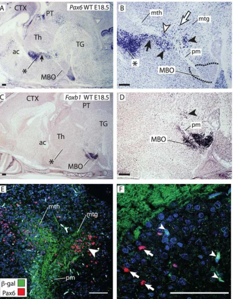
The pm branching point finds itself in the posterior hypothal-amus (ventral diencephalon), dorsal to the mammillary body, and approximately in register with the boundary between two dorsal diencephalic subdivisions classically named dorsal and ventral thalamus. Based on recent advances in our understanding of diencephalic development a new terminology is being introduced (see for instance [23–25]) in which the names prethalamus (formerly known as ventral thalamus) and thalamus (formerly known as dorsal thalamus) are preferred. In order to avoid confusion, we will call these two structures prethalamus/ventral thalamus (PTh/VTh, labeled in the Figures with an asterisk) and thalamus (Th). Transcription factor genePax6is a marker of PTh/ VTh [26,27] and we used it as the basis of our analysis. We found a trail ofPax6-positive cells joining the most ventral end of the PTh/VTh to the branching point (black arrow in Fig. 2A, B).
Closer examination (Fig. 2B) revealed an intriguing and complex distribution of Pax6-expressing cells around the mammillary axonal tree. ThePax6-expressing trail of cells was in contact with the mth and ended in a group of cells closely apposed to the branching point (black arrowheads in Fig. 2B).Pax6-positive cells were also present between the mth axons (white arrowhead in Fig. 2B) lending the first stretch of this tract its characteristic reticulate appearance [7,28]. Finally, numerousPax6-positive cells were found scattered in the area defined by the mtg and the mth (white arrow in Fig. 2B). Transcription factor Foxb1is a specific marker of the MBO (Fig. 2C, D) [7,29]. We detected a group of
Foxb1-expressing cells apposed to the caudal side of the branching point (arrowhead in Fig. 2D). To elucidate the relation between thePax6-expressing and theFoxb1-expressing cells around the pm branching point, we performed double immuno-staining for
beta-galactosidase and Pax6 on E18.5 Foxb1::tau-lacZ heterozygous brains [7] (beta-galactosidase detection indicatesFoxb1expression and the tau-beta-galactosidase fusion protein is localized to the corresponding axons) (Fig. 2E). The results showed a group of Pax6-positive cells and Foxb1-positive cells (arrowhead in Fig. 2E) in the caudal side of the branching point. Closer observation at higher magnification (Fig. 2F) revealed that marker expression was mutually exclusive—no green labeled cell somata (Foxb1-positive) (white arrowheads in Fig. 2F) had red nuclei (Pax6-positive) (white arrows in Fig. 2F).
TheFoxb1-expressing cells originate in the MBO
Since Foxb1-positive and Pax6-positive cells are distinct populations, we asked if they have different origins. Detection of
Foxb1expression on wild type embryonic brains at E10.5 (Fig. 3A)
Figure 2. A complex and specific cell aggregate around the bifurcation point.A–D) In situ hybridization forPax6(A, B) andFoxb1(C, D) on sagittal sections of wild type E18.5 brains (rostral to the left). (B) and (D) show high magnification details of (A) and (C). Black arrowheads, specific cell groups around the branching point. ac, anterior commissure; CTX, cortex; PT, pretectum. B)Pax6-expressing cells are also found forming a trail under the mth (black arrow) continuous with the PTh/VTh (asterisk), between the mth axons (white arrowhead), and in the area between mth and mtg (white arrow). E, F) Confocal pictures of antibody detection of Pax6 (red cell nuclei) and beta-galactosidase (green cell bodies; proxy forFoxb1
expression) on a sagittal section of an E18.5Foxb1-tau-lacZheterozygous brain. Blue labeling, DAPI nuclear staining. E) Double labeling of the branching point shows a compact group of Pax6- and Foxb1-positive cells (arrowhead). F)Foxb1-positive (arrowheads, green cell bodies) andPax6 -positive (arrows, red nuclei) cells are distinct from each other. Asterisk in A, B, C: PTh/VTh. Scale bars 100 micrometers.
revealed strong expression in the MBO [29] as well as in a ‘‘column’’ spreading dorsally from this nucleus (arrowheads in Fig. 3A) and in a more dorsal, looser group of cells (arrows in Fig. 3A). This column ofFoxb1-expressing cells expanded dorsally through E14.5 (Fig. 3B) and E16.5 (Fig. 3C), finally reaching the boundary between thalamus and PTh/VTh in the dorsal diencephalon (dotted line in Fig. 3C). The Foxb1-positive cell column seemed to be apposed to the lateral side of the pm axons (Fig. 3B, C).
Sagittal sections (Fig. 3D, E) confirmed that the labeled cells form a numerous group along the pm and mtg, and there is a looser group more dorsally positioned at the future branching point (arrow in Fig. 3D, E).
ThePax6-expressing cells originate in the PTh/VTh and are missing inPax6-deficient brains
To elucidate the origin of thePax6-expressing cells we used the
Pax6-lacZ mouse line, which carries a null mutation of Pax6
followed by lacZ as reporter [19] (see also Table 1). In E15.5 heterozygotes, a trail of beta-galactosidase-positive cells can be followed from the PTh/VTh to a specific point of the rostral side of the pm, where they aggregate (arrowhead in Fig. 4A). By E16.5, in heterozygous brains the trail of beta-galactosidase-positive cells connecting PTh/VTh and pm is still evident (Fig. 4B). In addition to the labeled cell group on the rostral side of the pm (arrowhead in Fig. 4B), a second group is forming on the caudal side (arrow in Fig. 4B). Because of the close proximity between the cells and the branching point, we hypothesized that they play a role in the branching process. Since mice deficient inPax6lack a PTh/VTh [27], we first asked if the branching point cells are also absent in these mutants. Homozygous brains at E15.5 showed only very few reporter-expressing cells in this region (arrowheads in Fig. 4C), and none of them reached the pm. Homozygotes at E16.5 showed
again few labeled cells and none of them was situated next to the pm (arrowhead in Fig. 4D). Other PTh/VTh marker gene,Arx
[30] (Fig. 4E) also labels the trail of cells between PTh/VTh and pm as well as a cell group around the pm branching point. Examination of the expression pattern database www.genepaint. org (in the public domain) in search for other markers of this region suggested that the cannabinoid receptorCnr1[31] could be a good candidate. Our in situs confirmed this, since Cnr1 is expressed likeArxandPax6in this region (Fig. 4G). BothArxand
Cnr1confirmed the lack of PTh/VTh cells around the pm in the mutant (Fig. 4F, H).
We concluded that the branching point cells are an extension of the PTh/VTh and that, like the rest of the PTh/VTh, they are absent in thePax6mutant.
Mammillary axons growing towards the thalamus in the
Pax6mutant
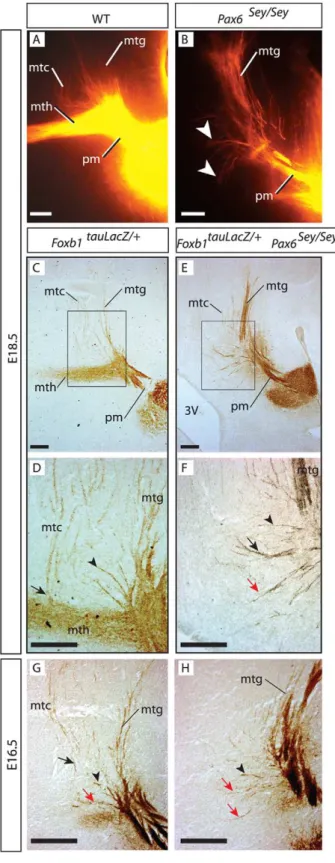
We then analyzed the mammillary axonal tree in wild type and in thePax6mutant by injecting DiI tracer into the MBO (Fig. 5A, B). In the wild type, the mth, mtg and mtc were easy to recognize (Fig. 5A). In the mutant diencephalon, the mth was absent. Instead, there was a number of axons apparently originated in the branching point and sometimes oriented towards the thalamus (arrowheads in Fig. 5B) which are however less in number and of shorter length than the axons of the wild type mth. They also lack the characteristic morphology of the early mth axons (thin, beaded axons weaving their way around local cell bodies that leave ‘‘holes’’ in an otherwise compact bundle) [7,28]. We termed them ‘‘problem axons’’ and set out to investigate their origin.
The problem axons develop earlier than the mth
To label the mammillary axons unambiguously, we crossed the
Pax6-deficient Small eye (Sey) mutant, carrying no reporter gene,
Figure 3.Foxb1-expressing cells migrate from the MBO along the pm.A)Foxb1expression on a transverse section of a wild type E10.5 brain. Arrowheads, column ofFoxb1-expressing cells originated in the MBO and migrating dorsally, preceded by a pioneer group (arrow). B, C)Foxb1
expression on transverse sections of wild type E14.5 (B) and E16.5 (C) brains. Left side shows Nissl counterstaining, right side shows dark field. Dotted line in C, E, external medullary lamina (zona limitans). D, E)Foxb1 expression in a sagittal section of an E14.5 wild type brain. (D) shows Nissl counterstaining, (E) shows dark field. Arrow, pioneer group ofFoxb1-expressing cells. Asterisk in C, D: PTh/VTh. Scale bars A, B, C: 50 micrometers; E: 25 micrometers.
[17,27] with theFoxb1-tau-lacZtransgenic line and used anti-beta-galactosidase antibody to compare the axons of Foxb1-tau-lacZ
heterozygous embryos (normal embryos) (Fig. 5C, D, G) (see also Table 1) to those of double mutant embryos (Foxb1-tau-lacZ
heterozygous/Seyhomozygous) (Fig. 5E, F, H).
Analysis ofFoxb1-tau-lacZheterozygotes at E18.5 showed that the mth and mtg separated from each other at right angles leaving a broad area between them occupied by mtc axons not forming an obvious bundle (Fig. 5C, D). Some mtc axons follow the mth for a short stretch to separate later at right angles, while others spread from the beginning over a wide area (Fig. 5C and see Fig. 6I below). Some of these loose axons spread over the ‘‘decision area’’ between mth and mtg, were oriented caudo-dorsally towards the tectum (Fig. 5D, arrowhead) while others followed originally a dorsal trajectory first, before turning sharply into the caudal direction (Fig. 5D, arrow). In thePax6mutant at E18.5 (Fig. 5E, F), some of the problem axons followed a caudal path similar to some of the non-bundled axons found in the wild type (Fig. 5F, black arrow and arrowhead). There was however a number of short axons extended in a dorsal and rostral direction towards the thalamus (Fig. 5F, red arrow). We asked if these short, thalamus-oriented axons were also present in the wild type, but hidden by the mth. To solve this question we analyzed mutants at an earlier age, E16.5, when there is no mth yet in the normal brain (Fig. 5G, H). Indeed the normal brain at that age showed also some axons growing in the direction of the thalamus (red arrow in Fig. 5G), and these appeared to be more numerous in thePax6mutant at the same age (red arrows in Fig. 5H).
We concluded that, in wild type as well as inPax6mutant brains there is a number of short mammillary axons extended in the direction of the thalamus as well as axons coursing dorsal/caudal before the mth is formed at all.
The mammillary axonal tree has three branches
The realization that there are some mammillary axons unaccounted for in the current descriptions of the mammillary
axonal tree, prompted us to examine the normal development of this fiber system using Foxb1-tau-lacZ heterozygotes. Foxb1 is specificallly expressed by neurons of the MBO as well as by the dorsal premammillary nucleus (DPM) [29]. The first pm axons can be seen at E10.5 growing towards the tegmentum (Fig. 6A, B) [32]. At E14.5 some axons from the pm start growing towards the tectum—they form the mtc (Fig. 6C, D). A pronounced bend in the pm is visible at E14.5 (Fig. 6C, D) and increases through E16.5 (Fig. 6E, F) and E18.5 (Fig. 6G, H). It is precisely in this bend that the mth develops. Although the first sprouts of the mth can be seen at E17.5 (not shown), its full extent however is only visible from E18.5 on (Fig. 6G, H), more than a full week later than the earliest pm axons (Fig. 6A, B). In agreement with the beta-galactosidase data and our previous results [7], at E18.5 three components of the mammillary axonal tree (mtc, mtg and mth) can be anterogradely visualized by injecting DiI tracer in the MBO (Fig. 6I). This confirms that the mammillary body generates not two but three axonal bundles (Fig. 6J, K).
The ‘‘problem axons’’ in thePax6mutant are probably misdirected mtc axons
Our observations suggested that the problem axons seen in the Pax6 mutant are not the product of pm branching, but simply an increased number of the mtc axons also found in normal animals. In that case, they would not be the product of a branching event but simply misdirected axons that set out in the wrong path and are unable to proceed (schematized in Fig. 7A). We reasoned that, if in thePax6mutant there is an increase in the number of mtc axons inappropriately navigating towards the thalamus, then there must be a smaller number of properly oriented mtc axons. We therefore counted the mtc axons and the problem axons inFoxb1-tau-lacZheterozygous and in double mutants (Foxb1-tau-lacZ heterozygous/Sey homozygous). To prevent some mtc axons from being hidden by the mth, we performed the countings at E16.5, when the mth has not yet been formed.
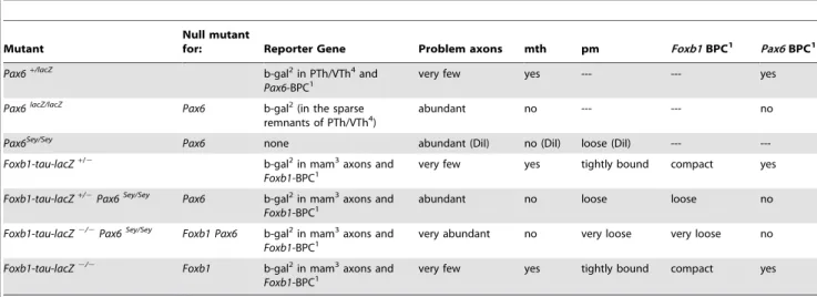
Table 1.Foxb1andPax6: Mutants and Phenotypes.
Mutant
Null mutant
for: Reporter Gene Problem axons mth pm Foxb1BPC1 Pax6BPC1
Pax6+/lacZ
b-gal2in PTh/VTh4and
Pax6-BPC1 very few yes --- --- yes
Pax6lacZ/lacZ
Pax6 b-gal2(in the sparse remnants of PTh/VTh4)
abundant no --- --- no
Pax6Sey/Sey
Pax6 none abundant (DiI) no (DiI) loose (DiI) ---
---Foxb1-tau-lacZ+/2 b-gal2in mam3axons and Foxb1-BPC1
very few yes tightly bound compact yes
Foxb1-tau-lacZ+/2Pax6Sey/Sey
Pax6 b-gal2in mam3axons and
Foxb1-BPC1 abundant no loose loose no
Foxb1-tau-lacZ2/2Pax6Sey/Sey
Foxb1 Pax6 b-gal2in mam3axons and Foxb1-BPC1
very abundant no very loose very loose no
Foxb1-tau-lacZ2/2 Foxb1 b-gal2in mam3axons and Foxb1-BPC1
very few yes tightly bound compact yes
The mutants are listed in the order they appear in the Results section. Two differentPax6mutants, with and without reporter were used. ThePax6-driven reporter (Pax6-lacZ) labels the PTh/VTh andPax6-BPC, but not the mammillary body, axons orFoxb1-BPC. ThePax6 Seymutant carries no reporter and its phenotype is analyzed by DiI axonal tracing. TheFoxb1-tau-lacZmouse carries aFoxb1-driven reporter labeling the mammillary body and axons and theFoxb1-BPC. The pm andFoxb1BPC have not been examined in thePax6-lacZmutant because they express neitherPax6nor thePax6-drivenlacZreporter.
1BPC: Branching Point Cells; 2b-gal: beta-galactosidase; 3mam: mammillary;
4PTh/VTh: Prethalamus/Ventral thalamus.
Our results show (Fig. 7B) that theFoxb1-tau-lacZ heterozy-gotes (i.e. normal animals) have a certain small number of problem axons [33], confirming our previous observation (Fig. 5G, red arrow). Sey/Sey mutants displayed significantly more problem axons than normal animals (Fig. 7B, compare white bars). Next we counted the mtc axons in the same samples and found that the Sey/Sey mutant had significantly less mtc axons than the normal animals (Fig. 7B, compare black bars), and that the difference in number approximately matched the difference found in problem axons. These results support the hypothesis that the problem axons are misdirected mtc axons and not the product of pm branching (schematized in Fig. 7C). SincePax6is not expressed by the MBO, the effect is non cell-autonomous and caused by the scattered cells, which control navigation in this area.
The pm does not branch inPax6-deficient mutants
In order to directly confirm that there is no morphological branching of the pm axons in theSey/Seymutant, we took resource to 3-D confocal microscopy imaging of our DiI data. In the wild type E18.5 brain, at low magnification, it was possible to observe images of axonal bifurcation (arrowheads in Fig. 7D) which could be confirmed at high magnification (arrowhead in Fig. 7E).
The same technique made obvious that the axons that can be seen inSey/Seyat right angles with the principal mammillary (red arrows in Fig. 7F) do not arise from bifurcations. In wild type brains, at earlier stages, the pm axons show swellings or varicosities from where the branches arise (not shown). Later, axons become thicker and the varicosities disappear (Fig. 7D, E). Interestingly, in theSey/Sey mutant brain, which does not have a mammillotha-lamic tract, the axonal varicosities were still present at this age, but no branches where visible (Fig. 7G). We concluded that thePax6
mutant shows a specific non-cell autonomous defect in pm branching.
Normal mth outgrowth in mutants with severe thalamic phenotypes and intact PTh/VTh
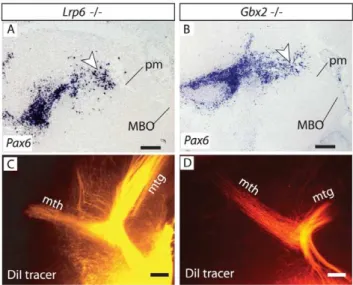
Pax6is expressed in the early dorsal thalamus, target of the mth. If target attraction was essential for pm branching, differentiation defects in thePax6-deficient thalamus [34] could contribute to the branching defect. If on the other hand target attraction was not essential for pm branching, mutant mouse brains showing an altered thalamus but preserving a normal PTh/VTh (together with branching point cells) should have a mth. The gene Lrp6
encodes an important co-receptor of Wnt ligands expressed in the thalamus [35,36]. Accordingly, the thalamus ofLrp6mutant mice is dramatically defective and unable to develop thalamocortical efferents [37]. Our analysis shows, however, that theLrp6mutant PTh/VTh expressesPax6, andPax6-expressing branching point cells are present in the appropriate position around the pm (Fig. 8A).Gbx2is a transcription factor gene essential for thalamus differentiation, and Gbx2 mutant mice show severely impaired thalamic development and absence of thalamocortical axons [38– 40].Pax6was expressed in theGbx2mutant PTh/VTh and there were Pax6-expressing cells in the cell groups around the pm branching point (Fig. 8B). Consistently, DiI tracings showed that the pm branches into a mth of normal appearance inLrp6mutants (Fig. 8C) and Gbx2 mutants (Fig. 8D). Together, these results suggest that an intact thalamus is not a precondition for the initial outgrowth of mth axons for as long as the local interactions (e.g. with thePax6-expressing branching point cells) are maintained.
Axonal fasciculation and cell aggregation impaired in the
Foxb1/Pax6double mutant
Foxb1::tau-lacZhomozygotes show a mth navigational phenotype that has been analyzed [7]. They showed however no alteration in mtg or mtc. Double homozygous brains forFoxb1::tau-lacZandSey, however, showed a slight increase in the number of misguided mtc axons (former ‘‘problem axons’’). This increase was statistically significant (Fig. 9A) (see also Table 1) and histologically visible (compare Fig. 5E, F with Fig. 9B, C red arrows) but not large enough to be reflected in a significant decrease of mtc axons (Fig. 9A). We then used sections along the dotted line in Fig. 9B to analyze the mtg. While in single Foxb1 homozygotes the mtg consisted of one compact axonal bundle (arrowhead in Fig. 9D), in
Foxb1 heterozygous/Sey homozygous brains the mtg was subdi-vided in a number of bundles (arrowheads in Fig. 9E). Double homozygotes showed an mtg disgregated into numerous smaller axonal fascicles (arrowheads in Fig. 9F). The Foxb1-expressing
Figure 4. Pax6-expressing cells are continuous with the PTh/ VTh and are missing in thePax6mutant.A–D) Beta-galactosidase antibody detection on sagittal sections, rostral to the left. Ages and genotypes as indicated. A trail ofPax6-expressing cells (arrowheads in A, B) from the PTh/VTh lands on the pm branching point at E15.5 (arrowhead in A). At E16.5 there is a second labeled cell group on the caudal side (arrow in B). In thePax6mutant these cells (arrowheads in C, D) are very scarce and do not contact the pm. E–H) In situ hybridization detection of PTh/VTh markers on sagittal sections. BothArx(E) andCnr1
(G) expression label the branching point cells continuous with the PTh/ VTh (arrowheads in E, G). Both markers are absent in thePax6-deficient diencephalon (F, H). Asterisk in A, B, E, G: PTh/VTh. Scale bars 100 micrometers.
branching point cells showed also progressively impaired aggre-gation (Fig. 9D through F).
We concluded that Foxb1 has a role in the control of cell adhesion and axonal fasciculation. This role could be non cell-autonomous (through loss-of-function in the Foxb1-expressing branching point cells) or cell autonomous (since the neurons originating the mtc and mtg axons express Foxb1). In this way
Figure 6. Stepwise development of the three components of the mammillary axonal tree. A, B) Beta-galactosidase activity detection in the flat-mounted right side of an E10.5 Foxb1::Cre x ROSA26Rheterozygous brain showing the first axons (arrwoheads in B) from the MBO navigating towards the tegmentum. Rostral to the left. (B) shows a high magnification detail of (A). ov, optic vesicle; TL, telencephalon. C–H) Antibody detection of beta-galactosidase on sagittal sections ofFoxb1-tau-lacZheterozygous brains. D, F, H show high magnification details of C, E, G, respectively. The dotted line in C, E, G marks the boundary between PTh/VTh and Th. C, D) The first mtc axons detach from the pm at E14.5. E, F) At E16.5 the pm acquires a pronounced bend marking the origin of the mtg. G, H) The mth appears at E18.5, branching from the bend in the pm observed at E16.5. I) DiI tracing shows the components of the mammillary axonal tree at E18.5. J) Diagram of MBO efferent connections to diencephalon and brainstem. K) Diagram of mammillary efferent axons. Grey, dorsal premammillary axons. Black, axons from the MBO proper. Asterisk in C, E, G: PTh/VTh. Scale bars: C, E, 25 micrometers; G, 50 micrometers; D, F, H, I, 100 micrometers.
doi:10.1371/journal.pone.0020315.g006 Figure 5. Thalamus-oriented axons in the Pax6-deficient
diencephalon.A, B) DiI tracing on sagittal sections of E18.5 wild type (A) andPax6homozygous (B) embryos. In the mutant, a few short axons (arrowheads in B) can be seen in place of a mth. C–H) Antibody detection of beta-galactosidase on sagittal sections, ages and genotypes as indicated. D, F are high magnification details the frames in C, E. C, D) Some mtc axons navigate directly towards the tectum (arrowhead in D) and others course towards the thalamus, then sharply change direction (arrow in D). E, F) In thePax6 mutant, similar mtc
axons can be seen changing course towards the tectum (black arrow in F), others grow straight dorsally (arrowhead in F) and finally others grow rostrally in the direction of the thalamus (red arrow in F). 3V, third ventricle. G, H) At E16.5, before the mth appears, there are mtc axons in
Pax6wild type (G) and mutant (H) (arrows and arrowheads as in F). Scale bars 100 micrometers.
Foxb1 cooperates with a non-cell autonomous role of Pax6 to guarantee the appropriate anatomy of the mammillary tree.
Discussion
How does the immediate cellular environment contribute to the formation and navigation of different fiber bundles in a complex, stereotyped axonal tree? Whilein vitroevidence suggests that the local environment could secret a variety of factors eliciting branching (see below), no example of a group of identified cells has been found which is essential for the formation of a specific axonal bundle by collateral branching in a certain system. We have identified an elaborate arrangement of specific cell populations migrating from different sources and converging around the branching point of a major forebrain axonal tract, the
pm. We offer several kinds of evidence (digital reconstruction of confocal images, axon counting, analysis of mutants with differential phenotypes) pointing to an indispensable role of these cells in collateral branching and navigation. This concept complements previous reports of axonal guidance supported by surrounding cells that serve as guideposts [41].
Several cell groups organize around the pm branching point
We show that migration from the PTh/VTh as well as from the hypothalamus results in several groups of cells arranged around the future pm branching point. Some of thePax6-expressing cells could arise in aPax6-expressing domain of the midbrain neuroepithelium [26]. Particularly curious is the cell group at the caudal side of the pm branching point, formed as the meeting point of Foxb1 -expressing andPax6-expressing cells originated respectively in the ventral and dorsal diencephalon. Although the diencephalon is the source of extensive non-radial migrations across dorso-ventral and rostro-caudal boundaries [16], formation of such cell groups of heterogeneous origin is not obvious from current paradigms of hypothalamic development [42–44]. Arranged along axonal bundles, some of thePax6-expressing cells could act as guideposts for mth axons as shown for other systems [45–47] and theFoxb1 -expressing cells could fulfill a similar role for pm axons.
Pax6-expressing cells and collateral branching
We show 1) that the Pax6-positive cells that surround the pm branching point are absent inPax6-deficient brains and 2) that this absence is ensued by major alterations in the axonal tree. We have investigated these alterations with a specific genetic marker of mammillary axons (the Foxb1::tau-lacZ allele) and digital recon-structions of confocal microscopy data to show unambiguously that the pm axons do not branch in the mutant. Previous descriptions of a number of pm collaterals inSey/Seybrains [33,48] probably result from unintentional co-labeling (DiI tracing or silver impregnation) of the mtc when attempting to label MBO projections.
Our results strongly suggest that close contact with thePax6 -expressing cells plays a role in fulfilling the potential of the
Figure 7. The problem axons in thePax6-deficient dienceph-alon are mammillotectal.A) Diagram showing the component axons of the MBO in wild type (top) andPax6mutant (bottom). In the wild type in blue, axons from the dorsal premammillary nucleus. In the mutant, problem axons are labeled by a question mark. B) Problem axons increase and mtc axons decrease in thePax6mutant. Mean+/2 SD; (**) P,0.01. C) Interpretation of the axon counting results in (B). The problem axons (red) are mtc axons initially directed dorsally and unable to turn caudally towards the tectum. D–G) 3D reconstruction of confocal images from DiI-traced pm branching point of wild type (D, E) andPax6-deficient (F, G) E18.5 brains at lower (D, F) and higher (E, G) magnification. D, E) Obviously bifurcated axons can be found in the wild type branching point. F, G) Mutant axons show the characteristic beads but no branching out of them.
doi:10.1371/journal.pone.0020315.g007
Figure 8. Mammillary branching is present in several thalamic mutants.A, B) Pax6 in situ hybridization shows that PTh/VTh and branching point cells are present in theLrp6mutant (A) and theGbx2
mutant (B) brains at E18.5. C, D) DiI tracing demonstrates presence of a mth in these mutants (C, D). Scale bars 100 micrometers.
initial branching bud. This agrees with previous work showing that direct physical contact with growth factor-soaked beads elicits branching in cultured axons [49,50] (see [51] for a review) and that contact with nearby dendrites enhances collateral branching of cortico-spinal axons [4]. That humoral factors, including those locally secreted, can elicit axonal branching is well established (see for instance [52]) and therefore an altered
Pax6-deficient thalamus [34,46,53,54] could cause the pm branching failure through lack of target attraction as shown in other models [2,55,56]. Our finding that a mth is present in mutants showing severe thalamic differentiation defects while preservingPax6-expressing cells around the pm branching point (Fig. 8) rather reinforces the notion that, in this model, branching and initial outgrowth depend on the local influence of a specific cell group.
Pax6,Foxb1 and adhesion
Adhesion proteins have a role in collateral branching [57] and specifically in mth development [58]. Abundant literature shows that a number of adhesion-related genes are downregulated inSey/ Seybrains:cadherin 4[45,59],L1cam[60],alpha 5 beta 1 integrin[61],
olfactomedin 3 (optimedin) [62], delta catenin [63], tenascin C [64],
semaphorin-3candsemaphorin-a5[65]. Intriguingly,Pax6seems to be involved in the pruning of inappropriate collateral branches of cortical pyramidal neurons [66]. Finally, previous analyses of
Pax6-deficient phenotypes support a function for this gene in contact guidance of pioneer axons in the forebrain [45–47,65,67].
In contrast,Foxtranscription factors have not been associated with adhesion gene expression [68,69], with the possible exceptions ofVcam1[70] andCdh7[71].
The mtc
An interesting observation made previously by us [7] and confirmed here is the existence of the mtc. Mammillo-tectal axons homologous to our mtc have been traced in the adult rat [8]. Specific expression ofFoxb1in the dorsal premammillary nucleus [7,29] as well as in the MBO evidences a molecular kinship of these nuclei and supports the proposed inclusion of the dorsal premammillary in an extended definition of the MBO [8]. The mtc bundle should be included in any discussion of the formation of the mammillary axonal tree. We show that non cell-autonomous
Pax6 expression is essential for mtc navigation. Intriguingly, although the abundantnetrin 1-expressing cells in the mammillary region do not expressPax6, their position around the mammillary axons is dramatically altered inPax6mutant brains [33,67]. This suggests that thePax6-expressing cells of the PTh/VTh secret a not yet identified factor contributing to the appropriate positioning of the netrin 1-expressing cells and, through this effect, they could influence also mammillary axonal navigation indirectly.
Foxb1andPax6in the control of mammillary axonal
organization
The double homozygotes demonstrate a Foxb1-regulated component in mtc and mtg navigation, probably mediated by
Figure 9. Axonal fasciculation and cell aggregation impaired in theFoxb1/Pax6double mutant.A) Slight increase in problem axons but no detectable change in mtc axons in thePax6mutant. White column, problem axons; black column, mtc axons. Mean+/2SD; (*) P,0.05; n.s. not significant. B, C) Beta galactosidase detection on sagittal section of doubleFoxb1-tauLacZ/Seyhomozygote. The problem axons (red arrow) seem more numerous as in singleSey homozygotes. The dotted line in (B) indicates the approximate plane of section of D, E, F. (C) shows a high magnification detail of the image in (B). D–F) Beta galactosidase detection on sections along the dotted line in (B) (left side is shown) through the branching point of E18.5 brains (genotypes as indicated). InFoxb1single homozygotes (D) there is a mth (branching takes place), the mtg is not subdivided into fascicles and theFoxb1branching point cells are tightly aggregated. In the double mutant (F), the problem axons (red arrow in E, F) are longer and more numerous, theFoxb1-expressing branching point cells (Foxb1BPC in Fig. 9D–F) are less compactly aggregated and the mtg is divided in more fascicles (arrowheads) as in theFoxb1heterozygote/Seyhomozygote (E). Scale bars 100 micrometers.
proteins involved in fasciculation. Since the Foxb1-expressing branching point cells are still present in theFoxb1-deficient brain, and the gene is also expressed by the neurons originating the affected axons, the role of this transcription factor could be or not cell-autonomous. A non cell-autonomous role has been suggested for the mth navigational phenotype found in theFoxb1mutant [7].
Conclusions
This work uncovers a series of complex cell migration events giving rise to several specific groups of cells of different origin essential for the formation and organization of an axonal crossroads linking hypothalamus, thalamus, midbrain and hind-brain. We show that many of these cells expressPax6, depend on expression of this gene for their origination, and are necessary for the formation of specific axonal collaterals through branching of the pm. Finally, we show cooperation of Pax6and Foxb1in mtc and mtg navigation. This work offers new insights into the
development of a specific cellular environment that favors the formation and navigation of specific axonal collaterals.
Acknowledgments
Andrea Wizenmann (University of Tu¨bingen) and Herbert Hildebrandt (University of Hannover) made valuable comments on the text. Michael Heide (University of Heidelberg) assisted with the confocal microscopy. The anti-Pax6 antibody, developed by A. Kawakami, was obtained from the Developmental Studies Hybridoma Bank developed under the auspices of the NICHD and maintained by The University of Iowa, Department of Biology, Iowa City, IA 52242.
Author Contributions
Conceived and designed the experiments: GA-B XZ. Performed the experiments: NS TZ MC XZ. Analyzed the data: NS TZ AS XZ GA-B. Contributed reagents/materials/analysis tools: AS XZ. Wrote the paper: NS TZ GA-B.
References
1. Harris WA, Holt CE, Bonhoeffer F (1987) Retinal axons with and without their somata, growing to and arborizing in the tectum of Xenopus embryos: a time-lapse video study of single fibres in vivo. Development 101: 123–133. 2. O’Leary DD, Terashima T (1988) Cortical axons branch to multiple subcortical
targets by interstitial axon budding: implications for target recognition and ‘‘waiting periods’’. Neuron 1: 901–910.
3. O’Leary DD, Bicknese AR, De Carlos JA, Heffner CD, Koester SE, et al. (1990) Target selection by cortical axons: alternative mechanisms to establish axonal connections in the developing brain. Cold Spring Harb Symp Quant Biol 55: 453–468.
4. Bastmeyer M, Daston MM, Possel H, O’Leary DD (1998) Collateral branch formation related to cellular structures in the axon tract during corticopontine target recognition. J Comp Neurol 392: 1–18.
5. Vann SD, Aggleton JP (2004) The mammillary bodies: two memory systems in one? Nat Rev Neurosci 5: 35–44.
6. Hayakawa T, Zyo K (1989) Retrograde double-labeling study of the mammillothalamic and the mammillotegmental projections in the rat. J Comp Neurol 284: 1–11.
7. Alvarez-Bolado G, Zhou X, Voss AK, Thomas T, Gruss P (2000) Winged helix transcription factor Foxb1 is essential for access of mammillothalamic axons to the thalamus. Development 127: 1029–1038.
8. Canteras NS, Swanson LW (1992) The dorsal premammillary nucleus: an unusual component of the mammillary body. Proc Natl Acad Sci U S A 89: 10089–10093.
9. Mombaerts P, Wang F, Dulac C, Chao SK, Nemes A, et al. (1996) Visualizing an olfactory sensory map. Cell 87: 675–686.
10. Kloetzli JM, Fontaine-Glover IA, Brown ER, Kuo M, Labosky PA (2001) The winged helix gene, Foxb1, controls development of mammary glands and regions of the CNS that regulate the milk-ejection reflex. Genesis 29: 60–71. 11. Labosky PA, Winnier GE, Jetton TL, Hargett L, Ryan AK, et al. (1997) The
winged helix gene, Mf3, is required for normal development of the diencephalon and midbrain, postnatal growth and the milk-ejection reflex. Development 124: 1263–1274.
12. Wehr R, Mansouri A, de Maeyer T, Gruss P (1997) Fkh5-deficient mice show dysgenesis in the caudal midbrain and hypothalamic mammillary body. Development 124: 4447–4456.
13. Kaestner KH, Schutz G, Monaghan AP (1996) Expression of the winged helix genes fkh-4 and fkh-5 defines domains in the central nervous system. Mech Dev 55: 221–230.
14. Zhao T, Zhou X, Szabo N, Leitges M, Alvarez-Bolado G (2007) Foxb1-driven Cre expression in somites and the neuroepithelium of diencephalon, brainstem, and spinal cord. Genesis 45: 781–787.
15. Soriano P (1999) Generalized lacZ expression with the ROSA26 Cre reporter strain. Nat Genet 21: 70–71.
16. Zhao T, Szabo N, Ma J, Luo L, Zhou X, et al. (2008) Genetic mapping of Foxb1-cell lineage shows migration from caudal diencephalon to telencephalon and lateral hypothalamus. Eur J Neurosci 28: 1941–1955.
17. Hill RE, Favor J, Hogan BL, Ton CC, Saunders GF, et al. (1991) Mouse small eye results from mutations in a paired-like homeobox-containing gene. Nature 354: 522–525.
18. Hogan BL, Horsburgh G, Cohen J, Hetherington CM, Fisher G, et al. (1986) Small eyes (Sey): a homozygous lethal mutation on chromosome 2 which affects the differentiation of both lens and nasal placodes in the mouse. J Embryol Exp Morphol 97: 95–110.
19. St-Onge L, Sosa-Pineda B, Chowdhury K, Mansouri A, Gruss P (1997) Pax6 is required for differentiation of glucagon-producing alpha-cells in mouse pancreas. Nature 387: 406–409.
20. Kokubu C, Heinzmann U, Kokubu T, Sakai N, Kubota T, et al. (2004) Skeletal defects in ringelschwanz mutant mice reveal that Lrp6 is required for proper somitogenesis and osteogenesis. Development 131: 5469–5480.
21. Simmons DM, Arriza JL, Swanson LW (1989) A complete protocol for in situ hybridization of messenger RNAs in brain and other tissues with radiolabeled single-stranded RNA probes. J Histotechnol 12.
22. Yaylaoglu MB, Titmus A, Visel A, Alvarez-Bolado G, Thaller C, et al. (2005) Comprehensive expression atlas of fibroblast growth factors and their receptors generated by a novel robotic in situ hybridization platform. Dev Dyn 234: 371–386.
23. Kiecker C, Lumsden A (2004) Hedgehog signaling from the ZLI regulates diencephalic regional identity. Nat Neurosci 7: 1242–1249.
24. Puelles L, Martı´nez S, Martı´nez-de-la-Torre M, Rubenstein JLR (2004) Gene Maps and Related Histogenetic Domains in the Forebrain and Midbrain. In: Paxinos G, ed. The Rat Nervous System. San DiegoCA: Academic Press. pp 3–25.
25. Scholpp S, Lumsden A (2010) Building a bridal chamber: development of the thalamus. Trends Neurosci 33: 373–380.
26. Stoykova A, Gruss P (1994) Roles of Pax-genes in developing and adult brain as suggested by expression patterns. J Neurosci 14: 1395–1412.
27. Stoykova A, Fritsch R, Walther C, Gruss P (1996) Forebrain patterning defects in Small eye mutant mice. Development 122: 3453–3465.
28. Alpeeva EV, Makarenko IG (2009) Perinatal development of the mammillotha-lamic tract and innervation of the anterior thamammillotha-lamic nuclei. Brain Res 1248: 1–13.
29. Alvarez-Bolado G, Zhou X, Cecconi F, Gruss P (2000) Expression of Foxb1 reveals two strategies for the formation of nuclei in the developing ventral diencephalon. Dev Neurosci 22: 197–206.
30. Miura H, Yanazawa M, Kato K, Kitamura K (1997) Expression of a novel aristaless related homeobox gene ‘Arx’ in the vertebrate telencephalon, diencephalon and floor plate. Mech Dev 65: 99–109.
31. Jelsing J, Larsen PJ, Vrang N (2008) Identification of cannabinoid type 1 receptor expressing cocaine amphetamine-regulated transcript neurons in the rat hypothalamus and brainstem using in situ hybridization and immunohisto-chemistry. Neuroscience 154: 641–652.
32. Easter SS, Jr., Ross LS, Frankfurter A (1993) Initial tract formation in the mouse brain. J Neurosci 13: 285–299.
33. Tsuchiya R, Takahashi K, Liu FC, Takahashi H (2009) Aberrant axonal projections from mammillary bodies in Pax6 mutant mice: possible roles of Netrin-1 and Slit 2 in mammillary projections. J Neurosci Res 87: 1620–1633. 34. Pratt T, Vitalis T, Warren N, Edgar JM, Mason JO, et al. (2000) A role for Pax6 in the normal development of dorsal thalamus and its cortical connections. Development 127: 5167–5178.
35. Bafico A, Liu G, Yaniv A, Gazit A, Aaronson SA (2001) Novel mechanism of Wnt signalling inhibition mediated by Dickkopf-1 interaction with LRP6/ Arrow. Nat Cell Biol 3: 683–686.
36. Brown SD, Twells RC, Hey PJ, Cox RD, Levy ER, et al. (1998) Isolation and characterization of LRP6, a novel member of the low density lipoprotein receptor gene family. Biochem Biophys Res Commun 248: 879–888. 37. Zhou CJ, Pinson KI, Pleasure SJ (2004) Severe defects in dorsal thalamic
development in low-density lipoprotein receptor-related protein-6 mutants. J Neurosci 24: 7632–7639.
38. Miyashita-Lin EM, Hevner R, Wassarman KM, Martinez S, Rubenstein JL (1999) Early neocortical regionalization in the absence of thalamic innervation. Science 285: 906–909.
40. Szabo NE, Zhao T, Zhou X, Alvarez-Bolado G (2009) The role of Sonic hedgehog of neural origin in thalamic differentiation in the mouse. J Neurosci 29: 2453–2466.
41. Zhou L, Bar I, Achouri Y, Campbell K, De Backer O, et al. (2008) Early forebrain wiring: genetic dissection using conditional Celsr3 mutant mice. Science 320: 946–949.
42. Altman J, Bayer SA (1986) The Development of the Rat Hypothalamus. Adv Anat Embryol Cell Biol 100: 1–178.
43. Bayer SA, Altman J (1995) Neurogenesis and Neuronal Migration. In: Paxinos G, ed. The Rat Nervous System. San Diego: Academic Press. pp 1041–1078. 44. Bayer SA, Altman J (1995) Principles of Neurogenesis, Neuronal Migration, and
Neural Circuit Formation. In: Paxinos G, ed. The Rat Nervous System. San Diego: Academic Press. pp 1079–1098.
45. Andrews GL, Mastick GS (2003) R-cadherin is a Pax6-regulated, growth-promoting cue for pioneer axons. J Neurosci 23: 9873–9880.
46. Mastick GS, Davis NM, Andrew GL, Easter SS, Jr. (1997) Pax-6 functions in boundary formation and axon guidance in the embryonic mouse forebrain. Development 124: 1985–1997.
47. Nural HF, Mastick GS (2004) Pax6 guides a relay of pioneer longitudinal axons in the embryonic mouse forebrain. J Comp Neurol 479: 399–409.
48. Valverde F, Garcia C, Lopez-Mascaraque L, De Carlos JA (2000) Development of the mammillothalamic tract in normal and Pax-6 mutant mice. J Comp Neurol 419: 485–504.
49. Gallo G, Letourneau PC (1999) Different contributions of microtubule dynamics and transport to the growth of axons and collateral sprouts. J Neurosci 19: 3860–3873.
50. Kalil K, Szebenyi G, Dent EW (2000) Common mechanisms underlying growth cone guidance and axon branching. J Neurobiol 44: 145–158.
51. Gibson DA, Ma L (2011) Developmental regulation of axon branching in the vertebrate nervous system. Development 138: 183–195.
52. Jeanneteau F, Deinhardt K, Miyoshi G, Bennett AM, Chao MV (2010) The MAP kinase phosphatase MKP-1 regulates BDNF-induced axon branching. Nat Neurosci.
53. Grindley JC, Hargett LK, Hill RE, Ross A, Hogan BL (1997) Disruption of PAX6 function in mice homozygous for the Pax6Sey-1Neu mutation produces abnormalities in the early development and regionalization of the diencephalon. Mech Dev 64: 111–126.
54. Mastick GS, Andrews GL (2001) Pax6 regulates the identity of embryonic diencephalic neurons. Mol Cell Neurosci 17: 190–207.
55. Bastmeyer M, O’Leary DD (1996) Dynamics of target recognition by interstitial axon branching along developing cortical axons. J Neurosci 16: 1450–1459. 56. Sato M, Lopez-Mascaraque L, Heffner CD, O’Leary DD (1994) Action of a
diffusible target-derived chemoattractant on cortical axon branch induction and directed growth. Neuron 13: 791–803.
57. Daston MM, Bastmeyer M, Rutishauser U, O’Leary DD (1996) Spatially restricted increase in polysialic acid enhances corticospinal axon branching related to target recognition and innervation. J Neurosci 16: 5488–5497. 58. Weinhold B, Seidenfaden R, Rockle I, Muhlenhoff M, Schertzinger F, et al.
(2005) Genetic ablation of polysialic acid causes severe neurodevelopmental defects rescued by deletion of the neural cell adhesion molecule. J Biol Chem 280: 42971–42977.
59. Stoykova A, Gotz M, Gruss P, Price J (1997) Pax6-dependent regulation of adhesive patterning, R-cadherin expression and boundary formation in developing forebrain. Development 124: 3765–3777.
60. Meech R, Kallunki P, Edelman GM, Jones FS (1999) A binding site for homeodomain and Pax proteins is necessary for L1 cell adhesion molecule gene expression by Pax-6 and bone morphogenetic proteins. Proc Natl Acad Sci U S A 96: 2420–2425.
61. Duncan MK, Kozmik Z, Cveklova K, Piatigorsky J, Cvekl A (2000) Overexpression of PAX6(5a) in lens fiber cells results in cataract and upregulation of (alpha)5(beta)1 integrin expression. J Cell Sci 113: 3173–3185. 62. Grinchuk O, Kozmik Z, Wu X, Tomarev S (2005) The Optimedin gene is a
downstream target of Pax6. J Biol Chem 280: 35228–35237.
63. Duparc RH, Boutemmine D, Champagne MP, Tetreault N, Bernier G (2006) Pax6 is required for delta-catenin/neurojugin expression during retinal, cerebellar and cortical development in mice. Dev Biol 300: 647–655. 64. von Holst A, Egbers U, Prochiantz A, Faissner A (2007) Neural stem/progenitor
cells express 20 tenascin C isoforms that are differentially regulated by Pax6. J Biol Chem 282: 9172–9181.
65. Jones L, Lopez-Bendito G, Gruss P, Stoykova A, Molnar Z (2002) Pax6 is required for the normal development of the forebrain axonal connections. Development 129: 5041–5052.
66. Pinon MC, Tuoc TC, Ashery-Padan R, Molnar Z, Stoykova A (2008) Altered molecular regionalization and normal thalamocortical connections in cortex-specific Pax6 knock-out mice. J Neurosci 28: 8724–8734.
67. Vitalis T, Cases O, Engelkamp D, Verney C, Price DJ (2000) Defect of tyrosine hydroxylase-immunoreactive neurons in the brains of mice lacking the transcription factor Pax6. J Neurosci 20: 6501–6516.
68. Tuteja G, Kaestner KH (2007) SnapShot: Forkhead Transcription Factors II. Cell 131: 192.
69. Tuteja G, Kaestner KH (2007) SnapShot: forkhead transcription factors I. Cell 130: 1160.
70. Mahlapuu M, Ormestad M, Enerback S, Carlsson P (2001) The forkhead transcription factor Foxf1 is required for differentiation of extra-embryonic and lateral plate mesoderm. Development 128: 155–166.