and Decreases Chronological Life Span in Fission Yeast
Alice Zuin1., Natalia Gabrielli1., Isabel A. Calvo1., Sarela Garcı´a-Santamarina1
, Kwang-Lae Hoe2, Dong
Uk Kim2, Han-Oh Park3, Jacqueline Hayles4, Jose´ Ayte´1, Elena Hidalgo1*
1Oxidative Stress and Cell Cycle Group, Universitat Pompeu Fabra, Barcelona, Spain,2Pombe Deletion Project, KRIBB, Yuseong-gu, Daejeon, Republic of Korea,3Bioneer Corporation, Daedeok-gu, Daejeon, Republic of Korea,4Cancer Research U. K., London, United Kingdom
Abstract
Background:Oxidative stress is a probable cause of aging and associated diseases. Reactive oxygen species (ROS) originate mainly from endogenous sources, namely the mitochondria.
Methodology/Principal Findings:We analyzed the effect of aerobic metabolism on oxidative damage in Schizosacchar-omyces pombeby global mapping of those genes that are required for growth on both respiratory-proficient media and hydrogen-peroxide-containing fermentable media. Out of a collection of approximately 2700 haploid yeast deletion mutants, 51 were sensitive to both conditions and 19 of these were related to mitochondrial function. Twelve deletion mutants lacked components of the electron transport chain. The growth defects of these mutants can be alleviated by the addition of antioxidants, which points to intrinsic oxidative stress as the origin of the phenotypes observed. These respiration-deficient mutants display elevated steady-state levels of ROS, probably due to enhanced electron leakage from their defective transport chains, which compromises the viability of chronologically-aged cells.
Conclusion/Significance: Individual mitochondrial dysfunctions have often been described as the cause of diseases or aging, and our global characterization emphasizes the primacy of oxidative stress in the etiology of such processes.
Citation:Zuin A, Gabrielli N, Calvo IA, Garcı´a-Santamarina S, Hoe K-L, et al. (2008) Mitochondrial Dysfunction Increases Oxidative Stress and Decreases Chronological Life Span in Fission Yeast. PLoS ONE 3(7): e2842. doi:10.1371/journal.pone.0002842
Editor:Stefan Wo¨lfl, Universita¨t Heidelberg, Germany
ReceivedApril 17, 2008;AcceptedJuly 7, 2008;PublishedJuly 30, 2008
Copyright:ß2008 Zuin et al. This is an open-access article distributed under the terms of the Creative Commons Attribution License, which permits unrestricted use, distribution, and reproduction in any medium, provided the original author and source are credited.
Funding:This work was supported by Direccio´n General de Investigacio´n of Spain Grant BFU2006-02610, and by the Spanish program Consolider-Ingenio 2010 Grant CSD 2007-0020 to E.H. The funders had no role in study design, data collection and analysis, decision to publish, or preparation of the manuscript.
Competing Interests:The authors have declared that no competing interests exist.
* E-mail: [email protected]
.These authors contributed equally to this work.
Introduction
Reactive oxygen species (ROS) homeostasis plays an important role in chronological aging processes and some degenerative diseases [1–5]. The main source of ROS in most cell types is the mitochondria, where they are formed upon incomplete reduction of oxygen at several sites on the electron transfer chain [6]. Physiological ROS levels are achieved by means of a complex collection of cellular activities such as superoxide dismutases, peroxiredoxins, glutathione peroxidases, or catalases. While ROS, such as superoxide or hydrogen peroxide (H2O2), are mediators of oxygen toxicity, they are also involved in intracellular signaling through the activation of response pathways (for reviews, see 7,8). Thus, in eukaryotic microbes, ROS sensors and other pathway components have been identified and characterized by treatment of cultures with extracellular peroxides, which rapidly increase the intracellular steady-state levels [8]. Thus, in Schizosaccharomyces pombe, the Pap1 and Sty1 pathways quickly respond to moderate and high extracellular concentrations of H2O2, respectively [9,10]. These cellular H2O2receptors are then able to trigger signaling pathways with the aim of engaging suitable cellular responses, which normally include peroxide scavengers and repair activities, and promote adaptation and survival [10–13].
levels [21] in the glucose-derepressed early stationary phase. Therefore, according to the literature,S. pombeseems to behave very similarly toS. cerevisiaeregarding glucose utilization, with low respiratory activities during the exponential phase of growth in the presence of fermentable glucose as the carbon source.
We decided to first characterize the metabolism of fission yeast when grown in the two most common glucose-containing laboratory media, defined medium and complex medium. We show that fission yeast grown in glucose-containing defined medium (2% glucose) not only ferments the sugar, but also respires it, as revealed by oxygen consumption measurements. In contrast, respiration is comparatively weaker in cells grown in complex medium, which contains a higher concentration of glucose (3%). To identify genes required for survival upon both exogenous and endogenous oxidative stress, we tested anS. pombe
collection of viable open reading frame deletion mutants with a double screening procedure. Yeast mutants that are sensitive to growth under respiratory-prone conditions (defined medium) and to H2O2on low-respiratory medium (complex medium) may carry mutations in genes that participate in the generation of respiration-linked basal oxidative stress. Out of a collection of approximately 2700 haploid yeast deletion mutants, 51 were sensitive to both conditions and 19 of these were related to mitochondrial function. The basis of double sensitivity to intrinsic and extrinsic oxidative stress of many genes coding for electron transfer chain components is unraveled here.
Results
Exponentially growing fission yeast cells respire in fermentable carbon sources at different rates depending on the growth medium
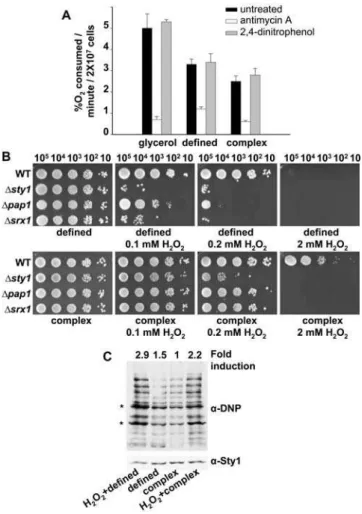
In order to verify whether glucose repression of respiration (the Crabtree effect) is severe in S. pombe grown in the two most common glucose-containing laboratory media, defined medium and complex medium [22], we measured the oxygen consumption (%O2, an indicator of respiratory rates) of cells growing exponentially in defined or complex media using an oxygen electrode, and compared the values to the oxygen consumption of cells growing in non-fermentable glycerol as a carbon source, where oxygen consumption has to be at a maximum. As observed in Figure 1A, exponentially growing S. pombe cells respire at different rates depending on the growth media. In all three cases, oxygen uptake was due to normal respiration, since it was abrogated by the complex III inhibitor antimycin A [23], but it was not affected by the uncoupler 2,4-dinitrophenol, which is not expected to decrease oxygen consumption [19] (Figure 1A). Thus, inS. pombeoxidative phosphorylation is the main source of energy in glycerol-containing defined medium, but it also occurs in cells growing in glucose-containing defined medium and, to a lesser extent, in complex medium.
Respiratory rates positively determine the level of intrinsic oxidative stress in fission yeast
The differences in respiratory rates suggested that cells grown in defined medium could have higher levels of intracellular ROS and, consequently, display exacerbated sensitivity to extracellular peroxides when compared with cells grown in complex medium. We measured cell survival against extracellular H2O2of wild-type and H2O2-sensitive cells (eg, cells lacking Pap1, Sty1 or Srx1, a protein essential for basal H2O2scavenging) [24] on definedversus complex media plates. As expected, cells are more sensitive to extracellular peroxides when grown in defined medium (compare the same concentrations of H2O2in complexvs. defined media,
Figure 1B). In fact, activation of the Pap1 pathway, which detects very low extracellular H2O2stress, requires lower concentrations of peroxide in cells grown in defined medium than in complex medium, as shown by the formation of the active, oxidized form of Pap1 (Figure S1A), and by Northern blot analysis of Pap1-dependent genes (Figure S1B). Furthermore, oxidative damage is Figure 1. Respiratory rates, intracellular ROS levels and ROS-mediated intrinsic oxidative stress depend on the type of growth media.(A) Oxygen consumption ofS. pombecells grown in different growth media. Wild-type strain 972 was grown on glycerol (defined medium with 2% glycerol and 0.2% glucose) (glycerol), defined medium (contains 2% glucose) (defined), and complex medium (contains 3% glucose) (complex), in the presence or absence of the mitochondrial inhibitors antimycin A or 2,4-dinitrophenol. Error bars of this figure represent the standard error measurement (SEM) of three replicates. (B) Survival of different strains in response to H2O2exposure
in definedvs.complex media plates. Strains 972 (WT), AV18 (Dsty1), AV25(Dpap1), and EA38 (Dsrx1) were grown in defined media, and the indicated number of cells were spotted onto defined or complex media plates with or without H2O2at the indicated concentrations. (C) Protein
carbonylation generated during growth in defined and complex media. Wild-type strain 972 was grown aerobically in defined or complex media, and cells were collected before or after treatment for 30 min with 2 mM H2O2at an OD600of 0.5. Protein carbonylation was detected
by reaction of carbonyl groups with DNPH, followed by SDS-PAGE and Western blot analysis by using anti-DNP (a-DNP, top panel) or anti-Sty1 antibodies as a loading control (a-Sty1, bottom panel). Fold induction numbers, obtained from the same blots, are the ratio of the absolute scan numbers for the indicated bands (*) and the corresponding amount of Sty1, and they relate to the values of the growth in complex medium. Similar results were obtained from 7 independent experi-ments.
stronger in wild-type cells grown in defined medium than in complex medium: protein carbonylation in extracts from cells grown in defined medium (Figure 1C, second lane), is 1.5-fold higher than protein carbonylation in extracts from cells grown in complex medium (Figure 1C, third lane).
Identification of the electron transfer chain as a major determinant of intrinsic oxidative stress
We then designed a double screen to isolate genes required for survival at elevated levels of intracellular ROS. As a first screening step, we checked anS. pombecollection of approximately 2700 haploid yeast deletion mutants for strains that were sensitive to growth on defined media plates, where respiratory rates are higher, but not on complex media plates. However, many of the selected mutants would be unable to grow on defined medium due to fundamental problems in biosynthetic pathways. Therefore, and to identify those genes whose inactivation confers sensitivity to intrinsic oxidative stress (eg, by enhancing ROS production or impairing ROS scavenging), our second screening step was the inability to grow on H2O2-containing complex media plates. We hypothesized that some mutant strains that were sensitive to extracellular peroxides in complex media would also be unable to cope with the oxidative metabolism of growth in defined medium and, therefore, that some genes would be equally necessary for survival in the face of intrinsic and extrinsic oxidative stress. From a total of 51 deletion strains isolated following the double screening procedure, 19 of the deleted genes were involved in different mitochondrial functions (Table 1), from which 12 coded directly for components of the electron transfer chain. We confirmed that none of these mutants could grow on glycerol where respiration is the only energy source (data not shown). Several genes that were unrelated to the mitochondrial category were also isolated in the screen and will be described elsewhere (Table S1). We selected some of the mitochondrial mutant strains for further analysis:Dcoq4and
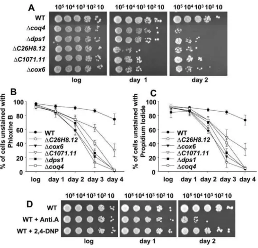
Ddps1(coding for proteins required for the biosynthesis of coenzyme Q),DC26H8.12(coding for a protein that catalyzes the cytochrome c-heme linkage), Dcox6(coding for the subunit VI of cytochrome c oxidase, or complex IV), and DC1071.11 (coding for a protein containing a flavin reductase-like domain; putative NADH oxidore-ductase). To confirm their double sensitivity to intrinsic and extrinsic oxidative stress, we compared the growth of these mutants and a wild-type strain in complex, defined, and H2O2-containing complex media plates (Figure 2A). To differing degrees, they all showed sensitivity to growth in defined media and in H2O2-containing complex media.
The growth defects of respiratory mutants in glucose-containing defined media are alleviated by antioxidants
The growth of these mutants in liquid defined media, but not in liquid complex media, was also severely impaired compared to a wild-type strain (Figure 2B and 2C). Addition of the glutathione-precursor agent cysteine (Figure 2C) or the antioxidant N-acetylcysteine (data not shown) to the liquid defined media enabled their growth to recover, indicating that ROS production may be enhanced in these mutants. Furthermore, incubation of defined media plates under anaerobic conditions partially or totally restored the growth of these mutants (Figure 2D). These data demonstrate that growth impairment of these mutants in defined media is due to intrinsic oxidative stress.
Mutations in electron transfer chain components increase the levels of intracellular ROS
The requirement of antioxidants or anaerobiosis for growth of the mitochondrial mutants on defined medium suggested that they
are more prone to produce ROS than a wild-type strain. Oxygen consumption, and therefore respiration, was severely inhibited in the mutants, as expected (Figure 3A). We tested the steady-state levels of ROS in these mutants by measuring oxidation of the dye 29,79-dichlorodihydrofluorescein diacetate (DCFH-DA) (Figure 3B). The levels of fluorescence, which are an indicator of the intracellular concentration of peroxides, during growth in complex media were between 1.3- and 1.7-fold higher in the respiration-deficient mutants than in the wild-type strain (Figure 3B). We propose that electron leakage is exacerbated in those mutants, as described when cells have been treated with electron transfer chain inhibitors such as antimycin A, where accidental ROS production can be enhanced, since the physiological transfer of electrons from one chain component to the other has been disturbed [25] (Figure 3B, WT+Anti.A). On the contrary, the uncoupler 2,4-dinitrophenol, which blocks respiration at the level of ATP synthesis, did not affect ROS production (Figure 3B, WT+2,4-DNP), as described else-where [25].
We then tested the effect of intrinsic oxidative stress on the survival rates of chronologically aged cells. We compared the viability of wild-type and mitochondrial mutant cultures 24 and 48 hr after reaching stationary phase by spotting cells in complex media plates (Figure 4A), and the results indicate that the elevated levels of ROS in these mutants are clearly deleterious for cell survival. Measurement of the percentage of metabolically inactive
Table 1.Mitochondrial genes required for survival upon extrinsic (extracellular H2O2) and intrinsic (growth on defined medium) oxidative stress.
NAME FUNCTION
C26H8.12 Covalently links the heme group to the apoprotein of cytochrome c
C1071.11 NADH dependent oxidoreductase; contains a flavin like domain
rip1 Subunit of cytochrome bc1, also known as respiratory complex III
sco1 Copper chaperone protein, essential for complex IV assembly
C1672.04c High similarity toS. cerevisiaeCox19p, which is a metal transporter for complex IV assembly
dps1 Decaprenyl diphosphate synthase, required for ubiquinone biosynthesis
cox6 Heme A-containing chain of cytochrome c oxidase
coq2 Required for ubiquinone biosynthesis
coq3 Hexaprenyldihydroxybenzoate methyltransferase. Ubiquinone biosynthesis
coq4 Ubiquinone biosynthesis protein
coq5 C-methyltransferase, ubiquinone biosynthetic process and aerobic respiration
coq10 Electron transport and cellular respiration; ubiquinone biosynthesis
C336.13c Removal of transit peptides for the targeting of proteins from the mitochondrial matrix
tom70 Receptor that accelerates the import of all mitochondrial precursor proteins
C8C9.06c Mitochondrial translation regulator, PPR domains
mss1 GTPase involved in the 5-carboxymethylaminomethyl modification of mitochondrial tRNAs
C2G2.07c Mitochondrial ribosomal protein (small subunit)
C25B2.04c Mitochondrial ribosome assembly protein
C1610.02c Mitochondrial ribosomal protein L1
cells, unable to efflux fluorescent dyes such as phloxine B (Figure 4B) or propidium iodide (Figure 4C), yielded similar results: short after entering stationary phase, the mitochondrial mutants display high percentages of metabolically inactive cells, when compared to wild-type cultures. The fitness of early stationary phase cultures of the mitochondrial mutants, prior to death, was also compromised, as determined by measuring protein carbonylation of logarithmic or stationary phase cultures (Figure S2). As indicated above, the use of electron transfer chain inhibitors such as antimycin A also decreases oxygen consumption and enhances electron leakage and ROS production (Figure 3B). Concomitantly, wild-type cells grown in the presence of antimycin A showed a decreased life span (Figure 4D; WT+Anti.A). The uncoupler 2,4-dinitrophenol, however, did not impair cell survival of wild-type cells (Figure 4D; WT+2,4-DNP), since it blocks ATP synthesis without an enhancement of ROS production (Figure 3B).
Discussion
Exposure to ROS is an inevitable part of aerobic metabolism. Endogenous ROS is believed to be a source of chronic damage in aerobic organisms, since they can harm all biomolecules. Previous reports have shown that cells lacking sufficient levels of scavenging enzymes (eg, superoxide dismutases or peroxiredoxins) can display growth defects under aerobic conditions, since respiration of nonfermentable carbon sources would then raise ROS to toxic levels [26,27]. We show here that increased production of ROS can also compromise growth in cells not devoid of antioxidant activities. We have isolated several strains lacking mitochondrial components that showed inhibited respiration and enhanced ROS production. Therefore, intrinsic oxidative stress can be achieved by genetic or drug-mediated disassembly of mitochondrial components and, specifically, by disruption of the mitochondrial electron transfer chain.
Double selection screening has proven useful when elucidating which of the strains with growth defects on defined medium suffer from oxidative stress and not from nutritional auxotrophies, since only the former group of strains would also display growth defects in complex medium in the presence of extracellular H2O2. Further proof that mitochondrial mutants suffer from oxidative stress is that they can grow in defined medium under anaerobic conditions, when respiration is minimal, or when cysteine or N-acetylcysteine are added. However, the growth defects of mitochondrial mutants are not fully reversed by adding exogenous cysteine to the cell cultures: lower cellular densities are reached during the stationary phase than with wild-type cells. This indicates that these mutants also suffer from inefficient utilization of glucose (only fermentation can take place). In fact, the deletion collection includes only a few other strains that are deficient in respiration and lack enzymes of the tricarboxylic acid pathway or mitochondrial ATPase. These strains do not show any deficiency during growth in defined media or complex media with extracellular H2O2, but they do reach lower cellular densities during the stationary phase and are unable to grow on nonfermentable carbon source glycerol (data not shown).
Electron leakage from the transport chain is probably enhanced, even though total oxygen consumption and ATP generation is severely inhibited in all the mitochondrial mutants. A similar effect has been described with electron transport chain inhibitors such as cyanide (inhibitor of complex IV) or antimycin A (blocks complex III) (for a review, see 28): by inhibiting the function of an electron carrier, these drugs cause the preceding carriers to accumulate in their reduced forms, which increases their rate of autoxidation and electron leakage [25]. However, we cannot rule out the possibility that mitochondrial dysfunction could trigger ROS formation at other cellular sites. Six of the 12 isolated strains lacking different components of the electron transfer chain are defective in ubiquinone synthesis (dps1, coq2–5, coq10), which has been reported to have a role as a lipid-soluble antioxidant [29,30]. In fact, these mutants that are defective in ubiquinone synthesis are more sensitive to intrinsic and extrinsic oxidative stress than other mitochondrial mutants (Figure 2A), which could be explained if ubiquinone deficiency both enhances ROS production and impairs cellular antioxidant capacity.
Our global screening, and the fact that the isolated mitochon-drial mutants display increased ROS production, enhanced levels of oxidative stress, and reduced life span has important implications. Many studies suggest that the mitochondria has a basic role in aging and age-related neurodegenerative conditions such as Alzheimer’s disease, Parkinson’s disease, amyotrophic lateral sclerosis, and Huntington’s disease [4,31]. In many of these diseases, there is evidence that a decline in mitochondrial function Figure 2. Growth defects of mitochondrial mutants in defined
medium are alleviated by the use of cysteine or the absence of oxygen.(A) The growth of some mitochondrial mutants is inhibited by both extracellular H2O2and defined medium. Wild-type strain 972 (WT),
and the mitochondrial mutants Dcoq4 and Ddps1 (ubiquinone),
DC26H8.12 (cyt c), Dcox6 (complex IV), and DC1071.11 (NADH DH) (see Text S1 for details), were grown in liquid complex media, and 10– 105cells were spotted onto defined or complex media plates with or
without 2 mM H2O2. (B) Growth curves of wild-type and mitochondrial
mutants in liquid complex medium. Strains such as those used in Figure 2A were grown in complex medium and the OD600 were
recorded at the times indicated. (C) Maximum OD600reached by
wild-type and mutant strains at stationary phase in defined medium in the presence or absence of cysteine. Wild-type strain 972 (WT) and the strains Dcoq4, Ddps1 and DC1071.11 were grown in defined media supplemented or not with 0.1 mg/ml cysteine (Cys) and the OD600was
recorded at 38 hours for each culture. Error bars of this figure represent the SEM of five replicates. (D) Survival of wild-type and mitochondrial mutant strains under aerobicversusanaerobic conditions. Strains such as those used in Figure 2A were grown in complex medium, and 10–105 cells were spotted onto plates of complex medium, defined medium, or defined medium under anaerobic conditions.
occurs early in the course. Furthermore, studies using mutator mice, expressing proofreading-deficient versions of the mitochon-drial DNA polymerase gamma, have shown that an increased rate of mitochondrial DNA mutations leads to decreased respiratory enzymatic activities and reduced ATP production, reduced lifespan, and premature onset of aging-related phenotypes, maybe due to enhanced ROS production (for a review, see 32). Furthermore, patients with nuclear-inherited isolated complex I deficiencies show a decrease in the amount of catalytically active complex I and increased rates of ROS production [33,34], and a mutation in a component of complex II of Caenorhabditis elegans
causes oxidative stress and aging [35]. However, whereas some of these reports emphasize the importance of reduced phosphoryla-tion in the etiology of some of these diseases, others emphasize the primacy of oxidative stress (for a review, see 31). Our report provides a wider, more general view of the fact that a deficient electron transfer chain leads to basal oxidative stress and shorter life span in all aerobic cell types.
Materials and Methods
Yeast strains and growth conditions
We used the wild-type strains 972 (h2), AV18 (h2sty1::kanMX6) [36], AV25 (h2 pap1::kanMX6) [36] and EA38 (h2 leu1 srx1::kanMX6) [24]. Cells were grown in complex medium (also known as YE, with 3% glucose) or in defined medium (also known as synthetic minimal medium or MM; contains 2% glucose). When indicated, the 2% glucose of the defined medium was substituted by glycerol (in fact, 2% glycerol and 0.2% glucose). All these culture media were prepared as described elsewhere [22]. When indicated, 0.1 mg/ml cysteine or 1.6 mg/ml N-acetylcys-teine was added as an antioxidant to liquid defined medium.
Measurement of oxygen consumption
For oxygen consumption experiments, the cell cultures were first grown in their respective media to reach stationary phase, then diluted into fresh media at a concentration of 1.56105cells/ml for cells grown in medium with glucose and of 76105cells/ml for cells grown in medium with glycerol. Growth at 30uC continued for
approximately 15–17 hr until cultures reached a final OD600of 1. Two hours before harvesting the cells, the cultures were treated or not with 0.135 mg/l antimycin A (Sigma-Aldrich) or 25 mg/l 2,4-dinitrophenol (Sigma-Aldrich). Cells were washed once with the respective fresh media and, for the oxygen consumption assay, cells were diluted 1:10 in defined media (2% glucose) to a final OD600 of 1. The measurements were made using an HI9146 oximeter with an Hl7640714 probe (Hanna Instruments), and readings were recorded every minute for 15 min.
H2O2sensitivity assay and anaerobic growth conditions It was performed as described elsewhere [27].
Preparation ofS. pombe extracts to measure protein carbonylation
It was performed as described elsewhere [27].
High-throughput sensitivity screen
A haploid deletion collection of approximately 2700 non-essential S. pombe genes was obtained and will be described elsewhere. The collection was grown in liquid complex media (96 well plates) containing kanamycin (100 mg/ml) at 30uC for 2 days without shaking. Cultures were replicated with a 96-pin metal replicator (Sigma-Aldrich) on 4 types of solid plates: complex media with or without H2O2(5 mM), defined media, or glycerol-containing media (defined media with 1% glycerol, 0.1% glucose). The plates were incubated at 30uC for 3–4 days.
Measurement of intracellular H2O2levels
Relative intracellular peroxide levels were analyzed using the redox sensitive fluorescent probe 29,59-dichlorofluorescein diace-tate (DCFH-DA; Molecular Probes). This dye produces green fluorescence in the presence of H2O2in both living and dead cells. To distinguish living cells from dead ones, we used a second indicator dye, propidium iodide (Sigma-Aldrich). Strains were grown in complex media to an OD600 of 0.3. When indicated, 0.135 mg/l antimycin A or 25 mg/l 2,4-dinitrophenol was added to the cell cultures two hours prior to harvesting. Cells were Figure 3. Mitochondrial mutants display reduced oxygen consumption and increased intracellular ROS levels. (A) Oxygen consumption of wild-type and mitochondrial mutants grown in complex media. Strains such as those in Figure 2A were grown in complex medium and oxygen consumption was measured. (B) Relative intracellular H2O2levels of wild-type and mitochondrial mutants. Wild-type and mutant strains
were grown in complex medium. When indicated, antimycin A (Anti.A) or 2,4-dinitrophenol (2,4-DNP) was added or not to the wild-type cultures. The cells were incubated with DCFH-DA for 40 min and analyzed by flow cytometry, as indicated in Materials and Methods. Significant differences between wild-type and drug-treated or mutant cells were determined by the Student’st-test. *p,0.05 compared with untreated wild-type cells. Data in both panels were obtained from three independent experiments and are expressed as mean6SEM.
centrifuged, washed twice with phosphate buffered saline (PBS; 137 mM NaCl, 10 mM phosphate, 2.7 mM KCl, pH 7.4), and incubated with 50mM DCFH-DA and 3mg/ml propidium iodide for 40 min on ice in darkness. Peroxide steady-state levels and cell viability were simultaneously analyzed by flow cytometry. Propidium iodide was monitored in channel FL3 (red fluores-cence-detecting), whereas DCFH-DA was monitored in channel FL1 (green fluorescence-detecting). Only cells negative for propidium iodide staining were analyzed for DCFH-DA-depen-dent green fluorescence. A total of 10,000 propidium iodide-negative cells were analyzed for each strain. For each cell culture, the absolute fluorescence numbers were normalized to cell size. Relative fluorescence values are indicated using the wild-type strain as a reference (with an assigned value of 1).
Determination of survival rates of chronologically aged cultures
For the experiment with the mitochondrial mutants, strains were grown in complex media. For the experiment with mitochondrial inhibitors, wild-type cells were grown in defined
medium, and the inhibitor was added or not to the liquid cultures at a final concentration of 0.135 mg/l antimycin A or 25 mg/l 2,4-dinitrophenol when cells were inoculated in fresh defined medium at an OD600of,0.05. Cultures were incubated at 30uC
until they reached stationary phase, at an approximate OD600of 5–8, depending on the strains and growth conditions. The same number of cells in 5ml were spotted on complex agar plates from cultures at the logarithmic phase (OD600of,0.3) or 24 and 48 hr
after reaching the stationary phase. The spots were allowed to dry and the plates were incubated at 30uC for 2–4 days. For viability tests using exclusion of fluorescent dyes as an indicator of metabolic activity, aliquots of the same cultures as above were stained with either propidium iodide or phloxine B. For propidium iodide staining, cells were centrifuged, washed twice with PBS, and incubated with 3mg/ml of the dye for 40 min on ice in darkness. Regarding phloxine B, cells were incubated with 5mg/ml of the dye for 2 h with shaking at 30uC in darkness, centrifuged and washed twice with PBS. For each sample, a total of 10,000 cells was analyzed by flow cytometry using channel FL3 for propidium iodide and channel FL2 for phloxine B.
Figure 4. Inhibition of the electron transfer chain causes a reduction of the life span.(A) Respiratory mutants have reduced viability at stationary phase. Strains such as those in Figure 2A were grown aerobically in complex medium. The same number of cells (10–105) were spotted
onto complex media plates during the logarithmic phase (log), and 24 (day 1) and 48 hr (day 2) after reaching the stationary phase. (B, C) Mitochondrial mutants display lower percentages of metabolically active cells at stationary phase than wild-type cells. Samples as in A were incubated with phloxine B (B) or propidium iodide (C). The percentage of unstained cells was determined by flow cytometry. Data in both panels were obtained from 3–5 independent experiments and are expressed as mean6SEM. (D) Inhibition of respiration by antimycin A reduces viability during the stationary phase. Strain 972 (WT) was grown aerobically in defined medium in the presence or absence of antimycin A (WT+Anti.A) or 2,4-dinitrophenol (WT+2,4-DNP). The same number of cells (10–105) were spotted onto defined media plates during the logarithmic phase (log), and at
Supporting Information
Text S1 Supplementary Materials and Methods. (Zuin et al.) Found at: doi:10.1371/journal.pone.0002842.s001 (0.03 MB DOC)
Table S1 (Zuin et al.)
Found at: doi:10.1371/journal.pone.0002842.s002 (0.07 MB DOC)
Figure S1 Activation of the Pap1 pathway requires lower H2O2 concentrations when the cells are grown in defined media. (A) Western blot analysis of the in vivo redox state of Pap1. Wild-type strain 972 was grown in complex or defined media and treated or not with H2O2 at the concentrations and times indicated. The redox state of Pap1 was analyzed by Western blot of TCA extracts (see Text S1). Reduced (inactive) and oxidized (active) Pap1 forms are indicated with arrows. (B) Northern blot analysis of the Pap1-dependent genestpx1and p25. Total RNA from wild-type strain 972 was obtained from cultures of cells grown in complex and defined media and treated with H2O2at the concentrations and times indicated (see Text S1), and probed againsttpx1, p25and
cdc2(loading control).
Found at: doi:10.1371/journal.pone.0002842.s003 (3.49 MB TIF)
Figure S2 Figure S2. Protein carbonylation of logarithmic and early stationary phase cultures of wild-type cells and mitochondrial mutants. Wild-type strain 666 (WT-666), and its derivative
deletion mutantsDcoq4,Ddps1,DC26H8.12,DC1071.11andDcox6
were grown aerobically in complex media. Cells were collected during the logarithmic phase (log) and 24 hr after reaching the stationary phase (day 1). Protein carbonylation was detected by reaction of carbonyl groups with DNPH, followed by SDS-PAGE and Western blot analysis by using anti-DNP antibody (a-DNP,
top panels). As a control of strong protein carbonylation, wild type strain 972 (WT-972), grown logarithmically in complex media, was treated (1) or not (2) with 2 mM H2O2 for 30 min prior to protein extraction. Coomassie staining of the gels is presented as loading controls (Coomassie; bottom panels).
Found at: doi:10.1371/journal.pone.0002842.s004 (4.78 MB TIF)
Acknowledgments
We thank Merce` Carmona for technical assistance, and other members of the laboratory for helpful discussions. We appreciate the advice from Elisa Cabiscol regarding protein oxygen consumption measurements, as well as Beatriz Gonza´lez-Flecha, Carlos Gancedo and Ton˜o Enrı´quez for very helpful comments on the manuscript.
Author Contributions
Conceived and designed the experiments: AZ NG IAC SGS JA EH. Performed the experiments: AZ NG IAC SGS. Analyzed the data: AZ NG IAC SGS JA EH. Contributed reagents/materials/analysis tools: KLH DUK HOP JH. Wrote the paper: EH.
References
1. Giorgio M, Trinei M, Migliaccio E, Pelicci PG (2007) Hydrogen peroxide: a metabolic by-product or a common mediator of ageing signals? Nat Rev Mol Cell Biol 8: 722–728.
2. Johnson FB, Sinclair DA, Guarente L (1999) Molecular biology of aging. Cell 96: 291–302.
3. Finkel T, Holbrook NJ (2000) Oxidants, oxidative stress and the biology of ageing. Nature 408: 239–247.
4. Lin MT, Beal MF (2006) Mitochondrial dysfunction and oxidative stress in neurodegenerative diseases. Nature 443: 787–795.
5. Dawson TM, Dawson VL (2003) Molecular pathways of neurodegeneration in Parkinson’s disease. Science 302: 819–822.
6. Fridovich I (2004) Mitochondria: are they the seat of senescence? Aging Cell 3: 13–16.
7. D’Autreaux B, Toledano MB (2007) ROS as signalling molecules: mechanisms that generate specificity in ROS homeostasis. Nat Rev Mol Cell Biol 8: 813–824. 8. Veal EA, Day AM, Morgan BA (2007) Hydrogen peroxide sensing and
signaling. Mol Cell 26: 1–14.
9. Vivancos AP, Jara M, Zuin A, Sanso M, Hidalgo E (2006) Oxidative stress in Schizosaccharomyces pombe: different H(2)O (2 )levels, different response pathways. Mol Genet Genomics 276: 495–502.
10. Chen D, Wilkinson CR, Watt S, Penkett CJ, Toone WM, et al. (2008) Multiple pathways differentially regulate global oxidative stress responses in fission yeast. Mol Biol Cell 19: 308–317.
11. Buck V, Quinn J, Soto PT, Martin H, Saldanha J, et al. (2001) Peroxide sensors for the fission yeast stress-activated mitogen- activated protein kinase pathway. MolBiolCell 12: 407–419.
12. Quinn J, Findlay VJ, Dawson K, Millar JB, Jones N, et al. (2002) Distinct Regulatory Proteins Control the Graded Transcriptional Response to Increasing H(2)O(2) Levels in Fission Yeast Schizosaccharomyces pombe. MolBiolCell 13: 805–816.
13. Chen D, Toone WM, Mata J, Lyne R, Burns G, et al. (2003) Global transcriptional responses of fission yeast to environmental stress. MolBiolCell 14: 214–229.
14. Crabtree HG (1929) Observations on the carbohydrate metabolism of tumours. Biochem J 23: 536–545.
15. Flores CL, Rodriguez C, Petit T, Gancedo C (2000) Carbohydrate and energy-yielding metabolism in non-conventional yeasts. FEMS Microbiol Rev 24: 507–529.
16. Bulder CJ (1964) Induction of Petite Mutation and Inhibition of Synthesis of Respiratory Enzymes in Various Yeasts. Antonie Van Leeuwenhoek 30: 1–9. 17. Bulder CJ (1964) Lethality of the Petite Mutation in Petite Negative Yeasts.
Antonie Van Leeuwenhoek 30: 442–454.
18. De Deken RH (1966) The Crabtree effects and its relation to the petite mutation. J Gen Microbiol 44: 157–165.
19. Heslot H, Goffeau A, Louis C (1970) Respiratory metabolism of a ‘‘petite negative’’yeast Schizosaccharomyces pombe 972h. J Bacteriol 104: 473–481.
20. de Jong-Gubbels P, van Dijken JP, Pronk JT (1996) Metabolic fluxes in chemostat cultures of Schizosaccharomyces pombe grown on mixtures of glucose and ethanol. Microbiology 142 ( Pt 6): 1399–1407.
21. Poole RK, Lloyd D (1973) Oscillations of enzyme activities during the cell-cycle of a glucose-repressed fission-yeast Schizosaccharomyces pombe 972h. Biochem J 136: 195–207.
22. Alfa C, Fantes P, Hyams J, McLeod M, Warbrick E (1993) Experiments with Fission Yeast: A Laboratory Course Manual. Cold Spring Harbor, N.Y.: Cold Spring Harbor Laboratory.
23. Bryla J, Kaniuga Z, Slater EC (1969) Studies on the mechanism of inhibition of the mitochondrial electron transport by antimycin. 3. Binding of antimycin to sub-mitochondrial particles and to complex 3. Biochim Biophys Acta 189: 327–336.
24. Vivancos AP, Castillo EA, Biteau B, Nicot C, Ayte J, et al. (2005) A cysteine-sulfinic acid in peroxiredoxin regulates H2O2-sensing by the antioxidant Pap1 pathway. Proc Natl Acad Sci U S A 102: 8875–8880.
25. Gonzalez-Flecha B, Demple B (1995) Metabolic sources of hydrogen peroxide in aerobically growing Escherichia coli. JBiolChem 270: 13681–13687. 26. Carlioz A, Touati D (1986) Isolation of superoxide dismutase mutants in
Escherichia coli: is superoxide dismutase necessary for aerobic life? Embo J 5: 623–630.
27. Jara M, Vivancos AP, Calvo IA, Moldon A, Sanso M, et al. (2007) The peroxiredoxin Tpx1 is essential as a H2O2 scavenger during aerobic growth in fission yeast. Mol Biol Cell 18: 2288–2295.
28. Slater EC (1973) The mechanism of action of the respiratory inhibitor, antimycin. Biochim Biophys Acta 301: 129–154.
29. Suzuki K, Okada K, Kamiya Y, Zhu XF, Nakagawa T, et al. (1997) Analysis of the decaprenyl diphosphate synthase (dps) gene in fission yeast suggests a role of ubiquinone as an antioxidant. J Biochem 121: 496–505.
30. Saiki R, Nagata A, Uchida N, Kainou T, Matsuda H, et al. (2003) Fission yeast decaprenyl diphosphate synthase consists of Dps1 and the newly characterized Dlp1 protein in a novel heterotetrameric structure. Eur J Biochem 270: 4113–4121.
31. Swerdlow RH (2007) Treating neurodegeneration by modifying mitochondria: potential solutions to a ‘‘complex’’ problem. Antioxid Redox Signal 9: 1591–1603.
32. Krishnan KJ, Greaves LC, Reeve AK, Turnbull DM (2007) Mitochondrial DNA mutations and aging. Ann N Y Acad Sci 1100: 227–240.
33. Iuso A, Scacco S, Piccoli C, Bellomo F, Petruzzella V, et al. (2006) Dysfunctions of cellular oxidative metabolism in patients with mutations in the NDUFS1 and NDUFS4 genes of complex I. J Biol Chem 281: 10374–10380.
35. Ishii N, Fujii M, Hartman PS, Tsuda M, Yasuda K, et al. (1998) A mutation in succinate dehydrogenase cytochrome b causes oxidative stress and ageing in nematodes. Nature 394: 694–697.