Tissue-Specific Gain of RTK Signalling
Uncovers Selective Cell Vulnerability during
Embryogenesis
Yannan Fan1☯, Sylvie Richelme1☯, Emilie Avazeri1☯, Stéphane Audebert2,
Françoise Helmbacher1, Rosanna Dono1‡, Flavio Maina1‡ *
1Aix-Marseille Université, CNRS, IBDM UMR 7288, Parc Scientifique de Luminy, Case 907, Marseille, France,2Aix-Marseille Université UM 105, CNRS UMR7258, Inserm U1068, CRCM, Institut Paoli-Calmettes, Marseille, France
☯These authors contributed equally to this work. ‡RD and FM are joint senior authors on this work.
Abstract
The successive events that cells experience throughout development shape their intrinsic capacity to respond and integrate RTK inputs. Cellular responses to RTKs rely on different mechanisms of regulation that establish proper levels of RTK activation, define duration of RTK action, and exert quantitative/qualitative signalling outcomes. The extent to which cells are competent to deal with fluctuations in RTK signalling is incompletely understood. Here, we employ a genetic system to enhance RTK signalling in a tissue-specific manner. The chosen RTK is the hepatocyte growth factor (HGF) receptor Met, an appropriate model due to its pleiotropic requirement in distinct developmental events. Ubiquitously enhanced Met in Cre/loxP-basedRosa26stopMetknock-in context (Del-R26Met) reveals that most tissues are capable of buffering enhanced Met-RTK signalling thus avoiding perturbation of devel-opmental programs. Nevertheless, this ubiquitous increase of Met does compromise selected programs such as myoblast migration. Using cell-type specific Cre drivers, we genetically showed that altered myoblast migration results from ectopic Met expression in limb mesenchyme rather than in migrating myoblasts themselves. qRT-PCR analyses show that ectopic Met in limbs causes molecular changes such as downregulation in the expression levels ofNotumandSyndecan4, two known regulators of morphogen gradi-ents. Molecular and functional studies revealed that ectopic Met expression in limb mesen-chyme does not alter HGF expression patterns and levels, but impairs HGF bioavailability. Together, our findings show that myoblasts, in which Met is endogenously expressed, are capable of buffering increased RTK levels, and identify mesenchymal cells as a cell type vulnerable to ectopic Met-RTK signalling. These results illustrate that embryonic cells are sensitive to alterations in the spatial distribution of RTK action, yet resilient to fluctuations in signalling levels of an RTK when occurring in its endogenous domain of activity.
OPEN ACCESS
Citation:Fan Y, Richelme S, Avazeri E, Audebert S, Helmbacher F, Dono R, et al. (2015) Tissue-Specific Gain of RTK Signalling Uncovers Selective Cell Vulnerability during Embryogenesis. PLoS Genet 11 (9): e1005533. doi:10.1371/journal.pgen.1005533
Editor:Nadia Dahmane, Perelman School of Medicine, University of Pennsylvania, UNITED STATES
Received:November 25, 2014
Accepted:August 25, 2015
Published:September 22, 2015
Copyright:© 2015 Fan et al. This is an open access article distributed under the terms of theCreative Commons Attribution License, which permits unrestricted use, distribution, and reproduction in any medium, provided the original author and source are credited.
Data Availability Statement:All relevant data are within the paper and its Supporting Information files.
(ANR-10-Author Summary
The need to achieve precise control of RTK activation is highlighted by human pathologies such as congenital malformations and cancers caused by aberrant RTK signalling. Identi-fying strategies to restrain RTK activity in cancer and/or to reactivate RTKs for counteract-ing degenerative processes is the focus of intense research efforts. We designed a genetic system to enhance RTK signalling during mouse embryogenesis in order to examine the competence of cells to deal with changes in RTK inputs. Our data reveal that most embry-onic cells are capable of: 1) handling moderate perturbations in Met-RTK expression lev-els, 2) imposing a threshold of intracellular signalling activation despite elevated Met-RTK inputs, and/or 3) integrating variable quantitative levels of Met-RTK signalling within bio-logical responses. Our results also establish that certain cell types, such as limb mesen-chyme, are particularly vulnerable to alterations of the spatial distribution of RTK expression. The vulnerability of limb mesenchyme to enhanced Met levels is illustrated by gene expression changes, by interference with HGF chemoattractant effects, and by loss of accessibility to incoming myoblasts, leading to limb muscle defects. These findings high-light how resilience versus vulnerability to RTK fluctuation is strictly linked to cell compe-tence and to the robustness of the developmental programs they undergo.
Introduction
Signalling by receptor tyrosine kinases (RTKs) coordinates developmental processes and ensures tissue homeostasis. Upon ligand stimulation, RTKs activate a number of intracellular signalling cascades to influence in cells identity acquisition, movement, survival, and prolifera-tion [1]. Cellular responses to RTKs rely on different mechanisms of regulation that establish proper levels of RTK activation, define timing of action, and exert quantitative and/or qualita-tive signalling outcomes. Mechanisms of RTK regulation involve transcriptional or post-tran-scriptional control, lateral membrane distribution, endocytosis, and intracellular
compartmentalisation, for example early endosomes in which RTKs activate distinct effectors [2,3]. The need to achieve precise control of RTK activation is highlighted by human patholo-gies caused by altered RTK signalling such as congenital malformations and cancer [2], and establishing strategies to restrain RTK function in disease cells is currently the focus of intense efforts [4]. Despite extensive studies, we are still far from understanding the fundamental prin-ciples of how cells perceive and integrate both quantitative and qualitative RTK signalling levels and how excess RTK signalling can perturb cellular and developmental homeostasis.
The vulnerability of cells to excess RTK signalling is conditioned by their competence to respond to an instructive RTK ligand by changing their behaviour. Such competence is influ-enced by the previous history of cells, during which their intrinsic capacity to respond to a given signal has been shaped through gene regulatory events leading to the acquisition or loss of molecular components necessary for the response (e.g. receptors, co-receptors, signalling modules, and transcription factors). Cellular responses to a given RTK are also conditioned by the simultaneous activation of other signalling inputs and by their synergistic or antagonistic nature [5,6]. Vulnerability of cells to an excess of a given RTK may also vary according to whether such excess results in an enhanced activation over endogenous levels, or whether this excess would result in ectopic activation in cells that would not normally express the RTK. Thus, cell vulnerability is most likely determined by the relative contribution of all these aspects and by the intensity of RTK signalling fluctuation.
INSB-04-01) for the Imaging facility, are also acknowledged. The funders had no role in study design, data collection and analysis, decision to publish, or preparation of the manuscript.
As highlighted by studies in cellular and animal models, constitutive activation of RTKs results in altered duration/intensity of intracellular signalling and/or participation of additional pathways not recruited under physiological conditions. Unsurprisingly, molecular pathways activated by aberrant RTKs are integrated within other signalling networks through crosstalk, causing signalling perturbations that in turn impact cell behaviour [7]. However, most of these studies have involved models in which enhanced activation of RTKs is achieved through strate-gies such as activating point mutations, oncogenic translocation, and gene amplification. These strategies lead to constitutive activated forms of RTKs that escape regulatory mechanisms, which normally modulate signalling intensity and duration. These strategies are therefore inap-propriate to assess whether cells are capable of: a) buffering subtle fluctuations in RTK activa-tion over physiological levels, and b) imposing a threshold of intracellular signalling activaactiva-tion despites the level of RTK input.
In order to provide insights into the capacity of cells to integrate enhanced RTK signalling above endogenous levels within developmental programs, we have engineered the first mouse model in which RTK signalling can be conditionally enhanced in a tissue-specific manner. Importantly, we opted for the wild-type version of the RTK to avoid perturbation of cellular mechanisms regulating signalling maintenance, extinction, and intracellular location. The cho-sen RTK is the hepatocyte growth factor (HGF) receptor Met, an appropriate model due to: a) its pleiotropic properties elicited in distinct cell types [8]; b) the variety of biological response regulated in different developmental programs and tissue homeostasis [9–11]; c) its impact on tumour evolution and resistance to anticancer therapies [8,12–14]. During embryogenesis, alteration of the HGF/Met system in mice causes liver, placenta, muscle, and neuronal defects [15–23]. Development of muscle in the limb represents a biological model of crosstalk between limb mesenchyme and myoblasts that migrate from the somites to colonize the limb buds. Whereas migrating myoblasts express Met, limb mesenchymal cells produce the HGF acting as a chemoattractant for incoming myoblasts [15,18,23]. Acquisition of motility by myoblast at limb levels is strictly dependent on HGF/Met, with no other redundant molecules capable of bypassing loss of Met signalling.
myoblast migration in a non-cell autonomous manner, leading to a drastic reduction of limb muscles. In contrast, enhanced Met signalling in myogenic cells themselves does not interfere with their migration or with limb muscle development. Vulnerability of limb mesenchyme is due to altered HGF bioavailability in the limbs, as assessed by functional studies. Together, our findings show that the accomplishment of developmental programs in a genetic setting with enhanced Met levels is ensured by the robustness of cell competence to buffer increased RTK activity and to conserve functional downstream signalling. These findings also indicate that developmental programs are sensitive to alterations in the spatial distribution of RTK action.
Results
Modelling enhanced wild-type Met signalling during mouse
embryogenesis
We have previously reported the generation of theRosa26LacZ-stop-Met(R26stopMet) mice, which allow Met levels to be enhanced in a temporal and spatial regulated manner, and have used them to demonstrate that boosting neuronal RTK signalling delays disease onset in an amyo-trophic lateral sclerosis animal model [24]. In the present study, we used theR26stopMetmice to explore genetically the robustness of developmental programs following enhanced wild-type RTK expression. To enhance Met-RTK signalling levels in all developing tissues, we crossed R26stopMetmice withTg(CMV-cre)1Cgn/Jtransgenics (namelyDeleter-Cre) [26] thus obtaining Deleter-Cre;R26Metmice/embryos (referred to asDel-R26Met) in order to excise the LacZ-stop cassette in every cells and allow ubiquitous expression of the Met transgene (Mettg;S1A Fig).β -galactosidase staining on whole mount or section of E10.5 embryos, used as readout of absence of recombination, showed a spectrum of recombination inDel-R26Metembryos ranging from cases with only residual staining compared toR26stopMetcontrols (defined as fully recombined Del-R26Metembryos) to cases with a mosaic and more pronounced staining (defined as par-tially recombinedDel-R26Metembryos;S1B–S1D Fig). These findings show that the Deleter-Creline permits excision of the LacZ-stop cassette in all embryonic cell types, although with a variable efficiency. This mosaicism is in part due to the random X-chromosome inactivation in females, as theCretransgene is X-linked [26]. In the present studies we only used fully recom-binedDel-R26Metembryos. Whole mount in situ hybridization (ISH) with a humanMetprobe, distinguishing the Mettgfrom the endogenous mouse Met (mMet), showed ubiquitous expres-sion of Mettgin fully recombinedDel-R26Metembryos compared to controls (S2 Fig). To fur-ther validate that this approach leads to enhanced Met signalling, we investigated the extent of Met activation in developmental tissues by following its phosphorylation state using anti-phos-pho-Met and anti-human Met antibodies in immunohistochemistry (IHC). Whereas Mettg appeared to be uniformly expressed in most organs inDel-R26Metembryos, phospho-Met staining was detected in restricted cell types (S3 Fig). These findings indicate the existence of a restricted“competence map”for Met activation.
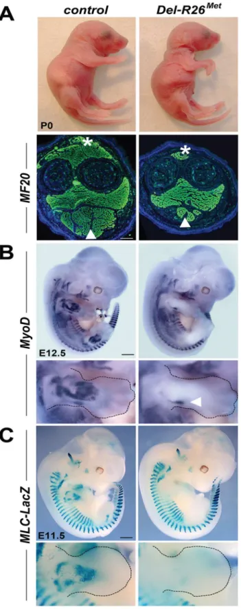
Fig 1. Ubiquitously excess wild-type Met in developing embryos results into hyperflexed forelimbs. (A) Top:Del-R26Metand control P0 mice showing hyperflexed limbs in mutants. Bottom: anti-myosin heavy chain II IHC using MF20 antibodies on forelimb transversal sections of P0Del-R26Metand control mice at the
enhancer [27]. In this mouse line,nLacZ(encoding nuclearβ-galactosidase) is expressed in all skeletal muscles throughout development, permitting visualisation of muscle formation in fully recombinedDel-R26Metembryos that no longer express the LacZ-stop cassette. Whole mount staining showed thatβ-galactosidase activity was drastically reduced inDel-R26Metdeveloping limbs compared to controls (Figs1CandS4).
Intriguingly, the limb phenotype of theDel-R26Metmice is reminiscent of the phenotype we reported inMetgrb2/grb2,Met2P/2P, andMet2S/2Sspecificity-switch signalling mutants [18,23,
28]. In theseMetsignalling mutants, Met-dependent migration of myoblasts is severely com-promised, leading to a reduction of limb muscles more pronounced in the dorsal than in the ventral limb compartment [18,23,28]. We therefore assessed whether the development of migrating myoblasts was also compromised inDel-R26Metmice. We addressed this issue using distinct markers of the myogenic program [29]. Whole mount ISH on E10.5 embryos using Lbx1andPax3probes revealed a drastic reduction of migrating myoblasts from the somites towards the limbs, with only few dispersed migrating cells left inDel-R26Metembryos com-pared to controls (Fig 2A and 2B). Quantification of the ISH signal throughout the limbs revealed approximately 90% reduction of migrating myoblasts inDel-R26Metforelimbs com-pared to controls (Fig 2C and 2D). Impaired migration of myoblasts towards the tongue was also found inDel-R26Metembryos (Fig 2A and 2B), indicating that ubiquitous expression of the Mettgalso compromises the developmental program of these migrating myoblasts. Together, these findings show that ubiquitously enhancing wild-type Met-RTK specifically interferes with the limb muscle developmental program by perturbing myoblast migration.
Enhanced wild-type Met expression is permissive in migrating
myoblasts, but not in limb mesenchyme
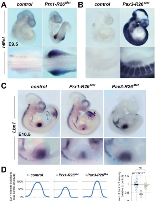
The intriguing similarity of the limb muscle phenotype in gain-of-functionDel-R26Metand in loss-of-functionMetspecificity-switch signalling mutants [18,23,28] could be interpreted in two ways. One scenario could be that in myoblasts, the amount of Met signalling must be quali-tatively and/or quantiquali-tatively maintained within a narrow range. If the signalling level is reduced (as inMetspecificity-switch signalling mutants) or enhanced (as inDel-R26Met mutants), the migration program may not occur. Alternatively, exogenous Met expression might act in limb mesenchymal cells by altering their permissiveness to invading myoblasts. To discriminate between these two possibilities, we selectively enhanced Met expression in either migrating myoblasts or in limb mesenchyme, using thePax3-Creknock-in or thePrx1-Cre transgenic lines, respectively [30,31]. Consistently, whole mount ISH inPrx1-R26Metand Pax3-R26Metmutants confirmed that expression of theMettgis adequately targeted toPrx1and Pax3expression domains, respectively (Fig 3A and 3B). Whole mount ISH withLbx1andPax3 probes unequivocally revealed a significant impairment in myoblast migration inPrx1-R26Met, but not inPax3-R26Metmutants (Figs3C,S5A and S5B). Quantification analyses revealed approximately 50% reduction of limb myoblasts inPrx1-R26Metmutants compared to Pax3-R26Metand control embryos (Figs3D,S5C and S5D). At E12.5, limb-forming muscles were reduced inPrx1-R26Met, but not inPax3-R26Metembryos, as revealed byMyoDISH (S5E Fig), even though the severity of this phenotype appeared less pronounced compared to earlier stages. Finally,Prx1-R26Metmice showed hyperflexed forelimbs and weak hindlimbs at birth
(arrowhead) muscle mass in mutants. (B, C) Whole mount ISH withMyoDprobe of E12.5 embryos (B) andβ -galactosidase staining of E11.5 embryos (C) showing that developing appendicular muscles are reduced in
Del-R26Metembryos (limbs are outlined in panels). The arrowhead in bottom panel B indicates developing ventral limb muscles (flexor). Scale: 500μm.
(S5E Fig; 90%, n = 16), whereasPax3-R26Metmutants were indistinguishable from controls. Together, these results disqualify myoblasts and identify limb mesenchymal cells as the cell type in which enhanced Met signalling acts to disrupt myoblast migration. This suggests that myoblast migration towards limb mesenchyme is impaired when exogenous Met expression overlaps with the endogenous source of HGF.
The myoblast migration defect inPrx1-R26Metmutants underlines the importance of spatial restriction of Met expression during development. We therefore asked whether alteration of the regionalised distribution of Met also interferes with the molecular program regulating limb patterning and skeletal morphogenesis. We followed expression ofShhandFgf8, two major regulators of early limb patterning and growth, and found no obvious defects inDel-R26Met mutants compared to controls at E10.5 (S6A Fig). Furthermore, skeletal staining analyses did not reveal any major defects in bone and cartilage formation inDel-R26Metforelimbs at birth
Fig 2. Myoblast migration is impaired inDel-R26Metmutants.(A, B) Whole mount ISH of E10.5 embryos withLbx1(A) andPax3(B) probes showing drastic reduction of migrating myoblasts towards the developing tongue (arrowhead), fore and hind limbs. Bottom panel reports an enlargement at forelimb levels. (C, D) Quantification analyses ofLbx1(C) andPax3(D) positive domains in forelimbs. Left panels: each plot represents the average signal distribution along the white line in forelimbs. Right panels: quantifications and statistical analyses of the sum of signal intensity based on intensity plots in left panels. Numbers of samples forLbx1: control, n = 13;Del-R26Met, n = 4; forPax3: control, n = 11;Del-R26Met, n = 8. The sum ofPax3signal intensity was calculated
between point A and B: A indicating a fixed position between the somites and the limb whereas B being placed at a fixed distance from A. Note almost lack of signal inDel-R26Metmutants. Scale: 500μm. Mann-Whitney and Student-ttest.
Fig 3. Ectopic Met in limb mesenchyme drastically reduces limb colonization by migrating myoblasts. (A, B) Whole mount ISH using the humanMetprobe showing the domain with Cre recombinase activity in
Prx1-R26Met(A) andPax3-R26Met(B) embryos. Note that: a) inPrx1-R26Metembryos, the expression of the
Mettg(detected byhMetprobe) is restricted in forelimbs; b) inPax3-R26Metembryos,Mettgexpression is
found inPax3-positive territories. (C) Whole mount ISH of E10.5 embryos with theLbx1probe showing drastic reduction of migrating myoblasts in the forelimbs ofPrx1-R26Met, but not ofPax3-R26Metmutants. Note that intact migration of myoblasts towards the forming tongue (arrowhead) inPrx1-R26Metmutants
correlates with the restricted expression of enhanced Met in limb mesenchyme. Bottom panel reports an enlargement at forelimb levels. (D) Quantification analyses ofLbx1positive domain in forelimbs. Left panels: each plot represents the average signal distribution along the white line in forelimbs. Right panel:
quantifications and statistical analyses of the sum of signal intensity based on intensity plots in left panels. Numbers of samples forLbx1: control, n = 13;Prx1-R26Met, n = 4;Pax3-R26Met, n = 5. Note the reducedLbx1
level inPrx1-R26Metmutants. Mann-Whitney and Student-ttest.
(S6B Fig). Together, these findings highlight the robustness of limb skeletal patterning pro-gram, ensured by the intercalation of multiple signalling components, which does not permit interference by ectopic Met.
We next analysed whether alteration of the regionalised distribution of Met leads to molecu-lar changes in limb buds by following mainly the expression levels of cell surface proteins such as heparan-sulfate proteoglycans (HSPGs), glypicans [32–34] and syndecans [35], which act as gatekeepers of cellular responses by modulating extracellular signal distribution and perception by targeted cells. We also analysed the expression levels of HSPG modifiers, such asNotum, Hst2st1, andHst3st1[36–38]. Among these candidates, some have been previously reported to also modulate HGF signalling [39–41] or to be regulated by HGF/Met [42–44]. We also ana-lysedSdf1levels for its cooperative function with Met signalling to control myoblast migration [45]. A total of 12 genes were screened: the 6Glypicans,Syndecan3and4,Hst2st1,Hst3st1, Notum, andSdf1. To analyse the expression levels of these genes, E10.5 forelimbs dissected fromDel-R26Metmutants and controls were used for qRT-PCR. Studies were restricted to embryos that showed low levels ofβ-galactosidase activity in the whole body as a read-out of efficient activation of the Mettg(seeS1 Fig). We first assessed the sensitivity of this approach in detecting molecular changes possibly occurring in a limited number of cells within the limb. In particular, we quantified transcript levels ofPax3and mouseMet, both expressed in migrating myoblasts, and consistently found a drastic reduction in their mRNA levels inDel-R26Met limbs compared to controls (Fig 4). Second, we confirmed a switch inLacZversusMettg expression in control andDel-R26Metlimbs (S7A Fig). We next screened the 12 selected candi-dates by comparing their expression levels in control versusDel-R26Metlimbs. No significant changes were observed in the expression levels of allGlypicans,of Syndecan3,Hst2st1,Hst3st1, andSdf1(S7B and S7C Fig). In contrast, we found that expression levels ofNotumand Synde-can4were significantly reduced inDel-R26Metversus control limbs (Fig 4). We next asked whether such molecular differences inDel-R26Metmutants resulted from the depletion of limb myoblasts, or reflected expression changes in limb mesenchyme. To address this question, we took advantage ofMetloss-of-function mutants (MetLacZ/d(neo)), in which limbs are also devoid of migrating myoblasts (S8 Fig), but where no specific changes in limb mesenchyme can be expected [18,22]. Consistently,Pax3and mouseMetmyoblast-specific transcripts were also absent fromMetLacZ/d(neo)mutant limbs (Fig 4). In contrast, the expression levels ofNotumand Syndecan4(changed inDel-R26Metlimbs) were similar inMetLacZ/d(neo)and control limbs (Fig 4). Altogether, these results exclude that downregulation ofNotumandSyndecan4in Del-R26Metlimbs is a consequence of lack of migrating myoblasts, and identify them as molecular changes caused by Met expression in limb mesenchyme.
Met-expressing myoblasts are capable of buffering enhanced wild-type
Met levels
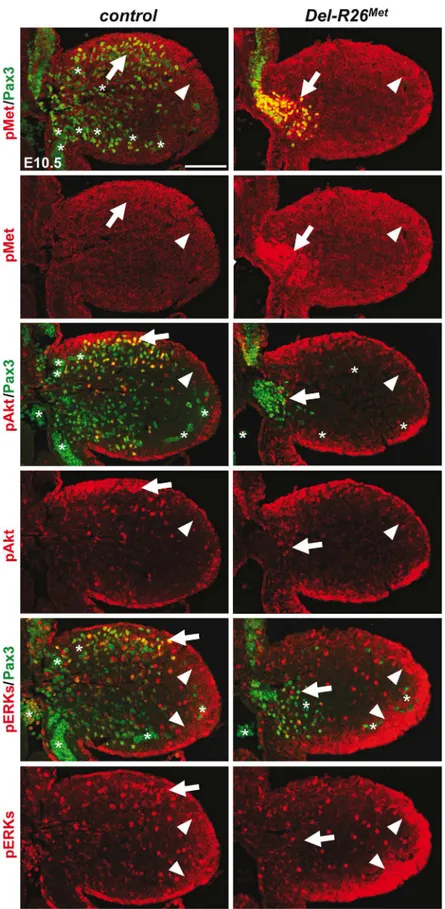
The intact myoblast migration inPax3-R26Metmutants could reflect either the robustness of the migration response to increased activation levels of Met intracellular effectors or an intrin-sic capability of cells to restrain Met-RTK input. By performing IHC on embryo sections, we followed Met signalling levels in control and mutant forelimbs at E10.5, when myoblast migra-tion occurs. In myoblasts, levels of pTyr1234–1235-Met were enhanced inDel-R26Met
(non-migrating cells blocked at the base of the limb) andPax3-R26Met(migrating cells progressing within the limb) compared to controls, whereas no major changes were observed in
andPrx1-R26Metmyoblasts, despite high levels of phospho-Met inDel-R26Metand Pax3-R26Met(arrows inFig 5andFig 6B) Together, these results show that myoblasts have the competence to buffer enhanced Met levels. In contrast, in limb mesenchymal cells levels of phospho-Met were slightly increased inDel-R26MetandPrx1-R26Metwhen compared to con-trol andPax3-R26Metembryos (Figs5and6A). This is best seen by comparing levels in areas devoid of myoblasts even in control embryos (arrowheads inFig 5and“distal mes”boxes in
Fig 6A). This was accompanied by an increase in the phosphorylation levels of Akt and more moderately of ERKs (Figs5and6B). These findings indicate that limb mesenchymal cells lacks the capacity to buffer an ectopic Met activation, which in turn causes defects in myoblast migration.
Restriction of enhanced Met signalling occurs at distinct levels as
revealed by biochemical studies in embryonic hepatocytes
We next explored how buffering of enhanced Met signalling occurs in cells. Because of techni-cal difficulties in isolating and establishing primary myoblast cultures from E10.5 embryos for quantitative western blot studies, we addressed this issue using primary embryonic hepato-cytes in which endogenous Met is expressed and required for their survival, as shown through our earlier studies ofMetsignalling mutants [18,28,46–48]. Primary embryonic hepatocyte
Fig 4. Ectopic Met in limb mesenchyme down-regulates the expression levels ofNotumandSyndecan4.qRT-PCR analysis of transcript levels of mouseMet(mMet),Pax3,Notum, andSyndecan4(Sdc4) in controls (ctrl; n = 11),Del-R26Met(Del-Met; n = 11),MetLacZ/d(neo)(KO; n = 7). Each dots
corresponds to transcript levels in forelimbs of E10.5 individual embryos (done in triplicate). Columns correspond to the average value, expressed as mean±s.
e.m. Note: downregulation ofmMetandPax3inDel-R26MetandMetLacZ/d(neo)mutants compared to control, consistent with lack of migrating myoblasts;
downregulation ofNotumandSyndecan4inDel-R26Metmutants compared to control, whereas no significant changes were found inMetLacZ/d(neo)mutants. Mann-Whitney and Student-ttest.
Fig 5. Enhanced Met expression levels inDel-R26Met
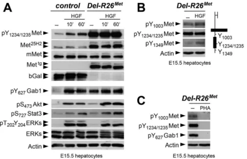
cultures were established from E15.5R26stopMetandDel-R26Metembryos and used to follow biochemical changes in the Met signalling cascade. As expected,Del-R26Metembryonic hepato-cytes express the Mettg, but not theβ-galactosidase (in the stop cassette;Fig 7A). Total Met pro-tein levels inR26stopMetandDel-R26Metprimary embryonic hepatocytes were assessed by using the Met25H2antibodies generated against a synthetic peptide containing the amino acids sur-rounding the Tyr1234–1235within the kinase domain (recognising human and mouse Met). Quantification of the Met25H2levels revealed an approximately 4-fold increase of total Met pro-tein amount inDel-R26MetversusR26stopMetcontrols. Increased levels of Met proteins correlate with a concomitant increase in Met phosphorylation levels on Tyr1234–1235(critical for Met kinase activation; visualized with an antibody recognising mouse and human phosphorylated Met), which are not further enhanced upon HGF stimulation (Fig 7A). We found two intrigu-ing aspects of enhanced Met expression on downstream components of the Met signallintrigu-ing cas-cade. Concerning Gab1, a cytoplasmic protein directly recruited by Met and functioning as a platform for Met effectors, we found that: a) it was already phosphorylated inDel-R26Metcells to a level comparable to that of control cells upon HGF stimulation, b) its phosphorylation lev-els inDel-R26Metcells was not further enhanced upon HGF stimulation (Fig 7A). Concerning further downstream Met effectors such as Akt, Stat3, and ERKs, we found that their phosphor-ylation levels increased upon HGF stimulation with comparable kinetic profiles inDel-R26Met and control cells (Fig 7A). These findings indicate that the constitutive enhancement of Met expression and phosphorylation on Tyr1234–1235in hepatocytes does not result in a constitutive activation of downstream signalling components.
To get insights into how enhanced Met signalling is restricted in embryonic cells, we ana-lysed the phosphorylation levels of two other tyrosine residues implicated in receptor signalling and endocytosis. Intriguingly, we found basal levels of Met phosphorylation on Tyr1003(critical for Met protein ubiquitination and degradation) and on Tyr1349(one of the two multifunc-tional docking sites required for Met signalling), which were enhanced upon HGF stimulation (Fig 7B). Consistently, Met and Gab1 phosphorylation was impaired in the presence of the Met inhibitor PHA665752 (Fig 7C). Thus, basal levels of Met phosphorylation in mutant embry-onic hepatocytes appear sufficient to initiate signalling activation (e.g. Gab1 phosphorylation on Tyr627), but not to propagate signalling activation to downstream pathways (e.g. Akt, Stat3, ERKs). Together, these findings indicate that the Mettgachieves its full signalling competence following a burst of HGF stimulation.
Gain of wild-type Met expression in limb mesenchyme does not perturb
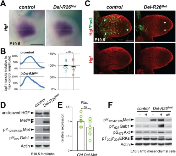
expression pattern and levels of its ligand HGF, but its bioavailability
As the domain of Prx1-cre activity in the limb encompasses the source of HGF exerting che-moattraction on migrating myoblasts, we next asked whether ectopic Met in these cells alters the bioavailability of HGF to migrating cells. ISH and IHC analyses revealed no significant dif-ferences on HGF expression patterns or levels betweenDel-R26Metand control embryos (Fig 8A, 8B and 8C), ruling out the possibility that ectopic Met altersHgfgene expression and HGF protein production in limb mesenchyme. Moreover, we found comparable protein levels of uncleaved HGF in E10.5Del-R26Metand control forelimbs (Fig 8D) as well as unchanged
showing the distribution of phospho-Met (on Tyr1234–1235), phospho-Akt, phospho-ERKs (red) and of Pax3
protein (green) in myoblasts. Note ectopic phospho-Met in limb mesenchyme (arrowheads) and in non-migrating myoblasts (arrows) inDel-R26Metmutants. Asterisks indicate non-specific staining in blood cells. Scale: 100μm.
Fig 6. Immunohistochemical analysis showing that myoblasts possess a buffering competence to enhanced Met levels.(A) Limb transverse sections of E10.5 control,Prx1-R26Met, andPax3-R26Met
embryos showing the phospho-Met (red) and Pax3-positive myoblasts (green). Note ectopic phospho-Met in limb mesenchyme ofPrx1-R26Metand in migrating myoblasts ofPax3-R26Metmutants. Few migrating myoblasts are present in limb mesenchyme ofPrx1-R26Metmutants. White boxes indicate the position of
enlargements shown in panel B. Yellow boxes indicate distal mesenchyme (distal mes) area devoid of myoblasts where enhanced phospho-Met signal is detected inPrx1-R26Metmutants. (B) High magnification of limb transverse sections showing phosphorylation levels of Met, Akt, and ERKs (red) in migrating
myoblasts (Pax3-positive; green) of E10.5 control,Prx1-R26Met, andPax3-R26Metembryos. Note that despite
the high phospho-Met levels inPax3-R26Metmutants, no major changes are observed in phospho-Akt and
mRNA levels of urokinase-type plasminogen activator (Plau), an enzyme involved in HGF pro-cessing (Fig 8E).
Next, we investigated whether Met expression in mesenchymal territories would alter HGF bioavailability. For this purpose, we established primary embryonic mesenchymal cell cultures from E10.5 forelimbs and biochemically assessed their competence to respond to HGF stimula-tion. As expected, control limb mesenchymal cells did not respond to HGF stimulation, consis-tent with the fact that these cells do not express the receptor (Fig 8F). In contrast, we found high levels of Met phosphorylation on Tyr1234–1235inDel-R26Metlimb mesenchymal cells (Fig
8F). HGF stimulation induced Gab1 phosphorylation and enhanced the basal levels of Akt and ERKs phosphorylation (Fig 8F). Furthermore, treatment with anti-HGF blocking antibodies drastically reduced the phosphorylation levels of Met inDel-R26Metlimb mesenchymal cells, thus showing that HGF is indeed produced by these cells and that basal Met phosphorylation is due to endogenous HGF in these culture conditions (Fig 8F). Finally, we biochemically ana-lysed the levels of HGF in conditioned media ofDel-R26Metlimb mesenchymal cells and found that levels of uncleaved HGF were comparable to those of control cells (S9A Fig).
To explore whether the HGF released by control andDel-R26Metcultures was bioactive, we co-cultured MDCK cells with E10.5 dissociated limb mesenchymal cells as a source of HGF
Fig 7. Biochemical analyses in embryonic hepatocytes show restriction of enhanced Met signalling at distinct levels.(A) Western blot analyses of total protein extracts of E15.5 primary embryonic hepatocytes derived fromR26stopMet(containing the LacZ-stop cassette therefore expressing theβ-galactosidase) and
fromDel-R26Met(after deletion of the LacZ-stop cassette, therefore expressing the transgenic Met detected
by anti-human Met antibodies) embryos. Analyses was performed before and after HGF stimulation (50ng/ml). Note: a) similar levels of mouse Met inR26stopMetandDel-R26Metcells; b) high levels of Met
phosphorylation on Tyr1234–1235inDel-R26Metcells independently of HGF stimulation; c) comparable Gab1,
Akt, Stat3, and ERK phosphorylation levels upon HGF stimulation inR26stopMetandDel-R26Metcells; d)
despite Gab1 phosphorylation in untreatedDel-R26Metcells in contrast to controlR26stopMetcells, the phosphorylation levels of Akt, Stat3, and ERK is unchanged. Actin protein levels were used as loading controls in all western blot analyses. (B) Left: western blot analyses of total protein extracts of E15.5 primary embryonic hepatocytes derived fromDel-R26Metembryos. Note basal levels of Met phosphorylation on Tyr1003and on Tyr1349; phosphorylation levels are further increase upon HGF stimulation (50ng/ml). Right:
schematic representation of Met indicating the different tyrosine residues analysed by western blots. (C) Western blot analysis of total protein extracts of E15.5 primary embryonic hepatocytes derived from Del-R26Metembryos showing impaired phosphorylation of Met and Gab1 in the presence of the Met inhibitor PHA665752 (PHA; 1μM).
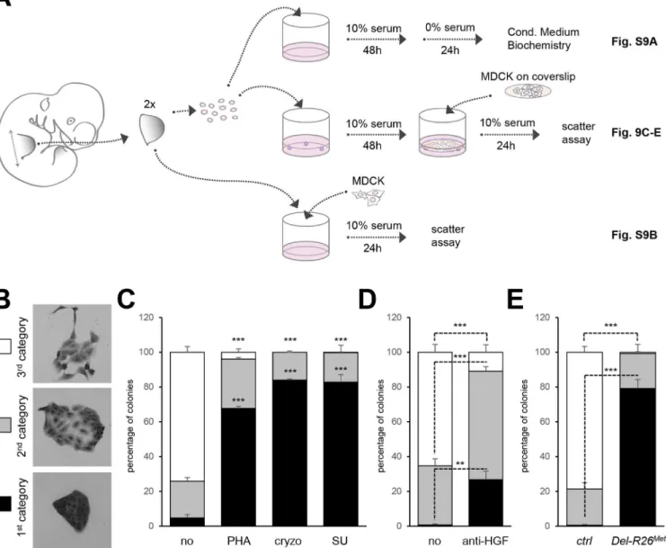
(Fig 9A). MDCK cells acquire a“scattered phenotype”upon HGF stimulation [49,50], thus providing an excellent readout of HGF bioavailability in conditioned media by limb mesenchy-mal cells. To evaluate HGF bioavailability, we established an experimental setting in which MDCK cells were co-cultured either with dissociated limb mesenchymal cells or with dissected limbs (Fig 9A). HGF activity was quantified by scoring the percentage of MDCK clones exhib-iting a dense phenotype (category I), an expanded phenotype (category II), or a scattered phe-notype with some or several cells detached from the clone (category III;Fig 9B). A condition with mesenchymal cells from two control forelimbs consistently triggered an efficient MDCK
Fig 8. Ectopic Met does not change the expression profile and levels of HGF in limb mesenchyme.(A) Whole mount ISH of E10.5 control andDel-R26Metembryos withHgfprobe showing comparable expression
domain in developing forelimbs. (B) Quantification analyses ofHgfpositive domains in forelimbs. Left panels: each plot represents the average signal distribution along the white line in forelimbs. Right panels:
quantifications and statistical analyses of the sum of signal intensity based on intensity plots in left panels. Numbers of samples: control, n = 6;Del-R26Met, n = 6. Note no significant changes of signal in controls and
Del-R26Metmutants. (C) Immunofluorescence analysis of HGF (red) and Pax3 (green) proteins in transversal
sections of E10.5 control andDel-R26Metembryos at forelimb levels. Note the impaired myoblast migration
(Pax3-positive cells) inDel-R26Metmutants, whereas no significant changes are detected in HGF protein levels. Asterisks indicate non-specific staining in blood cells. Scale: 100μm. (D) Western blot analyses of total protein extracts of E10.5 control andDel-R26Metdissected forelimbs. Note comparable levels of uncleaved
HGF inDel-R26Metmutants despite the ectopic expression of Mettgand the enhanced phosphorylation levels
of Met and Gab1. (E) qRT-PCR analysis showing comparablePlautranscript levels in E10.5 dissected forelimbs of control (ctrl; n = 11) andDel-R26Met(Del-Met; n = 11) embryos. (F) Western blot analysis of total
protein extracts of E10.5 forelimb mesenchymal cell cultures showing phosphorylation levels of Met, Gab1, Akt, and ERKs in control andDel-R26Metcultures, in cells untreated, stimulated with HGF (H) or in the
presence of anti-HGF blocking antibodies (αH).
scattering response (Fig 9C). MDCK scattering response triggered by conditioned media from control limb mesenchymal cells was drastically reduced in the presence of Met inhibitors (PHA665752, cryzotinib, or SU11274: 1μM;Fig 9C) or of anti-HGF blocking antibodies
(anti-HGF: 30μg/ml;Fig 9D). In contrast, we found a drastic reduction in the percentage of MDCK
colonies with a scattered phenotype using mesenchymal cells from twoDel-R26Metforelimbs (Fig 9E). Similar results were obtained using MDCK cells directly co-cultured with E10.5
Fig 9. Ectopic Met in limb mesenchyme alters HGF bioavailability.(A) Scheme illustrating the experimental procedure employed for evaluating through MDCK cell scattering the bioavailability of HGF from control andDel-R26Metmutant limb mesenchymal cells or from dissected forelimbs. The scheme indicates the experimental procedure applied for collecting media conditioned by limb mesenchymal cells for biochemical analysis (top; shown inS9A Fig), for MDCK scattering assays using co-cultures with limb mesenchymal cells (middle; shown in Fig 9C, 9D and 9E) or with dissected limbs (bottom; shown in
S9B Fig). (B) Pictures of MDCK colonies showing the three categories that were defined to determine the extent of cell contact and spreading for quantification studies of scattering response. (C) Quantitative analysis of MDCK cell scattering in co-cultures with control limb mesenchymal cells in the absence (no) and in the presence of the Met inhibitor PHA665752 (PHA; 1μM), cryzotinib (Cryzo; 1μM), or SU11274 (SU; 1μM). (D) Quantitative analysis of MDCK cell scattering in co-cultures with control limb mesenchymal cells in the absence (no) and in the presence of the anti-HGF blocking antibodies (anti-HGF; 30μg/ml). (E) Quantitative analysis of MDCK cell scattering in co-cultures with control orDel-R26Metlimb mesenchymal cells. Note a drastic reduction
in the scattering response when MDCK cells are co-cultured withDel-R26Metmutant cells (control: n = 4;Del-R26Met: n = 3). Mann-Whitney and Student-t
test.
forelimb explants (S9B Fig). Together, these findings indicate that although ectopic Met expression in limb mesenchyme does not alter HGF expression pattern or levels, it impairs bio-availability of HGF from mesenchymal cells.
Discussion
The events that cells experience overtime shape progressively their intrinsic capability to respond and to integrate RTK inputs. Our data illustrate that enhanced wild-type RTK signal-ling is differentially interpreted by cells according to their competence and the robustness of developmental programs they undergo. Most developing cell types appear insensitive to changes in Met expression either because enhanced Met protein levels is not followed by its activation (e.g. in the absence of ligand), or because it is neutralized by the action of other sig-nals acting on cells. This is not the case of migrating myoblasts that, although insensitive them-selves to enhanced Met expression, failed to migrate towards the limb buds as a result of ectopic Met expression in mesenchymal cells. Ectopic Met in the mesenchyme perturbs tran-scriptional regulation of some genes and interferes with HGF bioavailability, thus rendering the mesenchyme non accessible to myoblasts (Fig 10). Our results demonstrate that whereas most cell types during development are capable of handling moderate perturbations in RTK signalling levels, certain cell types are vulnerable to alterations in changes of spatial RTK action.
Cell competence to Met activation
The strategy of using the wild-type Met-RTK, rather than constitutive active forms, to generate Del-R26Metmice has enabled expression of the receptor at more physiological levels. A moder-ate increase in Met levels during development drastically affects limb muscle formation by per-turbing myoblast migration. Although we cannot exclude subtle phenotypes in other
developing organs, these results highlight the restricted sensitivity of the myoblast migration process to ectopic Met in limbs. We genetically demonstrate that myoblasts themselves are not susceptible to enhanced Met signalling levels for their migration, when gain of expression is achieved in the myogenic lineage withPax3-Cre. This context contrasts with a genetic setting in whichPax3gain-of-function triggers ectopic myoblast delamination and migration at non-limb somite levels, caused by Met over-expression [51]. It is therefore tempting to propose the existence of a threshold level of sensitivity to enhanced Met signalling for migrating myoblasts: Met signalling is enhanced below this level inDel-R26Met, thus failing to elicit any qualitative change in myoblast behaviour, whereas Met signalling is enhanced above this threshold in Pax3gain-of-function mutants, thus triggering ectopic delamination. However, it is also possi-ble that the effects ofPax3gain-of-function mutants involve missregulation of otherPax3 tar-get genes. TheMetspecificity-switch mutants have previously revealed that myoblast
migration is impaired by lowering qualitative and/or quantitative Met signalling levels [18,28]. Thus, findings from the present studies together with those discussed here highlight the exis-tence of a precise window of Met signalling compatible with proper myoblast migration: within this window, cells can buffer fluctuations in RTK signalling input to avoid changes in qualita-tive output. This also implies that myoblasts are competent to read Met signalling for proper migration within a defined quantitative range.
Modulating HGF bioavailability in limb mesenchyme for muscle
patterning
local source of HGF exerting a chemoattractant effect on myoblasts that migrate from the somites. In a wild-type context, migrating myoblasts experience a territory devoid of Met-posi-tive cells along their path. Our results ruled out that ectopic Met would act by altering the HGF mRNA and protein expression levels in limb mesenchyme. Instead, they provide evidence that
Fig 10. Schematic representation summarizing the different molecular and phenotypic effects of enhanced Met expression in myoblasts and limb mesenchymal cells.In a wild-type context (top), limb mesenchymal cells secrete HGF required for migration of myoblasts towards the limb buds. Enhanced expression of Met in myoblasts (as assessed inPax3-R26Metembryos; middle) does not alter their migration due to a buffering event: the activation levels of signalling effectors such as ERKs and Akt are restrained despite enhanced Met phosphorylation. The size of each signal is representative of their
phosphorylation levels. Limb mesenchymal cells are vulnerable to ectopic Met expression (as assessed inPrx1-R26Metembryos; bottom), illustrated by
changes in gene expression, by failure of HGF bioavailability, and by myoblast migration defects. Alteration of HGF bioavailability can be due to: 1) upregulation of a negative interactor that would interfere with the capacity of HGF to bind/activate Met (indicated as“HGF inhibitor”), 2) downregulation of a HGF interactor acting as enhancer of its bioactivity (indicated as“HGF activator”), 3) expression of a chemorepellent factor that renders limb mesenchyme inaccessible to migrating myoblasts, or 4) HGF titration by ectopic Met in mesenchymal cells (indicated as“HGF trapping”).
limb mesenchyme must be devoid of Met expressing cells to ensure normal HGF bioavailabil-ity. Although we cannot fully exclude that ectopic Met acts by titrating the HGF secreted by limb mesenchyme, a series of experimental arguments make this possibility unlikely. Compara-ble levels of HGF protein were found in conditioned media from control andDel-R26Metlimb mesenchymal cells, in spite of reduced scattering-induced activity, indicating that HGF secre-tion and release is not impaired by the occurrence of autocrine HGF/Met binding. Moreover, an event of HGF trapping by Met should recapitulate all developmental defects observed in Metloss-of-function mutants. Although the molecular mechanism remains to be identified, it is tempting to speculate that defects in HGF bioavailability could be linked to transcriptional and/or posttranscriptional changes caused by ectopic Met in limbs. This possibility is sup-ported by our data showing signalling (e.g. phospho-Met, phospho-Gab, and phospho-ERKs upregulation) and transcriptional (e.g.NotumandSyndecan4downregulation) changes occur-ring in mutant limbs compared to controls. Thus, ectopic Met expression and activation in limb mesenchyme triggers a number of qualitative responses that cannot be buffered by cells. Our co-culture studies highlight a difference in HGF bioavailability, which could result from a change in expression levels of a modulator of HGF, such as either the downregulation of a HGF interactor acting as enhancer of HGF bioactivity, or the upregulation of a negative inter-actor that would interfere with the capacity of HGF to bind/activate Met (Fig 10). Among posi-tive modulators, it has been shown that Glypican1 regulates HGF/Met-triggered migration of C2C12myoblasts [52], Syndecan4 is expressed together with Met in adult satellite cells where it contributes to muscle regeneration [53,54], and Glypican4 sustains HGF-mediated branching morphogenesis [39]. Alternatively, mutant cells could express a chemorepellent factor that would render the limb mesenchyme inaccessible to migrating myoblasts (Fig 10). The identifi-cation and the functional validation of the molecular mechanism(s) responsible for the non-cell autonomous defect of myoblast migration inDel-R26Metmutants is highly challenging, as: 1) a putative HGF modulator could be altered at the level of transcription, post-transcription, and/or secretion; 2) modulators of chemoattractants do not exert an“on or off”action, but rather fine tune signalling mechanisms, and may act redundantly with one another; 3) distinct mechanisms could cooperate to cause this qualitative phenotype, each of them participating although with a variable quantitative extent.
Robustness versus sensitiveness of developmental programs to
fluctuation on RTK signalling levels
The present study further expands our knowledge on robustness versus sensitiveness of cells in developmental processes with respect to subtle increases in RTK signalling. Besides the sen-sitivity of myoblasts to changes in Met signalling in limb mesenchyme, most other develop-mental processes appear robust and capable of buffering excess Met-RTK signalling (Fig 10). How can most cells deal with RTK signalling perturbation? For cells with endogenous Met, one possibility is that they can buffer the additional burst of RTK signalling by“considering”it as a negligible quantity with respect to the overall signalling network operating in cells. Such buffer-ing competence—or resilience—would be possible as long as this additional burst of RTK occurs within a defined quantitative window. Our biochemical studies show that effectiveness of the enhanced Met expression and activation is limited at different intracellular levels. A first point of restriction occurs at the level of the receptor itself: although Mettgis fully phosphory-lated on Tyr1234–1235that are critical for its kinase activation, it only becomes fully signalling competent after HGF stimulation, as shown by HGF-induced increased phosphorylation levels on Tyr1003and Tyr1349implicated in receptor signalling and endocytosis and by activation of Akt, STAT3, and ERKs. Thus, inDel-R26Metcells, HGF switches Met from a subthreshold sig-nalling status into a fully sigsig-nalling competent form. These results also show that theR26stopMet mice represent a valuable genetic model to explore the consequences of moderate perturbations of Met as its full signalling competence still depends on ligand stimulation. A second point of restriction occurs at the levels of Gab1, which is phosphorylated at comparable levels in control andDel-R26Metcells upon HGF stimulation. Notably, in the absence of HGF stimulation Gab1 is phosphorylated inDel-R26Methepatocytes (also expressing endogenous Met), but not in Del-R26Metmesenchymal cells (not expressing endogenous Met), indicating that the competence of cells to restrict signalling of enhanced Met is different according to the cell type, and that this competence can be influenced by factors such as expression levels of the receptor. Another restriction occurs further downstream, as revealed by the comparable phosphorylation levels of Akt, STAT3, and ERKs observed in control andDel-R26Methepatocytes (by biochemical stud-ies in culture) and myoblasts (by IHC in vivo). Such restrictions imply that different cell types possess distinct mechanisms that sense and calibrate the level of activation required for and compatible with biological programs. Overall, our studies provide additional insights into the existence of an exquisite monitoring of signalling levels within cells to attenuate enhanced RTK levels.
For cells that do not express endogenous Met, the issue of buffering an ectopic Met is only relevant provided that cells possess the adequate co-factors and downstream effectors. When they do, buffering may occur through mechanisms similar to those in cells with endogenous Met. Whereas most territories/cell types with ectopic Met appear capable of resilience, the limb mesenchyme is vulnerable to this signalling perturbation, illustrated by changes in gene expres-sion, by failure of HGF bioavailability, and by resulting myoblast migration defects.
Conditional
R26
stopMetmouse model to enhance wild-type Met signalling
in skeletal muscle
[56]. Loss ofFat1in mice causes muscle shape defects resulting from altered migration polarity of a selective group of muscles matching those affected in FSHD [56].Fat1expression is also regulated by HGF/Met, by Pax3-FKHR, and by Lbx1 [42,45,57]. Alteration of Met signalling can also impact muscle homeostasis by causing atrophy [58] and tumour formation. For exam-ple, Met is overexpressed in rhabdomyosarcoma, where it contributes to invasive growth [57], andMetgene amplification is frequently associated with sarcoma susceptibility in muscular dystrophy mouse models [51]. Therefore,R26stopMetmice can be instrumental for future evalu-ation of how Met signalling impacts on muscle physiology, regenerevalu-ation, and/or pathologies.
Conclusion
TheR26stopMetmice modelling conditional gain of RTK signalling exemplify robustness versus sensitiveness of cells to signalling perturbations in order to ensure reproducible developmental outcomes. The genetic approach we employed permits subtle changes in RTK signalling, in contrast to others where constitutive RTK activation, either by point mutations or ligand over-expression, leads to dramatic biological consequences. It is therefore not surprising that the Del-R26Metmice do not recapitulate defects reported in other transgenics with an over-activa-tion of the HGF/Met system. It is the case of mice in which HGF over-expression causes aber-rant myoblast and neural crest migration, leading to ectopic muscle formation and melanosis in the central nervous system [59,60]. A number of transgenic mice have been instrumental to demonstrate the capability of oncogenic forms of RTKs to trigger neoplasia. However, these mice do not permit assessment of how cells perceive and handle moderate changes in RTK sig-nalling overtime. TheR26stopMetgenetic setting represents therefore a suitable system to explore the in vivo robustness of cells to subtle increase of RTKs signalling during tissue homeostasis as well as during development and may disclose unexpected switches in cell sensitivity from development to adulthood.
Materials and Methods
Ethics statement
All procedures involving the use of animals were performed in accordance with the European Community Council Directive of 24 November 1986 on the protection of animals used for experimental purposes (86/609/EEC). The experimental protocols were carried out in compli-ance with institutional Ethical Committee guidelines for animal research (comité d’éthique pour l’expérimentation animale–Comité d’éthique de Marseille; agreement number D13-055-21 by the Direction départementale des services vétérinaires–Préfecture des Bouches du Rhône).
Transgenic lines and genotype analysis
Ethical Committee guidelines for animal research (comité d’éthique pour l’expérimentation animale–Comité d’éthique de Marseille; agreement number D13-055-21 by the Direction départementale des services vétérinaires–Préfecture des Bouches du Rhône).
RNA in situ hybridisation and histological analysis
For ISH, embryos were collected in phosphate buffered Saline (PBS) and fixed in 4% parafor-maldehyde (PFA) overnight. Whole mount RNA ISH done by using the relevant digoxigenin-labeled RNA probes. X-Gal and Salmon-Gal staining were performed as previously described [23,24,61]. For IHC of P0 limbs, newborns were embedded in cold glycol methacrylate (Tech-novit 8100) for cryosections. Limbs were cut transversally (16μm) and processed for antibodies
staining as described [24]. For IHC at E10.5, embryos were embedded in paraffin, cut transver-sally (10μm), and processed for antibodies staining as described [62].
MDCK scattering assays
Procedures were performed as described [49,50]. Briefly, MDCK cells (ATCC, Rockville, MD) were seeded in 96-well plates (for forelimb co-cultures; Corning, Acton, MA) or on coverslips (for forelimb mesenchymal cells co-cultures) at 1250 cells/cm2in DMEM containing 10% (v/v) fetal bovine serum (FBS), 100U/mL penicillin, 100μg/mL streptomycin, 4mM L-glutamine,
and 1mM of sodium pyruvate. Cells were first incubated for 24 h at 37°C, in 5% CO2to allow attachment and colony formation. For forelimb co-cultures, plates were processed as followed. In the first group, the media was changed with fresh media containing increasing concentra-tions of recombinant human HGF (0.1, 0.3, 1, 3, 10, 30ng/ml; R&D) to estimate the bioactivity dose of HGF released by wild-type limbs (comparable to 5ng/ml of recombinant human HGF). In the second group, media was replaced and added together with freshly dissected 2 forelimbs derived from E10.5 embryos of different genotypes (done by PCR using remaining tissue). Twenty-four hours later, the MDCK cells were fixed with 4% PFA in PBS, then cells were stained with Crystal violet. For quantification, at least 10 images with several cell colonies were analysed. Three different categories were defined: category 1 corresponds to not-scattered colo-nies; category 2 corresponds to colonies in which cells start losing their contact; category 3 cor-responds to colonies with visible scattered cells.
Limb mesenchymal cell cultures
Forelimbs from E10.5 embryos were dissected in HBSS supplemented with 100U/mL penicil-lin, 100μg/mL streptomycin, and 7mM Hepes (Life Technologies) and incubated for 5 min in
2% trypsin (Sigma). Limbs from individual embryos were processed separately and a piece of the body was used for genotyping. Then, trypsin was inactivated with DMEM supplemented with 10% (v/v) FBS, 100U/mL penicillin, 100μg/mL streptomycin, 4mM L-glutamine, 1mM of
R&D) where added prior to co-culture with MDCK cells on coverslips. After 24 hrs co-cultures, coverslips were fixed and processed as described above.
Western blots
Culture of primary embryonic hepatocytes were performed as previously described [28,46–
48]. Protein extracts from embryonic hepatocytes and limb mesenchymal cells were prepared and western blot (WB) analysis was performed as previously described [63,64]. For biochemi-cal analysis, conditioned media was collected, spin down to remove cellular debris, and then incubated with either heparin-beads or lectin-beads (Amersham).
Antibodies
Antibodies used were from Cell Signaling: anti-Met 25H2 (1:2000 for WB), anti-phospho-Tyr1234/1235-Met (1:2000 for WB; 1:50 for IHC), anti-phospho-Tyr1003-Met (1:2000 for WB), phospho-Tyr1349-Met (1:1000 for WB), phospho-Tyr627-Gab1 (1:2000 for WB), anti-phospho-Ser473-Akt (1:2000 for WB; 1:20 for IHC), anti-phospho-Ser727-Stat3 (1:2000 for WB), Tyr204-ERKs (#9106; 1:10000 for WB), anti-phospho-Thr202-Tyr204-ERKs (#4376; 1:150 for IHC), anti-ERKs (1:10000 for WB); from Santa-Cruz Biotech-nology: anti-mouse Met (1:200 for WB), anti-human Met (1:1000 for WB), anti-HGF (1:500 for WB); from Abcam: anti-β-galactosidase (1:2000 for WB); from R&D: anti-mouseHGF (1:50 for IHC); from Sigma-Aldrich: actin (1:12000 for WB); from Assay Designs: anti-human Met (1:500 for IHC); from hybridoma bank: Pax3 (1:10 for IHC), anti-myosin heavy chain II (MF20; 1:50 for IHC); from Jackson: anti-rabbit peroxidase or anti-mouse IgG-peroxidase (1:4000 for WB), anti-mouse fluorescent-coupled secondary antibodies (1:400 for IHC), anti-mouse or rabbit biotin-coupled secondary antibodies (1:500 for IHC).
Quantitative RT-PCR analysis
Total RNA was isolated from embryos using the RNeasy Mini Kit (Qiagen, Valencia, CA) according to manufacture instruction. cDNA was generated using the Reverse Transcription Kit (Biorad). cDNA (30ng) was amplified by real time PCR using 4μL SYBR Green qPCR
SuperMix-UDG with Rox (Biorad) and 2μL of forward and reverse primers (0.1μM). The
anal-ysis was performed on each sample in triplicates using Applied Biosystems (Foster City, CA). Relative transcript levels were calculated using the comparative Ct method and normalized to the housekeeping gene GAPDH. Primer sequences are listed inS1 Table.
Skeletal staining
New-born mice were scarified and placed in water overnight at 4°C, then eviscerated, and skin was removed. Samples were fixed in 96% ethanol overnight. Cartilage staining was performed using alcian blue solution for 24 hours (0,15mg/ml alcian blue from Sigma in 1:4 volumes of acetic acid glacial and 96% ethanol). Samples were then rinsed in ethanol 96% for 1 hour and clearing was done for 6 hours in 2% KOH. Bone staining was performed using alizarin red solution for 24 hours (0,07mg/ml alizarin red from Sigma in KOH 1%). Samples were treated with 1% KOH/20% glycerol, then stored in glycerol/ethanol (1:1 volume).
Image processing and analyses
intensity was measured along a horizontal line of a given pixel length (matching the forelimb). After background and threshold subtraction, the values were averaged between several samples of each genotype to generate an average signal distribution plot (considering left and right limbs separately). The total signal intensity was also calculated for each sample and plotted individually.
For Met25H2and HGF protein levels, quantification of signal intensity was done on western blots using the image J software. Images were process as described above. Values were averaged between different samples.
Statistical analysis
Results were expressed as the mean ±SEM. Statistical significant differences were estimated by applying unpaired t-Student test for data showing normal distribution and by Mann-Whitney test otherwise. P values are indicated in Figs.
Supporting Information
S1 Fig. Strategy to ubiquitously enhance wild-type Met in developing embryos.(A) Sche-matic representation of transgenic mice carrying the LacZ-stop cassette followed by chimeric Met before (Rosa26LacZ-stop-Met, namelyR26stopMet) and after Cre-mediated recombination ( tis-sue-specific-R26Met). (B, C) Whole mount (B) or transverse section (C)β-galactosidase staining of E10.5R26stopMetandDel-R26Metembryos. Note that different degree of Cre-mediated recombination results inDel-R26Metmutants with high (right) or low (middle)β-galactosidase activity. hb: hindbrain; ov: optic vesicles; sc: spinal cord; di: diencephalon. (D) Genotype analy-sis of embryos showing the mutant allele before and after Cre recombination. Note that the efficiency of Cre-mediated recombination results intoDel-R26Metembryos with total (right) or partial (middle) deletion of the LacZ-stop cassette. Scale: 500μm.
(TIF)
S2 Fig. Expression pattern of humanMetand mouseMetin control andDel-R26Met embryos.Whole mount ISH with humanMet(hMet) and mouse Met (mMet) probes in E10.5 control andDel-R26Metembryos. Scale: 500μm.
(TIF)
S3 Fig. Immunohistochemical analysis of phospho-Met and transgenic Met in E16.5 con-trol andDel-R26Metembryos.(A) InDel-R26Metembryos, whereas Mettg(detected by human Met antibodies; hMet) is expressed by most cell types in all tissues, phospho-Met is present only in a restricted number of cell types. Middle and bottom panels show enlargement of top panels at the levels of different organs. (B) Schematic representation of E16.5 embryos showing the level of sections reported in panel A and C. (C) Immunohistochemical analysis of phos-pho-Met and human Met (hMet) in control embryos showing background levels. (D) Table summarizing organs positive or not for phospho-Met. Scale: 200μm.
(TIF)
S4 Fig. Genetic analysis of muscle development using theMLC-LacZtransgenics.(A) Whole mountβ-galactosidase staining showing reduced developing appendicular muscles in E13.5Del-R26Metembryos compared to controls. Note residual cytoplasmicβ-galactosidase staining in theR26stopMetline due to a small proportion of cells in which the LacZ-stop cassette was not completely deleted. (B) Schematic representation of transgenic mice carrying the MLC-LacZtransgene alone (controls) or together with the transgenic Met inDel-R26Met embryos (limbs are outlined in bottom panels). Scale: 500μm.
S5 Fig. Genetic analysis of migrating myoblasts inPrx1-R26MetandPax3-R26Met condi-tional mutants.(A, B) Whole mount ISH of embryos withPax3probe showing reduced migrating myoblasts in the forelimbs ofPrx1-R26Met(A), but not ofPax3-R26Met(B) embryos. As inPax3-Creline theCregene is inserted in thePax3locus resulting also into a loss-of-func-tional allele, the adequate control embryos in panel B correspond toPax3-Creheterozygous (Pax3). (C, D) Quantification analyses ofPax3positive domain in forelimbs. Left panels: each plot represents the average signal distribution along the white line in forelimbs. Right panels: quantifications and statistical analyses of the sum of signal intensity based on intensity plots in left panels. Numbers of samples: control, n = 11;Prx1-R26Met, n = 6;Pax3, n = 4;Pax3-R26Met, n = 4. The sum ofPax3signal intensity was calculated between point A and B: A indicating a fixed position between the somites and the limb whereas B being placed at a fixed distance from A. Note the reducedPax3level inPrx1-R26Metmutants. (E) Left panels: Whole mount ISH of E12.5 embryos withMyoDprobe showing reduced developing appendicular muscles in Prx1-R26Metmutants (limbs are outlined in bottom panels). Right panel:Prx1-R26Metmice at birth show hyperflexed forelimbs. Scale: 500μm.
(TIF)
S6 Fig. Ectopic Met in developing limbs does not cause major patterning defects.(A) Whole mount ISH of E10.5 control andDel-R26Metembryos withShh(top) andFgf8(bottom) probes. Note thatShhandFgf8expression remain located in the posterior limb bud mesen-chyme and in the apical ectodermal ridge, respectively. Scale: 500μm. (B) Skeletal staining of
P0 control andDel-R26Metlimbs showing no major patterning defects. Note that the deltoid tuberosity in the humerus is significantly reduced inDel-R26Metmutants (100%: n = 7). As the deltoid tuberosity requires muscle mechanical forces at later stages besides initiating signals such as BMP4 [65], it is likely that this defect is a consequence of lack of muscles rather than a phenotype caused by ectopic Met in limb mesenchyme. Scale: 2mm.
(TIF)
S7 Fig. qRT-PCR analyses on the expression levels of candidate genes in control and Del-R26Metembryos.(A) qRT-PCR analysis of human Met (hMet) andLacZtranscripts in E10.5
control (n = 11) andDel-R26Met(n = 11) limbs. Each dots corresponds to transcript levels in forelimbs of E10.5 individual embryos (done in triplicate). Columns correspond to the average value, expressed as mean. Note that upregulation ofMettg(hMet)is paralleled by downregula-tion of theLacZtranscripts inDel-R26Metembryos. (B, C) qRT-PCR analysis of candidate genes in E10.5 control andDel-R26Metmutant limbs. No significant changes are observed. Hs2st1: heparan sulphate 2-O sulfotransferase 1.Hs3st1: heparan sulphate 3-O sulfotransferase 1.Sdf1: stromal cell-derived factor 1. Columns correspond to the average value, expressed as mean ± s.e.m. Mann-Whitney and Student-ttest.
(TIF)
S8 Fig. Met expression as visualized by the knock-in of the lacZ reporter in theMetlocus. (A, B) Whole mountβ-galactosidase staining showingMetdistribution in developing muscles of E10.5 (A) and E13.5 (B)MetLacZ/+andMetLacZ/d(neo)embryos. Note: a) the absence of mus-cles in limbs of E10.5 and E13.5MetLacZ/d(neo)embryos; b) thatMetexpression is restricted to a subgroups of developing muscles at E13.5; c) that developing limb tendons express alsoMetat E13.5 and the pattern of expression is not altered inMetLacZ/d(neo)mutants. Scale: 500μm.
(TIF)
heparin (left) or lectin (right) beads. Note no major differences in the level of uncleaved HGF between control and mutants. Numbers on the top of control and mutant lanes correspond to quantification analyses of uncleaved HGF. Quantification of the processed form of HGF was not performed because of the presence of a nonspecific band with the same molecular weight (ns). Lanes with human recombinant HGF (rec-hHGF: 5ng) and non-conditioned media (media) after purification. (B) Quantitative analysis of MDCK cell scattering using co-cultures with dissected forelimbs from control andDel-R26Metembryos with a recombination efficiency higher or lower than 60%. Quantification was performed according to categories defined inFig 9B. Note a significant increase of cell colonies corresponding to the 1stcategory using forelimbs from highly recombinedDel-R26Metmutants (control: n = 14;Del-R26Met>60%
recombina-tion: n = 5;Del-R26Met<60% recombination: n = 3). Mann-Whitney and Student-ttest.
(TIF)
S1 Table. Primer sequences used for qRT-PCR analyses. (DOCX)
Acknowledgments
We thank: R. Kelly and all members of our labs for helpful discussions and comments; Julie Leca and Egidio Caricati for their contribution at initial stages of the project; Julie Leca is also acknowledged for contribution toS3 Fig; Virginia Girod-David and Leo Jullien for excellent help with mouse husbandry at the IBDM, Mayyasa Rammah for precious advice for IHC.
Author Contributions
Conceived and designed the experiments: YF FH RD FM. Performed the experiments: YF EA SR SA FH RD FM. Analyzed the data: YF SA FH RD FM. Wrote the paper: FH RD FM.
References
1. Lemmon MA, Schlessinger J. Cell signaling by receptor tyrosine kinases. Cell. 2010; 141(7):1117–34. doi:10.1016/j.cell.2010.06.011PMID:20602996
2. Casaletto JB, McClatchey AI. Spatial regulation of receptor tyrosine kinases in development and can-cer. Nat Rev Cancan-cer. 2012; 12(6):387–400. doi:10.1038/nrc3277PMID:22622641
3. Bache KG, Slagsvold T, Stenmark H. Defective downregulation of receptor tyrosine kinases in cancer. Embo J. 2004; 23(14):2707–12. PMID:15229652
4. Maina F. Strategies to overcome drug resistance of receptor tyrosine kinaseaddicted cancer cells. Cur-rent medicinal chemistry. 2014; 21(14):1607–17. PMID:23992334
5. Flores GV, Duan H, Yan H, Nagaraj R, Fu W, Zou Y, et al. Combinatorial signaling in the specification of unique cell fates. Cell. 2000; 103(1):75–85. PMID:11051549
6. de Celis JF, Bray S, Garcia-Bellido A. Notch signalling regulates veinlet expression and establishes boundaries between veins and interveins in the Drosophila wing. Development. 1997; 124(10):1919– 28. PMID:9169839
7. Li J, Li WX. Drosophila gain-of-function mutant RTK torso triggers ectopic Dpp and STAT signaling. Genetics. 2003; 164(1):247–58. PMID:12750336
8. Trusolino L, Bertotti A, Comoglio PM. MET signalling: principles and functions in development, organ regeneration and cancer. Nat Rev Mol Cell Biol. 2010; 11(12):834–48. doi:10.1038/nrm3012PMID:
21102609
9. Huh CG, Factor VM, Sanchez A, Uchida K, Conner EA, Thorgeirsson SS. Hepatocyte growth factor/c-met signaling pathway is required for efficient liver regeneration and repair. Proc Natl Acad Sci U S A. 2004; 101(13):4477–82. PMID:15070743.
11. Chmielowiec J, Borowiak M, Morkel M, Stradal T, Munz B, Werner S, et al. c-Met is essential for wound healing in the skin. J Cell Biol. 2007; 177(1):151–62. PMID:17403932
12. Knudsen BS, Vande Woude G. Showering c-MET-dependent cancers with drugs. Curr Opin Genet Dev. 2008; 18(1):87–96. doi:10.1016/j.gde.2008.02.001PMID:18406132
13. Gherardi E, Birchmeier W, Birchmeier C, Vande Woude G. Targeting MET in cancer: rationale and progress. Nat Rev Cancer. 2012; 12(2):89–103. Epub 2012/01/25. doi:10.1038/nrc3205PMID:
22270953
14. Straussman R, Morikawa T, Shee K, Barzily-Rokni M, Qian ZR, Du J, et al. Tumour micro-environment elicits innate resistance to RAF inhibitors through HGF secretion. Nature. 2012; 487(7408):500–4. doi:
10.1038/nature11183PMID:22763439
15. Bladt F, Riethmacher D, Isenmann S, Aguzzi A, Birchmeier C. Essential role for the c-metreceptor in the migration of myogenic precursor cells into the limb bud. Nature. 1995; 376:768–71. PMID:7651534
16. Schmidt C, Bladt F, Goedecke S, Brinkmann V, Zschlesche W, Sharpe M, et al. Scatter factor/hepato-cyte growth factor is essential for liver development. Nature. 1995; 373:699–702. PMID:7854452
17. Uehara Y, Minowa O, Mori C, Shiota K, Kuno J, Noda T, et al. Placental defect and embryonic lethality in mice lacking hepatocyte growth factor/scatter factor. Nature. 1995; 373:702–5. PMID:7854453
18. Maina F, Casagranda F, Audero E, Simeone A, Comoglio P, Klein R, et al. Uncoupling of Grb2 from the Met receptor in vivo reveals complex roles in muscle development. Cell. 1996; 87:531–42. PMID:
8898205
19. Maina F, Hilton MC, Andres R, Wyatt S, Klein R, Davies AM. Multiple roles for hepatocyte growth factor in sympathetic neuron development. Neuron. 1998; 20:835–46. PMID:9620689
20. Maina F, Hilton MC, Ponzetto C, Davies AM, Klein R. Met receptor signaling is required for sensory nerve development and HGF promotes axonal growth and survival of sensory neurons. Genes and Development. 1997; 11:3341–50. PMID:9407027
21. Maina F, Klein R. Hepatocyte growth factor—a versatile signal for developing neurons. Nature Neuro-science. 1999; 2:213–7. PMID:10195212
22. Lamballe F, Genestine M, Caruso N, Arce V, Richelme S, Helmbacher F, et al. Pool-specific regulation of motor neuron survival by neurotrophic support. J Neurosci. 2011; 31(31):11144–58. doi:10.1523/ JNEUROSCI.2198-11.2011PMID:21813676
23. Caruso N, Herberth B, Lamballe F, Arce-Gorvel V, Maina F, Helmbacher F. Plasticity versus specificity in RTK signalling modalities for distinct biological outcomes in motor neurons. BMC Biol. 2014; 12 (1):56.
24. Genestine M, Caricati E, Fico A, Richelme S, Hassani H, Sunyach C, et al. Enhanced neuronal Met sig-nalling levels in ALS mice delay disease onset. Cell Death Dis. 2011; 2:e130. doi:10.1038/cddis.2011. 11PMID:21412276
25. Tonges L, Ostendorf T, Lamballe F, Genestine M, Dono R, Koch JC, et al. Hepatocyte growth factor protects retinal ganglion cells by increasing neuronal survival and axonal regeneration in vitro and in vivo. J Neurochem. 2011; 117(5):892–903. PMID:21443522. doi:10.1111/j.1471-4159.2011.07257.x
26. Schwenk F, Baron U, Rajewsky K. A cre-transgenic mouse strain for the ubiquitous deletion of loxP-flanked gene segments including deletion in germ cells. Nucleic Acids Res. 1995; 23:5080–1. PMID:
8559668
27. Kelly R, Alonso S, Tajbakhsh S, Cossu G, Buckingham M. Myosin light chain 3F regulatory sequences confer regionalized cardiac and skeletal muscle expression in transgenic mice. J Cell Biol. 1995; 129 (2):383–96. Epub 1995/04/01. PMID:7721942
28. Maina F, Pante G, Helmbacher F, Andres R, Porthin A, Davies AM, et al. Coupling Met to specific path-ways results in distinct developmental outcomes. Mol Cell. 2001; 7(6):1293–306. PMID:11430831
29. Buckingham M, Rigby PW. Gene regulatory networks and transcriptional mechanisms that control myo-genesis. Dev Cell. 2014; 28(3):225–38. doi:10.1016/j.devcel.2013.12.020PMID:24525185
30. Engleka KA, Gitler AD, Zhang M, Zhou DD, High FA, Epstein JA. Insertion of Cre into the Pax3 locus creates a new allele of Splotch and identifies unexpected Pax3 derivatives. Dev Biol. 2005; 280 (2):396–406. Epub 2005/05/11. PMID:15882581
31. Logan M, Martin JF, Nagy A, Lobe C, Olson EN, Tabin CJ. Expression of Cre Recombinase in the developing mouse limb bud driven by a Prxl enhancer. Genesis. 2002; 33(2):77–80. Epub 2002/07/12. PMID:12112875