Longevity through Steroid Hormone Signaling
Tracy M. Yamawaki¤a, Jennifer R. Berman¤b, Monika Suchanek-Kavipurapu, Mark McCormick¤c, Marta
Maria Gaglia¤d, Seung-Jae Lee¤e, Cynthia Kenyon*
Department of Biochemistry and Biophysics, University of California, San Francisco, San Francisco, California, United States of America
Abstract
InCaenorhabditis elegansandDrosophila melanogaster, removing the germline precursor cells increases lifespan. In worms, and possibly also in flies, this lifespan extension requires the presence of somatic reproductive tissues. How the somatic gonad signals other tissues to increase lifespan is not known. The lifespan increase triggered by loss of the germ cells is known to require sterol hormone signaling, as reducing the activity of the nuclear hormone receptor DAF-12, or genes required for synthesis of the DAF-12 ligand dafachronic acid, prevents germline loss from extending lifespan. In addition to sterol signaling, the FOXO transcription factor DAF-16 is required to extend lifespan in animals that lack germ cells. DAF-12/ NHR is known to assist with the nuclear accumulation of DAF-16/FOXO in these animals, yet we find that loss of DAF-12/ NHR has little or no effect on the expression of at least some DAF-16/FOXO target genes. In this study, we show that the DAF-12-sterol signaling pathway has a second function to activate a distinct set of genes and extend lifespan in response to the somatic reproductive tissues. When germline-deficient animals lacking somatic reproductive tissues are given dafachronic acid, their expression of DAF-12/NHR-dependent target genes is restored and their lifespan is increased. Together, our findings indicate that inC. eleganslacking germ cells, the somatic reproductive tissues promote longevity via steroid hormone signaling to DAF-12.
Citation:Yamawaki TM, Berman JR, Suchanek-Kavipurapu M, McCormick M, Gaglia MM, et al. (2010) The Somatic Reproductive Tissues ofC. elegansPromote Longevity through Steroid Hormone Signaling. PLoS Biol 8(8): e1000468. doi:10.1371/journal.pbio.1000468
Academic Editor:Marc Tatar, Brown University, United States of America ReceivedOctober 12, 2009;AcceptedJuly 20, 2010;PublishedAugust 31, 2010
Copyright:ß2010 Yamawaki et al. This is an open-access article distributed under the terms of the Creative Commons Attribution License, which permits unrestricted use, distribution, and reproduction in any medium, provided the original author and source are credited.
Funding:TMY was supported by a Larry Hillblom Foundation for Aging Research predoctoral grant. This work was funded by National Institute of Health grants RO1 AG020932 and RO1 AG032435 to CK, who is an American Cancer Society Research Professor, director of University of California San Francisco’s Hillblom Center for the Biology of Aging, and a founder of Elixir Pharmaceuticals. The funders had no role in study design, data collection and analysis, decision to publish, or preparation of the manuscript.
Competing Interests:The authors have declared that no competing interests exist. * E-mail: [email protected]
¤a Current address: Research Institute of Molecular Pathology, Vienna, Austria ¤b Current address: Self-employed, Mountain View, California, United States of America
¤c Current address: Department of Biochemistry, University of Washington, Seattle, Washington, United States of America
¤d Current address: Department of Plant and Microbial Biology, University of California, Berkeley, Berkeley, California, United States of America
¤e Current address: Division of Molecular and Life Sciences/I-BIO/World Class University Program IT Convergence Engineering, Pohang University of Science & Technology, Pohang, South Korea
Introduction
Aging and reproduction are two central aspects of an animal’s life history. Although evolutionary theorists have long hypothe-sized that an intrinsic cost to reproduction may shorten lifespan [1], many studies suggest that the relationship between the reproductive system and lifespan is more complex. Interestingly, studies in worms, flies, and mice have demonstrated that unknown signals emitted by the reproductive tissues can actively modulate lifespan [2–5]. Reproductive tissues are known to be important signaling centers. In humans, the reproductive tissues secrete a variety of hormones such as estrogens and testosterone, which have profound effects on development and behavior. However, little is known about how signals from the reproductive tissues can affect aging.
InC. elegans, the somatic reproductive tissues (also called the ‘‘somatic gonad’’) can have a dramatic lifespan-extending effect. When the germ cells of the worm are removed, the resulting sterile adult animals live 60% longer than they would with an intact germline. Removal of the somatic reproductive tissues along with
the germ cells suppresses this lifespan extension, suggesting that the somatic gonad dispatches lifespan-extending signals to other tissues when the germline is gone [2].
How does the reproductive system influence other tissues? In order for animals lacking the germline to live long, they require a signaling pathway involving the nuclear hormone receptor DAF-12, as well as genes that produce DAF-12 ligands (called ‘‘dafachronic acids’’), such as the cytochrome P450 gene daf-9 [2,6–8]. Thus, a steroid signaling pathway is embedded within this longevity system. This steroidal signaling pathway is a prime candidate for a pathway that allows the reproductive tissues to communicate with the rest of the animal.
DAF-16/FOXO is completely required for loss of the germ cells to increase lifespan, and expression ofdaf-16/FOXOexclusively in the intestine completely rescues the longevity of daf-16(2) mutants lacking a germline [10].
Previously, we demonstrated that daf-12/NHR and daf-9/ CYP450are partially required for DAF-16/FOXO to accumulate in intestinal nuclei when the germ cells are removed [11]. Furthermore, treatment of ligand-defectivedaf-9/CYP450mutants lacking germ cells with the DAF-12/NHR ligandD4
-dafachronic acid rescues DAF-16/FOXO nuclear localization [12]. Together, these findings indicate that DAF-9/CYP450 and DAF-12/NHR play a role in the nuclear localization of DAF-16/FOXO. However, interestingly, daf-12/NHR is still required for lifespan extension in animals carrying a mutant DAF-16/FOXO protein that localizes constitutively to nuclei [11]. Thus,daf-12/NHRhas another function, apart from regulation of DAF-16/FOXO nuclear localization, in the regulation of longevity by the reproductive system.
What is the other function of DAF-12/NHR? In this study we have asked whetherdaf-12/NHR might function in the signaling that takes place between the somatic reproductive tissues and the rest of the animal. Like DAF-12/NHR, the somatic gonad has a lifespan-extending function that does not involve DAF-16/FOXO nuclear localization. Germline-deficient animals that lack the somatic gonad do not live long even though DAF-16/FOXO accumulates in nuclei [13]. Here, we present data suggesting that this essential life-extending function of the somatic gonad is its ability to activate the DAF-12/dafachronic-acid signaling pathway.
Results
Exogenous Dafachronic Acid Can Restore the Longevity of Germline-Deficient Animals that Lack the Somatic Gonad
Worms that lack germ cells [germ cell (2)] have an extended lifespan that requires both the DAF-12 sterol-signaling pathway and the somatic gonad [2]. Therefore, we hypothesized that the somatic gonad might extend the lifespan of animals lacking germ cells by promoting the activity of DAF-12/NHR. In animals lacking germ cells, dafachronic acids are likely to stimulate DAF-12/NHR’s lifespan-extending activity because the extended life-span produced by germ-cell removal requires DAF-9/CYP450, which catalyzes the synthesis of dafachronic acids. If the somatic gonad extends lifespan by promoting dafachronic-acid signaling,
then it should be possible to bypass the requirement for the somatic gonad by providing germline-deficient animals with exogenous dafachronic acid.
To test this hypothesis, we supplemented the media of animals lacking both the somatic gonad and the germline [germ cell (2); somatic gonad (2)] withD4
-dafachronic acid, one of several isoforms of dafachronic acid hormones shown to bind DAF-12/NHR [8]. To generate adults that lack both the somatic gonad and germ cells, we ablated the two somatic gonad precursor cells (Z1 and Z4) in L1 larvae. The development of the germ cells requires the presence of a somatic gonad; therefore, killing the somatic gonad precursors eliminates the entire reproductive system. In three separate trials, we found that D4
-dafachronic acid was able to increase the lifespan of germline-deficient animals that also lacked the somatic gonad (Figure 1A, Table S1). In contrast, we saw little to no extension of lifespan when daf-12(rh61rh411) null mutants that lack the somatic gonad and germ cells were treated with
D4
-dafachronic acid (Figure 1B, Table S1). Thus, as expected,
D4
-dafachronic acid exerts its effects through the DAF-12 nuclear hormone receptor.
These findings are consistent with the hypothesis that the somatic gonad exerts its effect on lifespan by activating the DAF-12/dafachronic acid pathway. However, these data do not exclude the possibility that dafachronic acid extends lifespan through a parallel pathway that is not related to the reproductive system. Two experiments argue that this is not the case. First, if dafachronic acid extends lifespan via a pathway unrelated to the reproductive longevity system, then it would be expected to further extend the long lifespan of animals lacking germ cells. On the contrary, we found that D4
-dafachronic acid did not further extend the lifespan of germline-deficient (Z2/Z3-ablated) animals that contained the somatic reproductive tissues. In two out of three experiments, we saw no effect ofD4-dafachronic acid supplemen-tation (Figure 1C), as previously reported by Gerisch et al. [12]. In one of the three experiments, we observed a shortening of lifespan (Table S1). The fact that the effects of dafachronic acid and germline removal are not additive suggests that dafachronic acid is part of the reproductive longevity pathway. Moreover, it suggests that dafachronic acid is not a limiting factor for lifespan extension in germline-deficient animals. In addition, as predicted by the model that dafachronic acid can substitute for the somatic gonad in animals lacking a germline, the lifespan ofgerm cell (2);somatic gonad (2)animals treated with dafachronic acid was as long as that ofgerm cell (2)animals treated with dafachronic acid (Table S1). Second, if dafachronic acid extends lifespan via a pathway that is not related to the reproductive system, then it should extend the lifespan of normal, intact animals as well as that of animals that lack the reproductive system. However, Gerisch et al. reported previously that dafachronic acid does not increase the lifespan of intact animals. We repeated these experiments, measuring the lifespan of animals with an intact gonad maintained on
D4
-dafachronic acid plates, and also observed no change in lifespan (Figure 1D, Table S1). Thus, dafachronic acid only extends the lifespan of animals that lack both the germ cells and the somatic reproductive tissues. These results link the lifespan-extending effect of dafachronic acid to this reproductive signaling pathway in an interesting way: they suggest that loss of the germline is required for the somatic gonad to promote lifespan extension through the DAF-12/dafachronic acid pathway.
Unexpectedly, in the dafachronic-acid supplementation exper-iments, loss of the entire reproductive system in the absence of dafachronic acid shortened lifespan. We do not know what caused this effect in these experiments, however we note that the Antebi lab also observed a small but statistically significant shortening of
Author Summary
lifespan when the somatic gonad and germ cells were removed [6,7]. Nonetheless, dafachronic acid was able to overcome this lifespan shortening, as it caused animals lacking the entire reproductive system to live as long as long-lived germline-deficient animals grown in parallel under the same conditions.
In summary, supplementation of exogenous D4
-dafachronic acid increased the lifespan of animals lacking the entire reproductive system in a daf-12-dependent manner, but it did not increase the lifespan of animals lacking only the germline or the lifespan of animals with an intact gonad. Together, these findings are consistent with the idea that the somatic gonad transmits longevity signals to the rest of the animal through dafachronic acid signaling.
Increased Expression of DAF-9/CYP450 Increases the Lifespan of Germline-Deficient Animals that Lack the Somatic Gonad
Another way to investigate whether the dafachronic acid signaling pathway mediates the effect of the somatic gonad on lifespan was to ask whether overexpression of DAF-9/CYP450, which is predicted to increase the amount of endogenously generated dafachronic acids, extends the lifespan of germline-deficient animals lacking the somatic gonad. We removed the somatic gonad and germ cells of animals that carry a transgenic array containing multiple copies of a functional daf-9::GFP gene fusion driven by thedaf-9/CYP450 promoter. We found that in animals that overexpress DAF-9/CYP450, removal of the somatic gonad and germ cells extended lifespan (Figure 2A, Table S2), in contrast to the case in wild-type animals, where removal of the somatic gonad as well as the germ cells does not increase lifespan
[2]. Thedaf-9/CYP450promoter drivesdaf-9/CYP450expression in parts of the somatic gonad as well as in some non-reproductive tissues [6,14,15]. Thus, one might hypothesize that DAF-9/ CYP450 acts in the somatic gonad to promote longevity of germ-cell deficient animals. However, as overexpression of DAF-9/ CYP450 using its own promoter increased the lifespan of animals lacking the entire reproductive system, DAF-9/CYP450 can clearly function in tissues outside of the somatic gonad to promote longevity.
Besides being expressed in parts of the somatic gonad, daf-9/ CYP450is expressed in the neuroendocrine-like XXX cells and the hypodermis [6,14,15]. Thus, we asked whether limiting DAF-9/ CYP450 overexpression to just one of these tissues would be sufficient to restore the lifespan extension of germline-deficient animals lacking the somatic reproductive tissues. First, we used the XXX-cell-specific sdf-9 promoter to express daf-9/CYP450 in a hypomorphicdaf-9(e1406)mutant. We found that this construct was able to extend the lifespan ofgerm cell (2); somatic gonad (2) animals (Figure 2B, Table S2). Likewise, expressing DAF-9/ CYP450 using the hypodermal dpy-7 promoter, which is active during larval development, also extended the lifespan of animals lacking a reproductive system (Figure 2C, Table S2). Thus, DAF-9/CYP450 can function both in the hypodermis and XXX cells to extend lifespan ofgerm cell (2);somatic gonad (2)animals.
We also examined whether DAF-9/CYP450 could extend lifespan when expressed in cells that do not normally express daf-9/CYP450. We found that this was the case: expression of DAF-9/ CYP450 in ciliated sensory neurons using the cilium-specificche-2 promoter also extended the lifespan ofgerm cell (2); somatic gonad (2) animals (Figure 2D, Table S2). This finding implies that both the substrates of DAF-9/CYP450 and its products, the Figure 1. Dafachronic acid extends the lifespan ofgerm cell (2);somatic gonad (2)animals.(A) Laser ablation of the Z1 and Z4 somatic gonad precursor cells in young larve results in animals that lack both the germ cells and the somatic gonad, since development of the germline requires the somatic gonad.germ cell (2);somatic gonad (2)animals, obtained by ablation of Z1 and Z4, lived longer on media containingD4 -dafachronic acid (DA). Thus, increased -dafachronic acid can substitute for loss of the somatic gonad. (B) This lifespan increase requireddaf-12/NHR. (C) No further increase in lifespan was observed whengerm cell (2)animals, obtained by laser ablation of Z2 and Z3, the germline precursor cells of young larve, were grown on media containingD4-dafachronic acid. (D) Additionally, no increase was observed when intact-gonad animals were grown onD4-dafachronic acid containing media. This suggests that loss of the germ cells is required for dafachronic acid to extend lifespan. Details including means andpvalues for all experiments represented in this figure as well as replicates are listed in Table S1.
dafachronic acids, can travel between the various tissues ingerm cell (2)animals.
Just as with addition of exogenous D4
-dafachronic acid, increasing dafachronic acid levels by overexpression of DAF-9/ CYP450 had a greater effect on the lifespan ofgerm cell (2);somatic gonad (2)animals than it had on animals with intact gonads:germ cell (2); somatic gonad (2) animals lived longer than did intact animals in several strains carrying multi-copy daf-9/CYP450 transgene arrays. These findings, too, imply that dafachronic acid extends lifespan specifically in germ cell (2); somatic gonad (2) animals.
The Wild-Type Function of DAF-12/NHR in the Lifespan Extension Produced by Loss of Germ Cells
Reduction-of-function daf-9 alleles and the canonical daf-12(m20)allele both completely prevent loss of the germ cells from extending lifespan [2,6]. These findings led to the model that dafachronic acid extends the lifespan of germline-deficient animals simply by activating a lifespan extending activity of DAF-12/ NHR. Them20allele is predicted to eliminate the function of two isoforms of DAF-12/NHR while leaving a third isoform intact [16,17]. Therefore, it was interesting to ask how animals carrying a putative null DAF-12/NHR allele would respond to germline ablation. The doubledaf-12/NHRmutant rh61rh411is predicted to inactivate all DAF-12/NHR isoforms [17]. Consistent with this interpretation, mutants carrying this allele appear phenotypically to lack the developmental functions of DAF-12/NHR [17]. We
removed the germ cells ofdaf-12(rh61rh411)mutants and found, to our surprise, that they lived slightly longer than intact controls (Figure S1, Table S3). A similar small lifespan extension was reported in similar experiments carried out by the Antebi lab [12]. Consistent with the hypothesis that the somatic gonad exerts its lifespan-extending function through DAF-12/NHR, we found that this small lifespan extension was somatic-gonad independent (Figure S1, Table S3). Likewise, dafachronic acid had little or no effect on the lifespans of these animals (Figure 1B, Table S1). Because no lifespan increase was observed when the germline or entire gonad ofdaf-12(m20)animals was removed [2], it appears that a second, lifespan shortening function of DAF-12/NHR is revealed by the rh61rh411 allele. Together, these findings necessitate a revised model for the role of DAF-12/NHR in animals that lack germ cells (see Discussion).
The Somatic Gonad Is Required for the Expression of daf-12/NHR-Regulated Genes
To assess DAF-12/NHR activity at the level of downstream gene expression, we examined expression of genes directly or indirectly regulated by DAF-12/NHR in the presence and absence of the somatic gonad.
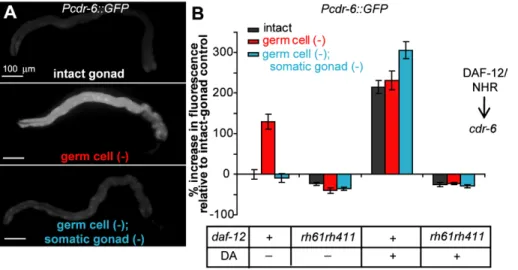
cdr-6, which encodes a homolog of a C. elegans cadmium-responsive gene, was identified in microarray experiments searching for genes regulated bydaf-12/NHRin animals that lack germ cells (MM and CK, in revision). We found that expression of GFP under the control of the cdr-6 promoter (Pcdr-6::GFP) was Figure 2. Overexpression of DAF-9/CYP450 in the XXX cells or hypodermis can extend the lifespan ofgerm cell (2);somatic gonad (2) animals.A GFP-tagged DAF-9/CYP450 protein, which catalyzes the final step of the synthesis of dafachronic acids, was overexpressed under the control of various promoters using multi-copy transgene arrays. Use of adaf-9(e1406)mutant background limited overexpression to specific tissue types. (A) Removal of the somatic gonad plus the germ cells of animals expressingdaf-9::GFPunder the control of thedaf-9promoter extended lifespan. Somatic-gonad plus germ cell removal also extended the lifespan of animals expressingdaf-9::GFPin the XXX cells using thesdf-9promoter (B) and in animals expressingdaf-9::GFPin the hypodermis using thedpy-7promoter (C). [However, expression of DAF-9::GFP in the hypodermis of germine-deficient animals lacking the somatic gonad using thecol-12promoter did not extend lifespan (Table S2). GFP fluorescence was visible in these animals, and the construct rescued the constitutive dauer formation phenotype ofdaf-9(e1406)animals [15] suggesting DAF-9/CYP450 was active in these transgenic animals. It is possible that the level of expression was not sufficient to rescue the longevity of these animals.] (D) Somatic gonad plus germ cell removal also extended the lifespan of animals expressingdaf-9::GFPunder the control of theche-2promoter, which drives expression in sensory neurons, which do not normally expressdaf-9/CYP450. Details including means andpvalues for all experiments are listed in Table S2.
up-regulated in the animal by germ cell ablation (Figure 3, Table S4). Thiscdr-6up-regulation requireddaf-12/NHR, as no increase in expression was seen when the germ cells were removed in a daf-12(rh61rh411) mutant (Figure 3B). We verified the daf-12/NHR dependent increase in cdr-6 expression in germ cell (2) animals using quantitative RT-PCR (qPCR) (Figure 4C, Table S9).
If signals from the somatic gonad cause DAF-12/NHR to up-regulate cdr-6 expression upon germline removal, then (i) the presence of the somatic gonad should be required for loss of the germline to increasePcdr-6::GFP expression and (ii) dafachronic acid should be able to substitute for the presence of the somatic gonad in the regulation of this gene. We found that both predictions were met. When we removed the somatic gonad as well as the germ cells, Pcdr-6::GFP expression was no longer elevated; instead, it was similar to the level observed in animals with an intact gonad (Figure 3, Table S4). Moreover, when these germ cell (2); somatic-gonad (2) animals were grown on plates containing D4-dafachronic acid, expression of Pcdr-6::GFP was restored to levels that were similar to those of Pcdr-6::GFP -transgenic animals lacking only the germline. As predicted, the increase in Pcdr-6::GFP expression caused by addition of dafachronic acid required a functional DAF-12/NHR protein. In adaf-12(rh61rh411) mutant, we observed no increase in cdr-6 expression upon D4
-dafachronic acid treatment (Figure 3B). Together, these findings support the interpretation that in germline-deficient animals, the somatic gonad activates a dafa-chronic-acid signaling pathway that turns oncdr-6gene expression through the activity of DAF-12/NHR.
Interestingly, D4
-dafachronic acid increased the expression of Pcdr-6::GFPin animals with an intact gonad (Figure 3B). This was noteworthy, as dafachronic acid does not increase the lifespan of animals with an intact gonad. This finding is consistent with the idea that loss of the germline has two effects. First, germline loss permits the somatic gonad to activate DAF-12/NHR via dafachronic acid signaling. Second, germline loss initiates additional events that are required for lifespan extension (see Discussion).
These findings were supported by our analysis of a second gene, dod-24, which we found to be negatively regulated bydaf-12/NHR in a different study (MMG, SJL, and CK, in preparation). The expression of a transcriptionalPdod-24::GFPgene fusion was variable; however, we found thatdod-24expression depended on the presence of the somatic gonad in adaf-12/NHRdependent fashion (Figure S2, Table S5), similar to results we obtained withcdr-6.
Expression ofdaf-16/FOXO-Dependent Genes Can Be Largely Independent ofdaf-12/NHR
Since DAF-12/NHR appeared to be regulated by the somatic gonad, we wondered if DAF-12/NHR activity could be responsible for all the effects of the somatic gonad. Previously we showed that the somatic gonad is required for the increased expression of a subset ofdaf-16/FOXO-regulated genes in germ-cell deficient animals [13]. We wondered ifdaf-12/NHRwas also required for the expression of these genes. We examined the expression ofsod-3, a direct target of DAF-16/FOXO [18–20]. When the germ cells are removed,sod-3expression increases, and this increased expression requires daf-16/FOXO [13]. We found that this increased expression was largely independent ofdaf-12/ NHR: in adaf-12(rh61rh411)mutant background, expression of a Psod-3::GFP fusion still increased upon germ cell removal. Furthermore, this increase in expression still required the somatic gonad (Figure 4A, Table S6). We observed a similar effect when we examined mRNA levels of sod-3 by quantitative RT-PCR (qPCR) in glp-1(e2141) mutants, which lack germ cells and are long-lived [5,21]. Whereas mutation ofdaf-16/FOXOresulted in a significant drop insod-3expression, mutation ofdaf-12/NHRdid not have a statistically significant effect (Figure 4C, Table S9). In intact animals, we also observed a decrease ofsod-3expression by qPCR upon mutation ofdaf-16/FOXO, but not whendaf-12/NHR was mutated (Table S9), consistent withsod-3’s being a daf-16/ FOXOregulated gene.
We note that, whereas in several experiments sod-3::GFP up-regulation in germ-cell ablated animals wasdaf-12/NHR
-indepen-Figure 3. The somatic gonad modulates the expression ofcdr-6in a DAF-12/NHR- dependent fashion.(A) Increasedcdr-6expression in germ cell (2)animals requires the somatic gonad. GFP driven by thecdr-6promoter was observed in the intestine of animals with intact gonads. In germ cell (2)animals, GFP levels increased in the intestine, whereas ingerm cell (2);somatic gonad (2)animals, the level of GFP dropped to levels similar to that of intact animals. Original images were rotated and placed on a flat black background. (B) The increase in expression ofcdr-6ingerm cell (2)animals requires functional DAF-12/NHR. No increase was observed indaf-12(rh61rh411)mutants. Activation of DAF-12/NHR by addition of dafachronic acid increased the expression ofcdr-6in intact andgerm cell (2);somatic gonad (2)to levels similar to those observed ingerm cell (2) animals in adaf-12/NHRdependent fashion. Means andpvalues as determined by the Student’sttest for the experiments shown in panel (B) as well as replicate experiments are listed in Table S4.
dent (Figure 4A), in one experiment, we did observe a slight decrease in the expression that was statistically significant (Table S6). Thus, DAF-12/NHR could potentially make a small, variable, contribution tosod-3expression.
Expression ofdaf-12/NHRRegulated Genes Can Be Largely Independent ofdaf-16/FOXO
As DAF-12/NHR had at best a modest effect on the expression of the DAF-16/FOXO-regulatedsod-3gene, we wondered about the converse situation—that is, whether DAF-16/FOXO might have a minor effect on the activity of DAF-12/NHR-regulated
genes. We therefore examined the expression of genes whose expression requires daf-12/NHR in a daf-16/FOXO mutant background.
First, we examined transgenic animals carrying thePcdr-6::GFP construct in a daf-16(mu86) null mutant and observed that expression ofPcdr-6::GFPstill increased relative to intact animals in response to loss of the germline (Figure 4B, Table S7). Furthermore, removal of the somatic gonad ingerm cell (2)animals lowered the expression ofPcdr-6::GFP. Thus, the somatic gonad was still able to modulate expression of cdr-6 when DAF-16/ FOXO was not functioning. Notably, the magnitude of up-Figure 4.daf-12/NHRanddaf-16/FOXOhave distinct effects ingerm cell (2)animals.daf-12/NHRanddaf-16/FOXOare both required for germ cell removal to extend lifespan. We examined whether the activity of one of these two transcription factors required the other. (A)daf-12/NHRis at most partially required for the transcriptional activity of DAF-16/FOXO. In adaf-12(rh61rh411)mutant, expression ofsod-3, a direct target of DAF-16/ FOXO still increased ingerm cell (2)animals and decreased when the somatic gonad was removed ingerm cell (2);somatic gonad (2)animals. In two experiments, expression ofPsod-3::GFPwas not lower ingerm cell (2) daf-12(rh61rh411)mutants compared togerm cell (2) daf-12(+) animals, whereas in a third experiment, its expression did decrease somewhat. Consistent with this decrease,daf-12/NHRwas identified as a ‘‘weak hit’’ in an unbiased screen looking for reducedPsod-3::GFPexpression ingerm cell (2)animals (MSK and CK, unpublished data). Means andpvalues are listed in Table S6. (B)daf-16/FOXOis partially required for the activity of DAF-12/NHR. In adaf-16(mu86)mutant, expression ofcdr-6still increased with germ cell removal, but to a lesser extent. Removal of both the somatic gonad and germ cells returned the level of expression to a similar level as that seen in intact animals. Means andpvalues for this experiment as well as additional replicates are listed in Table S7. (C) mRNA levels ofsod-3measured by quantitative RT-PCR inglp-1(e2141)mutants, which lack germ cells, decrease with mutation ofdaf-16/FOXO, but not with mutation ofdaf-12/NHR. Conversely, mRNA levels ofcdr-6decrease with mutation ofdaf-12/NHR, but not with mutation ofdaf-16/FOXO. ***p,0.0001, **p,0.001, *p,0.05, n.s.p.0.05. Means andpvalues for this experiment as well as additional replicates are listed in Table S9. (D) Activation of DAF-12/NHR byD4 -dafachronic acid treatment did not extend the lifespan ofdaf-16(mu86)mutants, suggesting that (E) DAF-12/NHR and DAF-16/FOXO are both required for the increased lifespan ofgerm cell (2)animals. Means andpvalues are listed in Table S1.
regulation of Pcdr-6::GFP in the daf-16(mu86) mutant was lower than in a wild-type background. We observed similar expression patterns when we measuredcdr-6mRNA levels by qPCR in glp-1(e2141)mutants, which lack germ cells. Mutation ofdaf-12/NHR resulted in a significant drop in the level ofcdr-6mRNA, whereas mutation of daf-16/FOXO did not affect cdr-6 mRNA levels (Figure 4C, Table S9). Together, these findings suggest that daf-16/FOXOis at most only partially required for loss of the germline to increase the expression of cdr-6. We also observed daf-16/ FOXO-indepdendent modulation of dod-24 expression by the presence of the somatic gonad (Figure S2, Table S8).
In summary, mutation ofdaf-16/FOXOonly slightly affects the transcription of thedaf-12/NHRregulated genescdr-6anddod-24, whereas mutation of daf-12/NHR only slightly affects the transcription of the directdaf-16/FOXOtarget,sod-3. Thus, while it remains possible that DAF-16/FOXO and DAF-12/NHR co-regulate yet-unidentified target genes, these two transcription factors regulate at least some of their target genes largely, but not completely, independently of one another.
daf-16/FOXOIs Required forD4-Dafachronic Acid Treatment to Extend Lifespan
As described above, dafachronic acid extends the lifespan of animals that lack the entire reproductive system. In contrast, dafachronic acid has no effect on the lifespan of intact animals. This finding suggests that the presence of the germline prevents dafachronic acid from extending lifespan. Loss of the germline activates the expression of multiple genes in a daf-16/FOXO -dependent fashion [13,22], and, as described above, at least some of these genes are activated largely independently ofdaf-12/NHR. It seemed possible that these daf-16/FOXO-dependent genes are collectively required for loss of the germline to extend lifespan. If many of these genes cannot be activated by DAF-12/NHR, then the addition of dafachronic acid would not be expected to extend lifespan in the presence of the germline. We tested this idea by removing the germ cells and the somatic gonad ofdaf-16/FOXO mutants, and then treating these animals with dafachronic acid. If DAF-16/FOXO-dependent genes are required for lifespan extension, then these animals should not live long. We found this was the case. Whereas in a wild-type, daf-16(+), background,
D4
-dafachronic acid extended the lifespan ofgerm-cell (2); somatic gonad (2) animals, in a daf-16(mu86) background, there was no change in lifespan (Figure 4D). Thus,daf-16/FOXOis still required for lifespan extension in animals with activated DAF-12/NHR. Together, these findings, along with our studies of germline-dependent gene expression, suggest that although there is some overlap, DAF-16/FOXO has an essential function in this lifespan extension pathway that is triggered mainly by germline loss, and DAF-12/NHR has another, distinct function that is activated by the somatic gonad when the germline is removed (Figure 4E).
Discussion
The Somatic Gonad Causes DAF-12/NHR to Increase Lifespan in Germline-Deficient Animals
The primary finding of this study is that the somatic gonad extends the lifespan of germline-deficient animals by activating a DAF-12/NHR-dependent sterol-signaling pathway. The somatic gonad and genes that produce dafachronic acid are both required for germ cell removal to extend lifespan, and their ability to influence lifespan requiresdaf-12/NHR. Furthermore, the somatic gonad is important for the proper activity of DAF-12/NHR, as the presence of the somatic gonad is required for the correct expression of daf-12/NHR-regulated genes such as cdr-6. Most
compelling, increasing the level of the DAF-12/NHR ligandD4 -dafachronic acid in animals that lack the somatic gonad is sufficient to rescue both lifespan extension and expression of daf-12/NHR-regulated genes, in a fashion that requiresdaf-12/NHR. One simple model to explain the longevity-promoting activity of the somatic gonad is that the somatic gonad stimulates dafachronic acid production when the germ cells are removed, which in turn affects DAF-12/NHR activity. Indeed, this model is not without precedent, as in humans the somatic reproductive tissues secrete steroid hormones such as androgens, which influence other tissues. In this model,germ cell (2);somatic gonad (2)animals fail to live long because they have insufficient dafachronic acid levels. We asked whether the somatic gonad might influence the level of daf-9/ CYP450gene expression, but this was not the case, as levels of DAF-9::GFP were not overtly different (unpublished data). However, the somatic gonad could potentially affect the level of dafachronic acid by alternate mechanisms; for example, by increasing the level of a biosynthetic precursor of dafachronic acid. It is also possible that the somatic gonad regulates the activity of DAF-12/NHR without affecting the total level of dafachronic acid. For example, the somatic gonad could influence the proportion of dafachronic acid in the animal that is available to bind to DAF-12/NHR. Another possibility is that the somatic gonad influences the levels or activities of DAF-12/NHR inhibitors or co-activators, though in this scenario, it is necessary to postulate that increased levels of dafachronic acid can overcome the effects of these co-regulators. It would be interesting to directly measure levels of dafachronic acids in animals that lack the germ cells or the entire reproductive system.
daf-9/CYP450is expressed in the somatic gonad (specifically, in the spermatheca [6,15]), so it was interesting to find that limiting DAF-9/CYP450 overexpression to one of several non-reproduc-tive tissues—the XXX cells, the hypodermis, or even to sensory neurons that do not normally express daf-9/CYP450—could increase the longevity of germ-cell defective animals that lack the somatic gonad. This finding indicates that tissues other than the somatic gonad and the intestine can participate in this reproductive signaling pathway by producing dafachronic acid. Perhaps loss of the germ cells stimulates the synthesis or release of a precursor of dafachronic acid, which in turn diffuses among the tissues. Alternatively, as mentioned above, various tissues could synthesize dafachronic acid independently of any input from the reproductive system if the somatic gonad controls other factors in the dafachronic acid/DAF-12 signaling pathway. Finally, it is possible that when the germline is removed but the somatic gonad is present, gonadal DAF-9/CYP450 also contributes to the pool of dafachronic acid in the animal. Indeed, whendaf-9/CYP450was overexpressed under the control of its endogenous promoter, which drives some expression in the somatic gonad, germline ablation caused a larger increase in lifespan than did loss of the entire gonad (Table S2). This was not the case whendaf-9/CYP450 was expressed only in non-gonadal tissues. This difference could be due to the loss of DAF-9/CYP450 in the somatic gonad.
determine how the somatic gonad modulates one aspect of DAF-12/NHR function, its ability to promote longevity, without affecting the other aspect, DAF-16/FOXO nuclear localization.
Removing the Germ Cells Is Necessary for DAF-12/NHR Activity to Promote Longevity
Giving dafachronic acids to animals with an intact gonad does not extend lifespan (Figure 1D and [12]). However, dafachronic acid does stimulate DAF-12/NHR to regulate germline-dependent genes in intact animals, since it produces adaf-12/NHR-dependent up-regulation ofcdr-6and adaf-12/NHR-dependent down-regula-tion ofdod-24in intact animals. Since activation of DAF-12/NHR is not sufficient to extend lifespan, other lifespan-promoting factors turned on in germline-deficient animals must be necessary for an increased lifespan. Because dafachronic acid extends lifespan in the absence of the somatic gonad, the somatic gonad itself is unlikely to provide these other lifespan-promoting factors. Instead, DAF-16/ FOXO is the most likely candidate for the factor activated by loss of the germ cells that is necessary, along with DAF-12/NHR and the somatic gonad, to increase lifespan. Consistent with this idea, genetic inactivation ofdaf-16/FOXO, like the presence of the germ cells, prevents dafachronic acid from extending lifespan.
The Complex Functions of DAF-12/NHR and Dafachronic Acid
An unexpected finding from this study was that DAF-12/NHR has a more complex role in this longevity pathway than previously appreciated. We found that when the germ cells are removed in animals containing a daf-12/NHR null mutation, lifespan is extended slightly. This daf-12-independent lifespan-extension pathway (referred to here as the ‘‘underlying pathway’’) does not require the somatic gonad and is not affected by dafachronic acid. Whendaf-12(+)activity is present but the somatic gonad is absent ordaf-9/CYP450is mutated, then germ-cell loss does not extend lifespan. This suggests that unliganded DAF-12/NHR prevents germ-cell loss from activating the underlying pathway. In contrast, liganded DAF-12/NHR extends lifespan in response to germ-cell loss, as daf-12(+)animals that have a daf-9(+)genotype plus the somatic gonad live long when the germ cells are removed. In the future, it will be interesting to explore the nature of the underlying daf-12-independent pathway and to learn howdaf-12/NHR(+)can affect its activity. Finally, we note that a dual ability of DAF-12/ NHR to extend and shorten lifespan is not without precedent. In intact animals, DAF-12/NHR extends lifespan in daf-9/CYP450 reduction-of-function mutants when animals are cultured at 15uC [6,14]. In contrast, at warmer temperature (25uC), DAF-12/NHR shortens lifespan in response to decreased daf-9/CYP450 in the absence of thermosensory neurons [23].
DAF-12/NHR and DAF-16/FOXO Have Distinct Transcriptional Effects in Germline-Deficient Animals
Both sterol signaling and DAF-16/FOXO are required for the long lifespan of germline-deficient animals. The relationship between DAF-12/NHR and DAF-16/FOXO in animals that lack the germ cells is not well understood. Previous work has demonstrated that DAF-12/NHR is partially (but not completely) required for the nuclear accumulation of DAF-16/FOXO in animals that lack the germ cells [11,12]. Consistent with this result, in this study we showed that indaf-12/NHRmutants, the DAF-16/ FOXO targetsod-3is still up-regulated (though perhaps to a lesser extent). Likewise, Wang and Ruvkun showed that the lipase gene K04A8.5is up-regulated by germline removal in adaf-16/FOXO -dependent butdaf-12/NHR-independent fashion [24]. These data
suggest that DAF-16/FOXO can promote the transcription of at least some of its target genes independently of DAF-12/NHR in animals that lack the germ cells. We have found that the converse also holds true. When daf-16/FOXO is mutated, DAF-12/NHR still retains the ability to affect transcription of genes such ascdr-6 and dod-24. However, DAF-16/FOXO affects the magnitude of this regulation, suggesting that DAF-16/FOXO could have a partial effect on the activity of DAF-12/NHR. In summary, based on the several genes we examined, it appears that DAF-16/FOXO and DAF-12/NHR have distinct effects on the transcriptome of germ-cell deficient animals, although each has minor effects on the activity of the other. This interpretation is supported by a genome-wide microarray analysis of germline-defectivedaf-16/FOXOand daf-12/NHRmutants (MM and CK, in revision).
Dafachronic Acid Signaling and DAF-16/FOXO Are Both Required for Germ-Cell Removal to Extend Lifespan
Although dafachronic-acid signaling and DAF-16/FOXO have distinct effects on gene transcription in animals that lack germ cells, both are required to extend lifespan. Furthermore, dafachronic acid does not override the requirement for DAF-16/ FOXO to extend longevity, and rendering DAF-16/FOXO constitutively nuclear does not override the requirement for DAF-12/NHR. These two pieces of data make it unlikely that DAF-12/NHR and DAF-16/FOXO operate in a simple linear pathway, where the transcriptional effects of mutation of one gene would be completely mimicked by the mutation of the other. Instead, the simplest interpretation is that DAF-12/NHR and DAF-16/FOXO function in parallel to promote longevity in animals without germ cells.
Therefore, we propose the following model (Figure 4E): germ-cell removal has two important effects: (i) DAF-16/FOXO accumulates in the nucleus, and (ii) DAF-12/NHR is indepen-dently stimulated to extend lifespan. In these germline-deficient animals, activated DAF-12/NHR and DAF-16/FOXO act in parallel on different target genes (for the most part) to promote lifespan extension. The presence of the somatic gonad in germ-cell deficient animals promotes the activation of DAF-12/NHR by ensuring sufficient levels of available dafachronic acids, possibly through an increase in their levels. When the somatic gonad is removed, DAF-12/NHR no longer extends lifespan, and the animals no longer live long.
Materials and Methods
C. elegansStrains
All strains used in this study were maintained under standard conditions [25]. The following strains were used:
N2
CF2479daf-12(rh61rh411)
daf-9(e1406); mgEx662[daf-9p::daf-9 genomic::GFP]
daf-9(e1406); mgEx670[sdf-9p::daf-9 cDNA::GFP;mec-7::GFP] daf-9(e1406); mgEx663[dpy-7p::daf-9 cDNA::GFP; mec-7::GFP] daf-9(e1406); mgEx666[che-2p::daf-9 cDNA::GFP; mec 7::GFP] daf-9(e1406); mgEx668[col-12p::daf-9 cDNA::GFP; mec 7::GFP] BC15369dpy-5(e907); sEx15369[Pcdr-6::GFP+pCeh361] CF3595sEx15369[Pcdr-6::GFP+pCeh361] obtained by outcross-ing BC15369 3 times to the laboratory N2
CF3596daf-12(rh61rh411); sEx15369[Pcdr-6::GFP+pCeh361] AU68agIs6[Pdod-24::GFP]
CF3556 agIs6[Pdod-24::GFP] obtained by outcrossing AU86 3 times to the laboratory N2
CF1553muIs84[Psod-3::GFP]
CF3604daf-12(rh61rh411); muIs84[Psod-3::GFP] CF3597daf-16(mu86); sEx15369[Pcdr-6::GFP+pCeh361] CF1903glp-1(e2141)
CF1880daf-16(mu86); glp-1(e2141) CF1658glp-1(e2141); daf-12(rh61rh411) CF1037daf-16(mu86)
Some nematode strains used in this study were provided by the Caenorhabditis Genetics Center, which is funded by the NIH National Center for Research Resources (NCRR). Construction of Psod-3::GFP was described previously in [10]. daf-9::GFP strains were provided by the Ruvkun Lab and were described previously in [15]. ThePdod-24::GFPstrain was kindly provided by D. Kim. Strains containing Pcdr-6::GFPwere obtained from the Genome British ColumbiaC. elegansGene Expression Consortium [26].
Laser Ablation
Germ-cell (Z2,Z3) or somatic-gonad (Z1,Z4) precursor cells of newly hatched L1 larvae were killed by laser ablation as described previously [2] using a VSL-337 nitrogen pumped dye laser (Laser Sciences, Inc.). At adulthood, absence of the gonad or germ cells was confirmed using a dissecting microscope. To obtain intact-gonad controls, un-ablated L1 larvae were anaesthetized and recovered from the same NaN3agarose pads as experimental animals.
Lifespan Analysis
Lifespan analysis was performed at 20uC as described previously [27,28] using OP50 bacteria. Lifespan analyses of animals grown on dafachronic acid were performed using 3 cm plates containing 5 mL of NG agarose media. Prior to use, 1ml of 1 mM dafachronic acid in ethanol was diluted in 100ml PBS and pipetted onto a plate containing a lawn of OP50 bacteria. As a control, 3 cm plates spotted with 1ml of ethanol diluted in 100ml PBS were used.
Animals were placed on dafachronic acid or control plates as L1 larvae directly after laser ablation. Statistical analysis was performed using Stata/IC 10.0 software (StataCorp LP). p values were determined using the log-rank (Mantel-Cox) method.
GFP Fluorescence Microscopy and Quantification On day 2 of adulthood, animals were anaesthetized on agarose pads containing 0.15 M NaN3. Images were taken using a Retiga
EXi Fast1394 CCD digital camera (QImaging) using the 106 objective on a Zeiss Axioplan 2 compound microscope (Zeiss Corporation). Each image was taken with the intestine in focus, since expression of the various transgenes was primarily in the intestine. For each trial, exposure time was calibrated to minimize the number of saturated pixels for that set of animals. Openlab 4.0.2 software (Improvision) was used to quantify the total intensity of fluorescence per worm as measured by intensity of each pixel in the selected area of a frame (i.e. the worm). Vulval expression of Psod-3::GFP, which was very bright, was excluded from quantifi-cation, since this structure is not present in animals lacking the gonad. Fluorescence of the entire animal was measured for all other GFP constructs. No expression of any of the constructs was visible in embryos prior to egg laying. Image processing for figures was performed using Adobe Photoshop 7.0 (Adobe).
Quantitative RT-PCR
Sterileglp-1(e2141ts) and wild-type N2 animals were raised at 25uC from L1 to day 1 of adulthood, then shifted to 20uC. On day 2 of adulthood, animals were collected for RNA extraction. RNA extraction, purification, and reverse transcription were carried out as described in [29].
qPCR was performed using the 7300 Real Time PCR System (Applied Biosystems) and data were analyzed using the Ct method (Applied Biosystems Prism 7700 Users Bulletin No. 2, http://docs. appliedbiosystems.com/pebiodocs/04303859.pdf). Data were gen-erated from at least two biological repeats. mRNA levels ofama-1, nhr-23, and Y45F10D.4 were used for normalization [29,30]. p values were determined using one-way ANOVA. Primer sequences are available upon request.
Supporting Information
Figure S1 Germ cell removal in a daf-12(rh61rh411) mutant slightly extends lifespan in a somatic gonad-independent fashion.Removal of the germ cells and the somatic gonad in animals carrying the putative nulldaf-12(rh61rh411)allele extends lifespan. However,daf-12(rh61rh411) germ cell (2)animals do not live as long asgerm cell (2)animals that carry the wild-type allele ofdaf-12/NHR. Means andpvalues are listed in Table S3. Found at: doi:10.1371/journal.pbio.1000468.s001 (0.07 MB TIF)
Figure S2 The somatic gonad represses the expression of dod-24. (A) dod-24 requires the somatic gonad for proper expression. The expression of the Pdod-24::GFP transgene was variable; however, across multiple experiments, we observed a consistent trend towards decreased intestinalPdod-24::GFP expres-sion when the germ cells were removed. (6 out of 11 experiments were statistically significant.) In germ cell (2); somatic gonad (2) animals,Pdod-24::GFPexpression levels increased relative to those of intact-gonad andgerm cell (2)animals. (8 out of 11 experiments were statistically significant.) Thus, the presence of the somatic gonad suppressesdod-24expression. (B) GFP driven by thedod-24 promoter was observed throughout the intestine of wild-type animals. Arrowheads indicate position of the head. Original images were rotated and placed on a flat black background. (C) The increase inPdod-24::GFPcaused by removal of the somatic gonad requireddaf-12/NHR. Expression of Pdod-24::GFPdid not increase indaf-12(rh61rh411)mutants in 4 out of 5 trials when the somatic gonad and germ cells were removed. Highly variable changes in overall expression ofPdod-24::GFPwere observed when daf-12/NHR was mutated, even in intact animals. (D) daf-16/ FOXOhad little effect on the ability of the somatic gonad to inhibit dod-24expression. Indaf-16(mu86)mutants lacking the germ cells, removal of the somatic gonad still resulted in increased expression ofPdod-24::GFP.pvalues for pair-wise comparisons to intact-gonad animals are indicated by: ***p,0.0001, **p,0.001, *p,0.05,ns p.0.05. Means andpvalues are listed in Tables S5 and S8. Found at: doi:10.1371/journal.pbio.1000468.s002 (0.73 MB TIF)
Table S1 Dafachronic acid extends the lifespan of animals that lack both the somatic gonad and germ cells.
Found at: doi:10.1371/journal.pbio.1000468.s003 (0.03 MB XLS)
Table S2 Overexpression of DAF-9/CYP450 extends the lifespan of animals that lack both the somatic gonad and germ cells.
Found at: doi:10.1371/journal.pbio.1000468.s004 (0.02 MB XLS)
Table S3 The daf-12(rh61rh411) allele uncovers a life-span shortening function of DAF-12/NHR in addition to its lifespan promoting activity ingerm cell (2)animals. Found at: doi:10.1371/journal.pbio.1000468.s005 (0.02 MB XLS)
Tabe S5 Pdod-24::GFPexpression changes in adaf-12/ NHR-dependent fashion when the germ cells and somatic gonad are removed.
Found at: doi:10.1371/journal.pbio.1000468.s007 (0.03 MB XLS)
Table S6 Psod-3::GFP up-regulation in animals that lack germ cells is largely independent ofdaf-12/NHR. Found at: doi:10.1371/journal.pbio.1000468.s008 (0.03 MB XLS)
Table S7 Pcdr-6::GFPup-regulation in animals that lack germ cells is largely independent ofdaf-16/FOXO. Found at: doi:10.1371/journal.pbio.1000468.s009 (0.03 MB XLS)
Table S8 Changes in Pdod-24::GFP expression in the absence of germ cells and somatic gonad do not require daf-16/FOXO.
Found at: doi:10.1371/journal.pbio.1000468.s010 (0.03 MB XLS)
Table S9 DAF-16/FOXO and DAF-12/NHR have
dis-tinct effects on gene expression in animals that lack germ cells.
Found at: doi:10.1371/journal.pbio.1000468.s011 (0.03 MB XLS)
Acknowledgments
We would like to thank Zhu Wang and David Mangelsdorf for generously providingD4
-dafachronic acid. We also thank Gary Ruvkun for providing thedaf-9/CYP450overexpression strains. We thank Sivan Korenblit for qPCR primers.
Author Contributions
The author(s) have made the following declarations about their contributions: Conceived and designed the experiments: TMY CK. Performed the experiments: TMY JRB. Analyzed the data: TMY CK. Contributed reagents/materials/analysis tools: MM MMG SJL. Wrote the paper: TMY CK. Initial work examining the daf-16 requirement for cdr-6 expression ingerm cell (2)animals: MSK.
References
1. Williams GC (1966) Natural selection, the costs of reproduction, and a refinement of lack’s principle. The American Naturalist 100: 687.
2. Hsin H, Kenyon C (1999) Signals from the reproductive system regulate the lifespan of C. elegans. Nature 399: 362–366.
3. Flatt T, Min KJ, D’Alterio C, Villa-Cuesta E, Cumbers J, et al. (2008) Drosophila germ-line modulation of insulin signaling and lifespan. Proc Natl Acad Sci U S A 105: 6368–6373.
4. Cargill SL, Carey JR, Muller HG, Anderson G (2003) Age of ovary determines remaining life expectancy in old ovariectomized mice. Aging Cell 2: 185– 190.
5. Arantes-Oliveira N, Apfeld J, Dillin A, Kenyon C (2002) Regulation of life-span by germ-line stem cells in Caenorhabditis elegans. Science 295: 502–505. 6. Gerisch B, Weitzel C, Kober-Eisermann C, Rottiers V, Antebi A (2001) A
hormonal signaling pathway influencing C. elegans metabolism, reproductive development, and life span. Dev Cell 1: 841–851.
7. Rottiers V, Motola DL, Gerisch B, Cummins CL, Nishiwaki K, et al. (2006) Hormonal control of C. elegans dauer formation and life span by a Rieske-like oxygenase. Dev Cell 10: 473–482.
8. Motola DL, Cummins CL, Rottiers V, Sharma KK, Li T, et al. (2006) Identification of ligands for DAF-12 that govern dauer formation and reproduction in C. elegans. Cell 124: 1209–1223.
9. Lin K, Hsin H, Libina N, Kenyon C (2001) Regulation of the Caenorhabditis elegans longevity protein DAF-16 by insulin/IGF-1 and germline signaling. Nat Genet 28: 139–145.
10. Libina N, Berman JR, Kenyon C (2003) Tissue-specific activities of C. elegans DAF-16 in the regulation of lifespan. Cell 115: 489–502.
11. Berman JR, Kenyon C (2006) Germ-cell loss extends C. elegans life span through regulation of DAF-16 by kri-1 and lipophilic-hormone signaling. Cell 124: 1055–1068.
12. Gerisch B, Rottiers V, Li D, Motola DL, Cummins CL, et al. (2007) A bile acid-like steroid modulates Caenorhabditis elegans lifespan through nuclear receptor signaling. Proc Natl Acad Sci U S A 104: 5014–5019.
13. Yamawaki TM, Arantes-Oliveira N, Berman JR, Zhang P, Kenyon C (2008) Distinct activities of the germline and somatic reproductive tissues in the regulation of Caenorhabditis elegans’ longevity. Genetics 178: 513–526. 14. Jia K, Albert PS, Riddle DL (2002) DAF-9, a cytochrome P450 regulating C.
elegans larval development and adult longevity. Development 129: 221–231. 15. Mak HY, Ruvkun G (2004) Intercellular signaling of reproductive
develop-ment by the C. elegans DAF-9 cytochrome P450. Developdevelop-ment 131: 1777– 1786.
16. Snow MI, Larsen PL (2000) Structure and expression of daf-12: a nuclear hormone receptor with three isoforms that are involved in development and aging in Caenorhabditis elegans. Biochim Biophys Acta 1494: 104–116. 17. Antebi A, Yeh WH, Tait D, Hedgecock EM, Riddle DL (2000) daf-12 encodes a
nuclear receptor that regulates the dauer diapause and developmental age in C. elegans. Genes Dev 14: 1512–1527.
18. Honda Y, Honda S (1999) The daf-2 gene network for longevity regulates oxidative stress resistance and Mn-superoxide dismutase gene expression in Caenorhabditis elegans. FASEB J 13: 1385–1393.
19. Furuyama T, Nakazawa T, Nakano I, Mori N (2000) Identification of the differential distribution patterns of mRNAs and consensus binding sequences for mouse DAF-16 homologues. Biochem J 349: 629–634.
20. Oh SW, Mukhopadhyay A, Dixit BL, Raha T, Green MR, et al. (2006) Identification of direct DAF-16 targets controlling longevity, metabolism and diapause by chromatin immunoprecipitation. Nat Genet 38: 251–257. 21. Austin J, Kimble J (1987) glp-1 is required in the germ line for regulation of the
decision between mitosis and meiosis in C. elegans. Cell 51: 589–599. 22. Ghazi A, Henis-Korenblit S, Kenyon C (2009) A transcription elongation
factor that links signals from the reproductive system to lifespan extension in Caenorhabditis elegans. PLoS Genet 5: e1000639. doi:10.1371/journal. pgen.1000639.
23. Lee SJ, Kenyon C (2009) Regulation of the longevity response to temperature by thermosensory neurons in Caenorhabditis elegans. Curr Biol 19: 715–722. 24. Wang MC, O’Rourke EJ, Ruvkun G (2008) Fat metabolism links germline stem
cells and longevity in C. elegans. Science 322: 957–960.
25. Brenner S (1974) The genetics of Caenorhabditis elegans. Genetics 77: 71–94. 26. McKay SJ, Johnsen R, Khattra J, Asano J, Baillie DL, et al. (2003) Gene expression profiling of cells, tissues, and developmental stages of the nematode C. elegans. Cold Spring Harb Symp Quant Biol 68: 159–169.
27. Kenyon C, Chang J, Gensch E, Rudner A, Tabtiang R (1993) A C. elegans mutant that lives twice as long as wild type. Nature 366: 461–464.
28. Arantes-Oliveira N, Berman JR, Kenyon C (2003) Healthy animals with extreme longevity. Science 302: 611.
29. Van Gilst MR, Hadjivassiliou H, Yamamoto KR (2005) A Caenorhabditis elegans nutrient response system partially dependent on nuclear receptor NHR-49. Proc Natl Acad Sci U S A 102: 13496–13501.