Functional Interaction between HEXIM and
Hedgehog Signaling during
Drosophila
Wing
Development
Duy Nguyen1¤, Olivier Fayol1, Nicolas Buisine2, Pierrette Lecorre1, Patricia Uguen1 *
1UMR-S1174, Univ. Paris-Sud, Inserm, Université Paris-Saclay, Bât. 440, 91405 Orsay, France,2MNHN, UMR CNRS 5166, 75231 Paris, France
¤ Current address: Department of biochemistry, Université de Sherbrooke, Sherbrooke, QC J1E 4K8, Canada
Abstract
Studying the dynamic of gene regulatory networks is essential in order to understand the spe-cific signals and factors that govern cell proliferation and differentiation during development. This also has direct implication in human health and cancer biology. The general transcrip-tional elongation regulator P-TEFb regulates the transcriptranscrip-tional status of many developmental genes. Its biological activity is controlled by an inhibitory complex composed of HEXIM and the 7SK snRNA. Here, we examine the function of HEXIM duringDrosophiladevelopment. Our key finding is that HEXIM affects the Hedgehog signaling pathway. HEXIM knockdown flies display strong phenotypes and organ failures. In the wing imaginal disc, HEXIM knock-down initially induces ectopic expression of Hedgehog (Hh) and its transcriptional effector Cubitus interuptus (Ci). In turn, deregulated Hedgehog signaling provokes apoptosis, which is continuously compensated by apoptosis-induced cell proliferation. Thus, the HEXIM knock-down mutant phenotype does not result from the apoptotic ablation of imaginal disc; but rather from the failure of dividing cells to commit to a proper developmental program due to Hedge-hog signaling defects. Furthermore, we show thatciis a genetic suppressor ofhexim. Thus, HEXIM ensures the integrity of Hedgehog signaling in wing imaginal disc, by a yet unknown mechanism. To our knowledge, this is the first time that the physiological function of HEXIM has been addressed in such detailsin vivo.
Introduction
Transcription of protein-coding genes is mediated by RNA polymerase II (RNA Pol II) whose processivity is tightly controlled by the positive transcription elongation factor b (P-TEFb)
after transcriptional initiation [1,2]. This kinase promotes productive transcription elongation
by catalyzing the phosphorylation of a number of regulatory factors, namely the Negative elon-gation factor (NELF), the DRB-sensitivity inducing factor (DSIF), as well as the C-terminal
domain (CTD) of RNA Pol II [3].
a11111
OPEN ACCESS
Citation:Nguyen D, Fayol O, Buisine N, Lecorre P, Uguen P (2016) Functional Interaction between HEXIM and Hedgehog Signaling duringDrosophila
Wing Development. PLoS ONE 11(5): e0155438. doi:10.1371/journal.pone.0155438
Editor:Andreas Bergmann, University of Massachusetts Medical School, UNITED STATES
Received:January 8, 2016
Accepted:April 28, 2016
Published:May 13, 2016
Copyright:© 2016 Nguyen et al. This is an open access article distributed under the terms of the
Creative Commons Attribution License, which permits unrestricted use, distribution, and reproduction in any medium, provided the original author and source are credited.
Data Availability Statement:All relevant data are within the paper and its Supporting Information files.
Funding:PU was funded by Agence Nationale de la Recherche (ANR-06-BLAN-0072).http://www. agence-nationale-recherche.fr. DN had a fellowship from Cancéropôle Ile de France.http://www. canceropole-idf.fr. The funders had no role in study design, data collection and analysis, decision to publish, or preparation of the manuscript.
In human cells, P-TEFb forms two alternative complexes, which differ in size, components,
and enzymatic activity [2,4]. A“small complex”(SC), composed of CyclinT and CDK9,
corre-sponds to the catalytically active P-TEFb. In contrast, P-TEFb is kept in a catalytically inactive
state and forms a“large complex”(LC) when bound by a macromolecular complex containing
the 7SK snRNA, Bicoid-interacting protein 3 (BCDIN3), La-related protein 7 (LARP7), and Hexamethylene bis-acetamide inducible protein 1 (HEXIM1). The formation of the LC is reversible and P-TEFb can switch back and forth between LC and SC in a very dynamic man-ner. Thus, HEXIM, together with other factors, acts as a sink of active P-TEFb which regulates
its biological availability at target genes [5] in response to the transcriptional demand of the
cell [6–9]. Although HEXIM target genes are not known, many lines of evidence strongly
sup-port a connection between developmental pathways or diseases and the control of transcription
by HEXIM [10].
Transcriptional pause was initially described in the late 80s for theDrosophilaHSP90 gene,
where transcription stalls shortly after the elongation start and RNA Pol II accumulates at the
5' end of the gene, which is thus poised for transcription [11]. It has been proposed that this
phenomenon may be more general, as virtually all developmental genes inDrosophila[12,13]
and approximately 20 to 30 percent of genes in human and mouse show similar properties
[14,15]. The release from pause and the transition to productive elongation is under the control
of the NELF factor [16], and so to P-TEFb, which is in turn controlled by HEXIM. Given that
these genes already completed transcriptional initiation and that mRNA synthesis started, release from pause allows for a very fast and synchronized transcriptional response with low transcriptional noise.
It has been proposed that sustained pause may be a potent mechanism to actually repress gene transcription. This leads to the apparent paradox where transcriptional repression
requires transcriptional initiation (reviewed by [17]). Therefore, knockdown of the
transcrip-tional pausing factor HEXIM would release transcription and reveal the regulation of poised genes.
HEXIM1 has been initially identified as a 359 aa protein whose expression is induced in human vascular smooth muscle cells (VSMCs) following treatment with hexamethylene
bis-acetamide (HMBA) which is a differentiating agent [18]. It is also called estrogen
down-regu-lated gene 1 (EDG1) due to its decreased expression by estrogen in breast cancer cells [19,20].
Ortholog of HEXIM1 in mice and chickens is activated in heart tissue during early
embryogen-esis, and was so named cardiac lineage protein 1 (CLP-1) [21,22]. HEXIM1 is involved in
many kinds of cancer, viral transcription of HIV-1, cardiac hypertrophy, and inflammation
[10]. Overall, HEXIM defects are strongly associated with imbalance in the control of
prolifera-tion and differentiaprolifera-tion. The CLP-1/HEXIM1 null mutaprolifera-tion is embryonic lethal in mice, and results in early cardiac hypertrophy. Heterozygous littermates are still affected but with a less
severe phenotype and survived up to adulthood [22–25]. Moreover, Mutation in the
carboxy-terminal domain of HEXIM1 causes severe defects during heart and vascular development by reducing the expression of vascular endothelial growth factor (VEGF), which is essential for
myocardial proliferation and survival [26–28]. Overexpression of HEXIM1 in breast epithelial
cells and mammary gland decreases estrogen-driven VEGF expression, whereas it is strongly increased in loss of function mutant. As reported recently, HEXIM1 expression is required for
enhancing the response to tamoxifen treatment in breast cancer patients [29]. In addition,
increased HEXIM1 expression correlates with a better prognosis and decreases probability of
breast cancer recurrence [20,29,30]. Additionally, terminal differentiation of murine
erythro-leukemia cells induced by HMBA or DMSO correlates with elevated levels of both HEXIM1 mRNA and protein. Furthermore, in neuroblastoma cells, HEXIM1 overexpression inhibits
transcription rate of NF-κB, an important regulator of apoptosis, cell proliferation,
differentia-tion, and inflammation [33]. However, despite theses advances, the dissection of HEXIM
func-tions was mostly approached on a biochemical basis, and to date, very little is known about its physiological and developmental relevance in an integrated model. In order to address this
important point, we developed anin vivomodel and recently showed that a similar P-TEFb
regulation pathway also exists inDrosophila, and that HEXIM is essential for proper
develop-ment [34].
InDrosophila, the Hedgehog (Hh) signaling pathway controls cell proliferation,
differentia-tion and embryo patterning [35–40]. The Hh activity is transduced to a single transcription
fac-tor, Cubitus interruptus (Ci) [38,41–44], theDrosophilahomolog of Gli [37,39]. Wing
imaginal discs can be subdivided into two compartments based on the presence of Hh protein.
The posterior compartment (P) expresses Engrailed (En), which activates Hh and repressesci
expression. The anterior compartment (A) expressesci. The full length Ci protein (called
Ci155) is constitutively cleaved into a truncated protein acting as a transcriptional repressor
(Ci75) ofhhand Decapentaplegic (dpp) genes. Hh inhibits the proteolytic cleavage of Ci, which
then acts as a transcriptional activator of a number of target genes (Patched (ptc) anddpp, to
name a few) [45]. Thus, Ci155is accumulated at the boundary between the A and P
compart-ments where there are high levels of Hh, and it is absent in P compartment [46]. Ci regulates
the expression of Hh target genes in a manner dependent on Hh levels [47]. In addition to
pro-teolytic cleavage, the biological activity of Ci is also modulated by phosphorylation and
nucleo-cytoplasmic partitioning [48–51]. The mis-regulation of any components of the Hh pathway
usually modifies the Ci155levels, and results in developmental defects [52,53].
In this paper, we examine the function of HEXIM duringDrosophiladevelopment. We
show that HEXIM knockdown disrupts organ formation. In the wing disc, this latter effect is mediated by a strong ectopic induction of Hh signaling followed by apoptosis. The death of proliferative cells is subsequently compensated by proliferation of the neighboring cells: this is
the mechanism of apoptosis-induced cell proliferation (see [54]). Ci, the transcriptional
effec-tor of Hh pathway, is highly accumulated at both mRNA and protein levels in cells where HEXIM is knocked-down. Thus, the severe phenotype of HEXIM mutants resulted from
Hh-related wing patterning defects. Furthermore, we also show thatciacts as a genetic suppressor
ofhexim, suggesting that HEXIM is an interacting factor of the Hh signaling pathway. To our knowledge, this is the first time that the physiological function of HEXIM has been addressed in a whole organism.
Materials and Methods
Fly stocks and transgenes
All stocks were maintained and raised under standard conditions. Fly crosses were performed at 29°C, unless otherwise indicated. For any phenotype, at least 80 progenies from 2 to 4 inde-pendent crosses were analysed, unless specified otherwise. The phenotype penetrance was
100%, unless specified otherwise. Two RNAi-mediatedHeximknockdown mutants were used,
one on chromosome II, the other on III (ID transformants 34633 and 34632, respectively,
ViennaDrosophilaResource Center). To date, the stock 34633 is not available anymore from
VDRC. The specificity of this RNAi-mediated knockdown has been confirmed, as previously
described [34], by rescuing HEXIM knockdown phenotypes with a UAS-HEXIM strain
over-expressing HEXIM. The RNAiHeximprobe sequence targets bothDrosophila HeximmRNA
isoforms [34]. Fly strains were obtained from different sources, as follows:UAS-Hex
(home-made transgene, BestGene),UAS-hid(gift of Hyung Don Ryoo),UAS-p35(gift of Jean-Philippe
GMR-Gal4,so-Gal4,ey-Gal4,rn-Gal4,Canton S,Dpp-lacZ,UAS-Hh-RNAi(number 31475)
(Bloomington stock center),Hh-lacZ(gift of Thomas Kornberg),Sp/Cyo;MKRS/TM6,Sp-Cyo/
SM5-TM6(gift of Jean-Philippe Parvi). Additional strains were constructed by association
approach:UAS-p35;rn-Gal4,UAS-p35;UAS-Hex-RNAi,UAS-Hex-RNAi;Hh-lacZ,
UAS-Hex-RNAi;Dpp-lacZ,UAS-Hex-RNAi;UAS-Ci-RNAi,Dpp-lacZ;rn-Gal4,UAS-Hex-RNAi;UAS-Hex, UAS-Hex-RNAi;UAS-Hh-RNAi.
Immunocytochemistry
For antibody staining, imaginal discs of third-instar larvae were dissected in PBS 1X, and fixed with fixation buffer (4% formaldehyde in PBS 1X) for 20 mins at room temperature. The discs were then blocked in blocking solution (PBS 1X, 0.1% BSA, 0.3% Triton X-100) for at least 1h, before being incubated with the primary antibody overnight at 4°C. We used the following pri-mary antibodies: mouse anti-GFP (1:2000, Sigma), rabbit anti-Caspase 3 (1:200, Cell Signal-ing), mouse anti-Delta (1:50, DSHB), rat anti-Serrate (1:50, gift of Irvine), mouse anti-Notch intra- and extra-cellular (1:100, DSHB), mouse anti-Patched (1:50, DSHB), mouse anti-Wing-less (1:500, DSHB), rat anti-Sal (1:300, gift of John F. de Celis), mouse anti-Cut (1:100, DSHB), mouse anti-Engrailed (1:50, DSHB), rat anti-Ci (1:50, DSHB), rabbit anti-P-H3 (1:200, Milli-pore), mouse anti-beta galactosidase (1:1000, Promega), rabbit anti-beta galactosidase (1:2000, gift from A. Plessis). The rabbit anti-dHEXIM (1:2000) was custom made by Genecust. After incubation with primary antibodies, the discs were washed four times with blocking buffer, and incubated with the appropriate fluorescent secondary antibody diluted in blocking buffer (1:200) for at least 2h at room temperature. The following secondary antibodies were used: anti rabbit, mouse, rat, and sheep antibodies (Alexa 488, 568, and Jackson Immunoresearch Cy3, Cy5). Finally, the discs were washed several times with washing buffer, and mounted in Glyc-erol 80%. Images were captured by Nikon Eclipse confocal microscopy and processed by Adobe Photoshop CS4 software. Immunocytochemistry was carried out on 5 to 10 indepen-dent wing discs and a representative disc is shown. In all experiments using double RNAi-mediated knockdown mutants, an immunocytochemistry of both knockdown proteins (i.e. HEXIM and Ci, or HEXIM and Ptc) were performed to control the RNAi efficiency.
EdU Click-iT
TMcell proliferation assay
The EdU assay was performed using the Click-iTTMcell proliferation kit (Invitrogen) following
instructions. Briefly, third-instar wing discs were dissected and incubated in 10μM EdU
incu-bation buffer for 1h. The wing discs were then fixed in fixation buffer and permeabilized by 0.5% Triton-X 100 for 20 mins, before washing several times in washing buffer (PBS 1X, 3%
BSA). Discs were then incubated in 200μl of Click-iT reaction cocktails in humid chamber, at
room temperature for 30 mins. Finally, discs were rinsed in washing buffer and processed to nuclear localization by DNA staining (DAPI, Roche) after mounting on slides.
TUNEL cell death assay
TUNEL assay was carried out using thein situcell death detection kit (Roche) following
proto-col of the producer with minor modifications. Briefly, wing discs of third-instar larvae were dis-sected, fixed, and permeabilized with fixation buffer (PBS 1X, Triton-X 100, PIPES, EDTA, 4% formaldehyde) in 20 mins. Fixed discs were then washed several times with washing buffer (PBS 1X, Triton-X 100), before incubated with TUNEL reaction mixture containing TdT enzyme and fluorescein-dUTP. The incubation process was performed in humid chamber, at
groups of broken DNAs was subsequently stopped by washing 4 times with washing buffer. Discs were finally mounted on slides in Glycerol 80%, and captured by fluorescent microscopy.
X-galactosidase staining
Wing discs were dissected, and fixed with fixation buffer (PBS 1X, MgCl2, 0.5% glutaralde-hyde). Fixed discs were then washed 4 times with PBS 1X, and incubated with coloration solu-tion (PBS 1X, 0.2% X-Gal, Ferri-ferrocyanide, MgCl2) for at least 2h at 37xxC, in dark chamber. The coloration reaction was stopped by washing several times with PBS 1X. Discs were finally mounted in Glycerol 80% for image processing.
Measure of wing discs and adult wings sizes
All flies were reared in similar growth conditions, and wing imaginal discs of corresponding third-instar larvae were analyzed. The images of wing discs were taken with a 10X objective lens after fixation with 4% formaldehyde in PBS 1X for 20 mins. The wing disc size was
quanti-fied by the Histogram function in Adobe Photoshop CS4, as previously described [55]. Adult
wings were dehydrated in ethanol and mounted before being imaged on a Leica MZ APO with a 10X objective lens. The relative area of the adult wing was manually outlined and determined using ImageJ software. Statistical significance was evaluated through a two-tailed, unpaired
Student’s t-test.
Quantitative real time PCR (RT-qPCR)
Specific primers used to amplifycigene are the followings, SensCi1: CTTGTTGTGCATATG
CGGCG, Asensci1: GGATACTCGCAAGTGTATGG. Total RNA were extracted from 80 wing discs using RNEasy kit (QiaGen), and cDNA were synthesized using Superscript II and random hexamer (Invitrogen) following supplier instructions. qPCR were performed on a
CHROMO 4 instrument (Biorad), according to manufacturer’s instructions. Data were
nor-malized over EIFgamma internal control and results were analysed according to the 2-ΔΔCT
method [56]. The statistical significance was evaluated through a two-tailed, unpaired Student’s
t-test.
Microarray analysis
Total RNAs were extracted from entire heads (~200 heads/sample) of wild type (W118) and
mutant (GMR-Gal4>UAS-Hex-RNAi) flies with the Qiagen Tissue-lyser and the RNEasy kits.
RNAs were hybridized on AffymetrixDrosophila_2 3' end microarrays at IGBMC (Illkirch,
France). Hybridizations were carried out with four biological replicates. Technical procedures were performed following the manufacturer's instructions. Data processing was carried out within the R environment. Signal was normalized with GCRMA and differential analysis was
performed with the LIMA package [57,58]. The data are available on GEO as GSE54590.
Results
We previously showed that HEXIM knockdown leads to a variety of developmental defects
[34]. Despite many attempts, we failed to generate loss-of-function alleles ofHexim. Since no
deficiency stock exists inHeximregion, we addressed the function of HEXIM during
develop-ment using RNAi-mediated gene knockdown coupled to the UAS-Gal4 enhancer trap system
HEXIM knockdown induces cell death and transient systemic
proliferation arrest
As previously reported, RNAi-mediated HEXIM knockdown targeted to the proliferative
region of wing disc severely impedes the development of the corresponding region [34] (Fig
1A). We thus suspected that these developmental failures can result either from the death of
cells in imaginal disc or a deregulation of developmental signaling pathways.
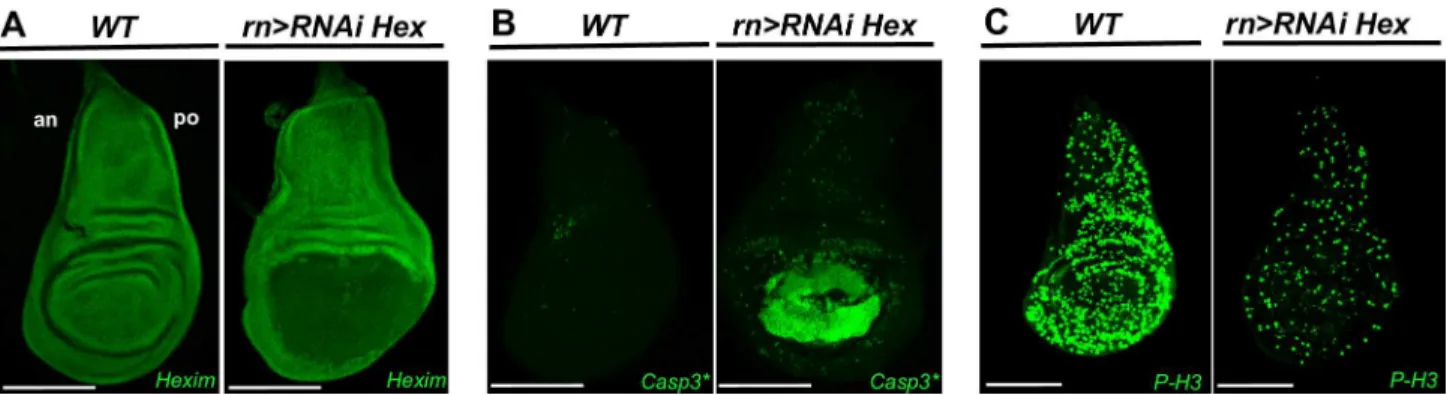
We first addressed whether these developmental defects were mediated by apoptosis by fol-lowing the activity of caspase 3, the mammalian ortholog of the effector caspase DriCE, in the
wing pouch disc, usingrotundGAL4 driver on RNAiHeximmutants (rn>RNAi Hex) (Fig
1B). As expected, the levels of developmentally programmed activated caspase 3 were
extremely low in the wild type (WT) discs. In contrast, they were strongly induced in the wing
pouch of the HEXIM knockdown flies (Fig 1B,rn>RNAi Hex). Levels of activated caspase 3
increased progressively during larval growth (S1A Fig). The latter was delayed by three to four
days. Cell death was further confirmed by TUNEL assay (S2 Fig). Thus we can suggest there is
a direct or indirect connection between HEXIM knockdown and apoptosis.
We next tested whether these developmental failures were due to proliferation defects by motoring the entry in mitosis and S-phase with phospho-histone 3 (P-H3) labeling and EdU incorporation assay, respectively. As expected, WT discs showed high levels of P-H3 positive
cells (Fig 1C) and strong incorporation of EdU (S3A Fig). In contrast, inrn>RNAi Hex
mutant, these markers showed poor labeling not only in the wing pouch, but also in the entire
wing disc (Fig 1CandS3A Fig,rn>RNAi Hex), where HEXIM expression is not affected by
RNAi-mediated expression (Fig 1A). This proliferation arrest is detected at both early and late
L3 stages (S3B Fig). Such transient and non-autonomous reduction of proliferation is known
to occur in damaged tissues after induction of apoptosis [60,61].
If the defects of the HEXIM knockdown were only limited to an induction of apoptosis and/ or a reduction of the cell proliferation rate, one would expect to rescue this phenotype by
co-expression with either the p35 inhibitor of caspase 3 [62,63] or enhancers of cell proliferation,
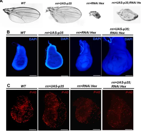
such as CycE or dMyc [64]. In fact, co-expression of p35 failed to rescue the HEXIM
knock-down phenotype. Of note, the corresponding flies usually died at pupal stage, except for a few
escapers (10.4% of the progeny) harboring a partial rescue (Fig 2A,rn-Gal4>UAS-p35;
rn-Gal4>RNAi Hex). Co-expression of CycE or dMyc rescued the wing phenotype for 57.5% of
the progeny with CycE and for 70.7% with dMyc.
Fig 1. HEXIM knockdown induces cell death and transient systemic proliferation arrest.(A) Expression of HEXIM in WT and rn-Gal4>RNAi Heximwing discs at early L3 stage. (B) Immunodetection of cleaved caspase 3 (Casp3*) and (C) Phospho-Histone 3 (P-H3) in WT andrn-Gal4>RNAi Heximwing discs at early L3 stage. The scale bar is for 100μm. In this and all subsequent figures, wing discs are orientated anterior (an) at left and posterior (po) at right.
Taken together, these experiments show that HEXIM knockdown results in a progressive induction of apoptosis and transient proliferation arrest. In order to characterize further the relative impact of proliferation and apoptosis on the HEXIM knockdown phenotypes, we addressed whether wing developmental pathways were affected.
HEXIM knockdown triggers apoptosis-induced compensatory
proliferation and affects cell fate commitment
Despite early proliferation arrest in HEXIM mutant, the mutant wing disc was bigger than the
WT one by the end of larval growth (Fig 2B,WTandrn>RNAi Hex). The latter is delayed by
an average of three to four days. At early L3 stage, the number of P-H3 positive cells is only
half that of WT flies (Fig 2C,WTandrn>RNAi Hex). But after one to two more days, the
pro-liferation rate increases before dropping down at the end of the delayed time (S3B Fig,WTand
rn>RNAi Hex). The increased wing size, together with delayed larval growth, phenocopies the
compensatory proliferation induced by apoptosis in the wing pouch [55,60,65]. Although the
global proliferation rate in HEXIM mutant is lower than the WT, it is compensated by a longer
Fig 2. Co-expression of p35 or dMyc/Cyclin E partially rescue HEXIM knockdown phenotype.(A) Adult wing, (B) DAPI stained wing disc and (C) Phospho-Histone 3 (P-H3) immunodetection at early L3 stage of WT, rn-Gal4>UAS-p35,rn-Gal4>RNAi Heximandrn-Gal4>UAS-p35;RNAi Heximflies.
growth time, finally leading to larger wing discs (x1.8-fold) (Fig 2BandS3C Fig,WTand rn>RNAi Hex). Furthermore, in HEXIM knockdown flies co-expressing p35, the wing disc
size is even larger (2.6-fold higher;Fig 2BandS3C Fig,rn>UAS-p35; RNAi Hex), due to many
“undead cells”continuously producing mitogenic signals [55,66,67]. Of note, overexpression of
p35 in WT background induces a slight overgrowth of wing disc (x1.4-fold;Fig 2B,rn>
UAS-p35) due to inhibition of developmental apoptosis, as previously described [67].
We next sought to identify the signaling pathways that are impaired during wing disc
devel-opment. We first examined the expression patterns of the Notch pathway’s components. We
showed that HEXIM knockdown effectively abrogated the expression of Delta (Dl) and Serrate (Ser) in the Ventral (V) and Dorsal (D) compartments, although expression is maintained in
the stripe between the D-V boundary (S4C and S4D Fig). In addition, the expression of Notch
(N) intra- and extra-cellular components was completely repressed in the wing pouch (S4I and
S4J Fig). The expression levels of other markers (Sal, Cut, and En) follow a similar trend (S4F and S4H Fig). Therefore, HEXIM knockdown strongly affects wing patterning. In addition, we also examined the expression levels of Ptc and the two morphogens Dpp and Wingless (Wg), which are induced in response to tissue damage and promote compensatory proliferation
[55,60–61,65,67–68]. In third instar WT wing discs, Wg is expressed along the D-V boundary
as well as in two concentric circles at the border and outside of the wing pouch [65] (S4E Fig).
In contrast, Dpp and Ptc are expressed in the A compartment, restricted at the A-P boundary, in which Dpp expression is further extended by a 7-cell-diameter region along the A-P stripe,
compared to Ptc (S4A and S4B Fig). In HEXIM knockdown mutants, the expression of both
Dpp and Ptc was increased (S4A and S4B Fig). Of note, the expression domain of Dpp was also
enlarged in the A-P stripe. Moreover, we found that HEXIM knockdown resulted in high accu-mulation of Wg in the cells surrounding the wing pouch, along with a weak, extended stripe at
the D-V boundary (S4E Fig). Thus, the expression patterns of Wg, Dpp and Ptc markers in
HEXIM mutant wing discs resemble the patterns observed when apoptotic cells promote
com-pensatory proliferation [65,68–69]. Taken together, our results show that HEXIM knockdown
affects cell fate commitment of the proliferative cells of the wing imaginal disc.
HEXIM knockdown deregulates Hh signaling and induces Ci expression
To precisely characterize Hedgehog signaling, we monitoredhhexpression in HEXIM
knock-down flies with ahh-lacZreporter. Strikingly, we found a profoundly alteredhhexpression,
which was expressed not only in the canonical anterior part but also in the posterior part of the
wing pouch (Fig 3A–3C). It is noteworthy that co-labeling of Hh-LacZ and En (a posterior
marker of wing disc) reveals aberrant definition of anterior-posterior territories in HEXIM
knockdown imaginal disc (Fig 3B–3C). Importantly, proliferative cells typically do not respond
to apoptosis signal by increasing Hh expression, as it is the case for differentiated cells [54].
Therefore, the ectopic expression of Hh in HEXIM knockdown mutants is not a simple response to the ongoing apoptosis of the wing disc. We also note that Hh levels drop down
non-autonomously in the notum part of the P compartment (Fig 3A, arrow), which is
surpris-ing because the notum is properly developed in the HEXIM mutant. This reduced level of Hh may be transient.
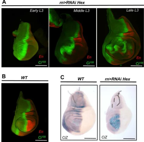
We next characterized the timing and expression profile of Ci, the transcriptional effector of
the Hh pathway [38,41–44]. In HEXIM mutants, we found a broader expression domain of
Ci155that expanded beyond the A-P stripe into the anterior part of the disc. This accumulation
of Ci155, which is highest at early L3, persisted throughout the extended phase of larval growth
Ci155(S1A and S1C Fig). Moreover, when apoptosis was blocked by co-expression of p35 in rn-Gal4>RNAi Heximmutant, Ci155protein level was still high at the A-P stripe (S5 Fig). This
result shows that Hh deregulation takes place before apoptosis. In addition, X-Gal staining of a ci-lacZreporter revealed an increased accumulation ofcitranscripts localized at the anterior
part of the wing pouch in HEXIM mutant (Fig 4C).cimRNA quantification by RT-qPCR
showed a 2-fold increase in HEXIM mutant compared to WT (S6 Fig). This indicates a
tran-scriptional regulation link betweenCiand HEXIM. Altogether, these data show that the altered
Fig 3. HEXIM knockdown deregulates Hh signaling pathway.(A) X-Gal staining ofhh-lacZreporter, in WT andrn-Gal4>RNAi Heximwing discs. For the mutant,β-galactosidase staining duration was reduced to limit signal saturation. En and Hh-LacZβ-galactosidase co-immunodectection in WT (B) and inrn-Gal4>RNAi Hexim(C) wing discs. Arrows indicate the non-autonomous down-regulation of Hh in the posterior
compartment of the notum part. The wing pouch is marked with dotted white line. The assays were performed at early L3 stage.
Ci155expression profile is not a simple consequence of apoptosis, but rather an early response to HEXIM knockdown, which in turn, strongly affects Hh signaling, apoptosis, compensatory proliferation and wing patterning.
We next probed the epistasis between HEXIM and some components of the Hh pathway.
We used a combination of single and double RNAi targeting HEXIM and Ci155expression in
the wing pouch to monitor the expression of two known Ci155target genes:dppandptc. In
adultrn>RNAi Ciflies, the wing blade presented aberrations between interveins 3 and 4 (Fig
5A,rn>RNAi Ci), as expected [70]. The specificity and efficiency of the Ci RNAi construct
were controlled by immunocytochemistry (Fig 5B–5D) and RT-qPCR, both showing a strong
reduction of Ci expression (2.6-fold reduction;S6 Fig). Thern>RNAi Cimutant flies display a
weak but significant decrease of Ptc level in the A-P stripe, especially at the cross of the A-P
and D-V boundaries (Fig 5D,rn>RNAi Ci, arrow), but the expression of both Dpp (data not
shown) and Wg (S7C Fig,rn>RNAi Ci) were unaffected compared to WT (S7A Fig,WT). This
agrees well with the known biology of the Hh pathway, in which Ptc expression requires more
Ci155than Dpp [71]. We then addressed the combined effect of both HEXIM and Ci155
knock-downs. Strikingly, in double mutant, the strong HEXIM phenotype was almost fully rescued since all flies developed wings similar to single Ci knockdown mutant, but with a missing
ante-rior crossvein and an altered vein 3 (Fig 5A,rn-Gal4>RNAi Ci; RNAi Hex,red arrows). In
dou-ble knockdown mutant, Ptc expression was abrogated in the wing pouch due to the absence of Ci (Fig 5, compare E,WTand F,rn-Gal4>RNAi Ci; RNAi Hex), whereas Dpp expression is
Fig 4. HEXIM knockdown deregulates Ci155expression at both protein and transcript levels.
Expression of Ci155and En inrn-Gal4
>RNAi Hexim(A) and WT (B) wing discs, at various L3 stages. (C) Transcription of theCi-lacZreporter in WT andrn-Gal4>RNAi Heximwing discs.
Fig 5. HEXIM is a regulator of Ci.(A) Wing phenotype in WT,rn-Gal4>RNAi Ciandrn-Gal4>RNAi Ci; RNAi Heximflies. Red arrows refer to vein or intervein defects. For single or double mutants, 100% of flies displayed the phenotype (over>200 mutants flies scored). Immuno-localization of Ci155and Ptc in the wing disc of WT (B),rn-Gal4>RNAi Hexim (C)andrn-Gal4>RNAi Ci(D) flies. The reduced levels of Ci155in the A-P stripe (white
arrow) causes aberrations at the intervein 3 and 4. Immuno-localization of Ci155, Ptc and HEXIM in WT (E) and
inrn-Gal4>RNAi Hexim; RNAi Cidouble mutant (F) wing discs. (G) Immuno-localization of Ci155and Dpp in WT
and inrn-Gal4>RNAi Hexim; RNAi Cidouble knockdown. (H) X-Gal staining ofdpp-lacZreporter in WT and in rn-Gal4>RNAi Hexim; RNAi Cimutant.
expended in the A compartment probably due to the absence of Ci75repressor (Fig 5G and 5H,
compareWTandDppZ;rn-Gal4>RNAi Hexim; RNAi Ci). Note that other markers of wing
development like Wg and Cut displayed a WT expression profile in this double mutant (S7A
and S7D Fig). Furthermore, only a modest activation of caspase 3 was detected in the wing
pouch of the double mutant (Fig 6A–6D), which is similar to single RNAi-mediated Ci mutant
(Fig 6C). Therefore, this result suggests thatciis a genetic suppressor ofheximduring wing development.
We then tested whether Hh ectopic overexpression caused by HEXIM knockdown could
trigger apoptosis, by using single and double RNAi against Hh and HEXIM. Singlern>RNAi
Hhmutant displayed reduced anterior crossvein and L3-L4 intervein area (S1 Table;Fig 7A),
as expected [72,73]. Consistently, wing discs show a strong decrease of Ci155and Ptc
immuno-staining (Fig 7B and 7C, compareWTwithrn>RNAi Hhstrains), with no apoptosis (Fig 6E)
and WT expression profile of Wg and Cut (S7E Fig). Similarly, doublern>RNAi Hexim; RNAi
Hhmutants also display reduction of Ci155and Ptc immuno-staining (Fig 7D), and modest
lev-els of caspase 3 (Fig 6F) compared to singlern>RNAi Heximmutant. Therefore, apoptosis
acti-vation in HEXIM mutant is indeed dependent on a high expression level of Hh. Of note, the
development ofUAS-Hex-RNAi; UAS-Hh-RNAimutant is blocked at pupal stage. In addition,
Wg and Cut expression profiles are highly affected (S7F Fig), in a manner similar torn>RNAi
Heximmutant (S7B Fig). This may not be surprising because it is already known that Hh
knockdown mutant may have a different phenotype from Ci mutant [74].
Altogether, our results strongly suggest that HEXIM is a functional (direct or indirect) inter-acting partner in the Hedgehog signaling pathway.
HEXIM knockdown similarly affects other differentiating and proliferating
tissues
We next addressed whether other differentiating or proliferating tissues respond in a similar manner to HEXIM knockdown. To this end, we focused on different parts of the eye imaginal disc. When targeted to the differentiating cells located in the posterior part of the eye discs, HEXIM knockdown resulted in a mild phenotype with rough and/or black-spotted eyes of
smaller size in adult flies (glass multiple reporter,GMR>RNAi Hex), without noticeable
alter-ation of the eye disc morphology [34] (Fig 8A and 8B). In some rare cases (4 over 983 flies), a
mass of overgrowing cells protrudes through the eyes (Fig 8B) suggesting a deregulation of cell
proliferation in this mutant. In contrast, when targeted to the anterior (proliferative) part of
the eye discs with theeyelessdriver (ey>RNAi Hex), the phenotype was much more
pro-nounced and resulted in the ablation of the entire eye-antenna discs (Fig 8A and 8D). The
cor-responding headless larvae died before hatching. Moreover, HEXIM knockdown driven bysine
oculisGAL4 driver(so-Gal4), which follows an increasing gradient from the anterior to the
posterior compartment of the eye discs [75] (Fig 8A), resulted in flies with an intermediate
phe-notype of rough and smaller eyes (25 to 40% smaller than WT;Fig 8CandS1 Table). These
data show that in the eye disc, HEXIM knockdown affects both proliferating and differentiating cells, at different degrees. Furthermore, HEXIM knockdown leads to apoptosis in the eye
imag-inal disc, as observed in the wing disc. Indeed, inGMR>RNAi Hexmutant activated caspase 3
is significantly accumulated in the differentiating region, posterior to the secondary mitotic
wave (S8 Fig).
In order to further characterize the molecular phenotype of HEXIM knockdown in
differen-tiating cells, we conducted Affymetrix microarrays gene expression analysis of theGMR>RNAi
Hexadult eyes (Fig 8B). We found approximately 200 genes differentially expressed, in which
owing to the necrosis in the eye territories, the expression levels of genes involved in the immune system were remarkably elevated (e.g. attacin, cecropin, semmelweis). The expression of several markers of eye terminal differentiation was reduced (e.g. rhodopsin, sepia, irregular chiasm C-roughest) in HEXIM knockdown mutants, while the expression of other genes was
Fig 6. Apoptosis is reduced in double RNAi-mediated knockdown mutant of HEXIM and Ci or HEXIM and Hh. Immunodetection of cleaved caspase 3 (Casp3*) and Ci155at early L3 stage of WT (A),rn-Gal4
>RNAi Hexim(B), rn-Gal4>RNAi Ci(C),rn-Gal4>RNAi Hexim; RNAi Ci(D),rn-Gal4>RNAi Hh(E) andrn-Gal4>RNAi Hexim; RNAi Hh(F) strains.
Fig 7. Reduction of Hh does not rescue HEXIM knockdown mutant.(A) Wing phenotype in WT andrn-Gal4>RNAi Hhflies. The distance between L3 and L4 veins are indicated with a red bar. Immuno-localization of Ci155and Ptc in WT (B),rn-Gal4
>RNAi Hh(C) and rn-Gal4>RNAi Hexim; RNAi Hhdouble mutant (C) wing discs. The reduced levels of Ptc in the A-P stripe (white arrow) are marked in single and double mutants.
doi:10.1371/journal.pone.0155438.g007
Fig 8. HEXIM knockdown affects both differentiating and proliferating tissues.(A) Schematic diagram of the eye-antenna disc summarizing the expression patterns of theGal4drivers:ey(red),GMR(green) and so(hatched). (B) WT andGMR-Gal4>RNAi Heximeyes. (C) WT andso-Gal4>RNAi Heximeyes. (D) WT and theey-Gal4>RNAi Hexim antenna imaginal discs (white circles) and brain. Note the absence of eye-antenna discs iney-Gal4>RNAi Hexim(white arrows).
induced (e.g. mre11, metabolic genes). These results show defects of cell fate commitment dur-ing eye disc development in HEXIM mutants.
Therefore, HEXIM knockdown affects cell fate commitment in both proliferating and dif-ferentiating cells, although the mechanism may be cell-specific and dependent on their ongoing developmental programs.
Discussion
RNAi-mediated HEXIM knockdown phenotype results from failure to
commit to a developmental program
Given that HEXIM is a general regulator of transcription elongation, the transcription machin-ery of mutant cells is eventually expected to be strongly affected that leads to cell death. One
would argue that thern>RNAi Hexmutant phenotype (undeveloped wing) is likely to be a
simple consequence of a severe demolition of the wing pouch. However, we clearly show that the whole tissue is not ablated, although HEXIM mutant displays significant levels of apoptosis. Indeed, dying cells are efficiently replaced by new ones through apoptosis-induced prolifera-tion (AIP) to such extent that the wing disc, including the wing pouch, increases strongly in size but still fails to promote the proper development of the wing. Thus, the phenotype is not a consequence of reduced size of the wing pouch, but rather cells fail to commit to a proper developmental program. The ectopic induction of Hh is one (among other) clear signature of abnormal development.
HEXIM knockdown profoundly affects Hh signaling
Two lines of evidence support a functional connection between HEXIM and Hedgehog
signal-ing: 1) Ci expression is induced early, and 2)ciis a genetic suppressor ofhexim.
Althought Hh is supposed to be mainly anti-apoptotic, there are a few reports indicating that it can promote apoptosis during development. For example when Ptc is deleted, there is
increasing apoptosis in hematopoietic cells [76] or Shh increases cell death in posterior limb
cells [77]. In our study, the induction of Hedgehog signaling is a primary event that precedes
the wave of apoptosis, in HEXIM knockdown mutants. Given that cells subject to patterning defects often undergo apoptosis, the ectopic expression of Hh is probably the molecular event that triggers apoptosis in the wing disc. Then, the subsequent AIP will produce new cells and fuel a self-reinforcing loop of Hh activation and apoptosis (since HEXIM expression is
contin-uously repressed). Accordingly, inrn>RNAi Hexmutant, cells undergoing AIP survive but fail
to differentiate. This is supported by previous reports where deregulation of Hedgehog
signal-ing, through modifications of Ci expression levels, leads to developmental defects [53,78,70].
The phenotype of double knockdown mutants of Ci and HEXIM can be simply explained as following: cells lack the ability to respond to Hedgehog signaling and become blind to Hh pat-terning defect, thus leading to a Ci-like phenotype.
Although we can not exclude that Ci155expression is directly affected by HEXIM, the
extended expression domain of Ci155inrn>RNAi Hexmutants may also indirectly result from
increased levels of Hh. Indeed, the breadth of the AP stripe is defined in part by a morphoge-netic gradient of Hh, with a decreasing concentration towards the anterior part of the wing
disc. Thus, the augmented levels of Hh induced inrn>RNAi Hexmutants could in principle
explain the broader Ci expression at the AP stripe. To summarize, HEXIM knockdown
increases Hh expression, potentially through regulation of P-TEFb complex [79], leading to
A genetic screen inDrosophilashowed that the two components of the small P-TEFb
com-plex, Cdk9 and Cyclin T, are strong activators of the Hh pathway [79], but so far, no evidence
directly connects HEXIM to Hh pathway. To this regard, our work clearly establishes this con-nection. It is then tempting to speculate that by knocking-down HEXIM, the levels of active P-TEFb will be eventually increased that leads to an ectopic activation of the Hh pathway. More work is needed to specifically address this mechanistic point.
Interestingly, when carried out in the eye discs,GMR>RNAi Hexmutants display an
extreme but rare phenotype with protuberances of proliferating cells piercing through the eyes. Although we could not characterize these few events any further, the parallel with the prolifer-ating cells, which fails to differentiate in the wing disc, is striking. Of note, the role of HEXIM
in the balance between proliferation and differentiation is not quite novel (seeIntroduction).
Indeed, HEXIM was previously reported to be up-regulated upon treatment of HMBA [18], a
well known inducer of differentiation. In this paper, we show that the regulatory role of HEXIM during development is mediated via controlling the Hedgehog signaling pathway. To
our knowledge, this is the first time that this has been addressedin vivoand in a
non-pathologi-cal context.
HEXIM: a regulator of transcription during development?
Among other functions, HEXIM acts as a regulator of the P-TEFb activity which is in turn a
general regulator of elongation [34]. The availability of the P-TEFb activity mediates
transcrip-tional pausing, a mechanism by which RNA pol II pauses shortly after transcription initiation and accumulates at the 5' end of genes. Transcription may then or may not resume, depending
on a number of inputs [80]. In these cases, RNA pol II appears 'stalled' at the 5' end of genes.
Release from transcriptional pausing is fast and allows a more homogeneous and synchronized transcription at the scale of an imaginal disc or organ. In the other hand, a lack of release from
transcriptional pausing is also a potent way to silence transcription [81]. Interestingly, genome
wide profiling of RNA pol II revealed a strong accumulation at the 5' end of 20 to 30% of the
genes, most of which involved in development, cell proliferation and differentiation [12–15].
In this context, HEXIM knockdown would be expected to have strong developmental defects. We clearly see such effects in all tissues tested so far.
The patterning of WT wing disc is set by a morphogenetic gradient of Hh, with high levels in the P compartments and no expression in the A compartment. It is therefore tempting to
Fig 9. Model of HEXIM-dependent regulation of wing disc development.(A) In WT wing pouch, HEXIM regulates hedgehog signaling and so its transcriptional effector Ci. (B) In HEXIM knockdown background, Hh is strongly induced and so Ci, and provokes apoptosis, which activates apoptosis-mediated compensatory proliferation. The resulting patterning defects prevent wing development despite the
compensatory proliferation.
speculate that the Hh coding gene would be in a transcriptionally paused state in the anterior part of the wing pouch, that would be released upon HEXIM knockdown. This simple molecu-lar mechanism, although speculative, would account for the induction of the ectopic expression of Hh in the anterior part of the wing pouch and the subsequent loops of apoptosis and AIP, ultimately leading to the wing developmental defects. We tried to ask whether the distribution
of RNA Pol II alonghhandciis compatible with a transcriptional pause by using a number of
RNA Pol II ChIP-Seq datasets that have been generated, together with RNA-Seq data, over the
past few years (e.g. [82–83]). We processed these datasets and computed the stalling index (SI)
for all genes, as previously defined [15] (data not shown). The SI is computed after mapping
ChIP-Seq reads on the reference genome and corresponds to the log ratio of the reads density at the 5' end of the gene over the reads density along the gene body. Although these datasets
clearly reveal a number of 'stalled' genes (>>100), we could not find evidence of paused RNA
Pol II forhhandci(SI value of order 0), which were instead being transcribed. We note,
how-ever, that these datasets have been generated from whole embryos and S2 cell line. Given that Hh and Ci define morphogenetic gradients, their expression (and their transcriptional status) is likely highly variable between cells located in the different sub-regions of a disc, which may therefore not be reflected in these datasets.
Concluding remarks
Apart from the developmental function of HEXIM that we address in this work and the con-nection between HEXIM and Hedgehog signaling, our results may also be of interest for human health studies. First, Hedgehog is a major signaling pathway that mediates liver
organo-genesis and adult liver regeneration after injury [84]. In a murine model of liver regeneration,
the Hedgehog pathway promotes replication of fully differentiated (mature) hepatocytes [84].
Thus, addressing whether a connection between HEXIM and Hh exists would provide a mech-anistic link between the control of gene expression and adult liver regeneration. Second, dereg-ulated Hedgehog signaling is a common feature of many human tumors, and is found in at least 25% of cancers. In addition, recent data showed that aberrant Hedgehog signaling acti-vates proliferation and increases resistance to apoptosis of neighboring cells and thus helps
cre-ate a micro-environment favorable for tumorigenesis [85]. Since its discovery, deregulated
HEXIM expression is often associated to cancers and other diseases [10]. Adding a new
con-nection between HEXIM and Hedgehog signaling will shed more light into the role of HEXIM in abnormal development and cancer.
Surprisingly, although the biochemical interactions between HEXIM and its partners have been thoroughly described, very little is known about its biological function. Thus, this is the first time that the functional impact of HEXIM has been addressed in an integrated system.
Supporting Information
S1 Fig. Immuno-localization of activated caspase 3 and Ci155in WT and during the delayed larval growth ofrn>RNAi Heximmutants.Expression of both Casp3(A) and Ci155(B) is
described inrn>RNAi Heximmutants at different stages of L3 (from early to late). A
magnifi-cation of middle L3 stage shows that Ci155positive cells are not apoptotic cells. Expression of
En and Ci155inrn>UAS-hidimaginal wing disc and together with Casp3(D) is also depicted.
(TIF)
S2 Fig. HEXIM knockdown induces cell death in imaginal wing discs.TUNEL assay in WT andrn-Gal4>RNAi Heximwing discs at early L3 stage.
S3 Fig. HEXIM knockdown with or without p35 leads to a transient proliferation arrest and to an increase of wing disc size.(A) EdU assay in WT andrn-Gal4>RNAi Heximwing
discs. (B) Number of P-H3 positive cell of WT (blue);RNAi Hexim(HEX-, green), and
UAS-p35;RNAi Hexim(HEX-+ P35, red) wing discs during delayed larval growth. (C)
Quantifica-tion of the wing disc size in WT (blue);RNAi Hexim(HEX-, green), andUAS-p35;RNAi
Hexim(HEX-+ P35, red) strains. Counting of PH3 positive cells was performed from 5 to 10
wing discs for each genotype. Wing size is the average from 8 individuals wings. (P<0.001;
error bars: standard deviation). (TIF)
S4 Fig. Comparison between WT andrn>RNAi Heximmutants of the immuno-staining of
several selector genes and morphogens known to be involved in imaginal wing disc devel-opment.(C-J) Immunocytochemistry of the selector genesDelta (Del),Serrate (Ser),Wingless (Wg),Spalt (Sal),Cut,Engrailed (En),Notch extra-cellular (Ne), andNotch intra-cellular (Ni)in
WT andrn-Gal4>RNAi Hexim(C’-J’) wing discs. (A,B) Immunocytochemistry of
morpho-gensDecapentaplegic (Dpp)andPatched (Ptc)in WT andrn-Gal4>RNAi Heximwing discs.
The breadth of Dpp and Ptc expression are indicated with a red scale on the figures (A,B). Immunocytochemistry were performed at early L3 stage.
(TIF)
S5 Fig. Patterns of Wg and Ci155inrn-Gal4>RNAi Heximwith or without co-expression of
p35.Immuno-staining at early L3 stage of Wg and Ci155in WT,rn-Gal4>RNAi Hexim, and
rn>UAS-p35; RNAi Hexim.
(TIF)
S6 Fig.cimRNA quantification by RT-qPCR inrn-Gal4>RNAi Hexim and rn-Gal4>RNAi Cimutants.mRNA quantifications are means from duplicate experiments and are compared
to WT condition (P<0.01; error bars: standard deviation).
(TIF)
S7 Fig. Patterns of Wg and Cut in various mutants.Immuno-staining of Wg and Cut in WT (A),rn-Gal4>RNAi Hexim(B),rn-Gal4>RNAi Ci(C),rn-Gal4>RNAi Hexim; RNAi Ci(D),
rn-Gal4>RNAi Hh(E) andrn-Gal4>RNAi Hexim; RNAi Hh(F).
(TIF)
S8 Fig. HEXIM knockdown induces cell death in eye discs.Immuno-localization at ealy L3
stage of Casp3in WT andGMR-Gal4>RNAi Heximeye discs. MF: Morphogenic Furrow.
SMW: Secondary Mitotic Wave. (TIF)
S1 Table. Phenotype of Hh or HEXIM RNAi mutants.
(PDF)
S2 Table. List of the differentially expressed genes in theGMR-Gal4>RNAi Heximmutants
compared to the WT adult heads.
(DOC)
Acknowledgments
We are grateful to John F. de Celis, Kenneth Irvine, Thomas B. Kornberg, Miura Masayuki,
Jean-Philippe Parvi, Anne Plessis, Hyung Don Ryoo, Carl S. Thummel, the Bloomington
reading of the manuscript, and we thank Niel Randsholt and Dario Coen for helpful discussions.
Author Contributions
Conceived and designed the experiments: DN PU. Performed the experiments: DN OF PU PL. Analyzed the data: DN NB PU. Wrote the paper: DN NB PU.
References
1. Peng J, Zhu Y, Milton JT, Price DH. Identification of multiple cyclin subunits of human P-TEFb. Genes Dev. 1998; 12: 755–762. PMID:9499409
2. Price DH. P-TEFb, a cyclin-dependent kinase controlling elongation by RNA polymerase II. Mol. Cell. Biol. 2000; 20: 2629–2634. PMID:10733565
3. Marshall NF, Peng J, Xie Z, Price DH. Control of RNA polymerase II elongation potential by a novel car-boxyl-terminal domain kinase. J. Biol. Chem. 1996; 271: 27176–27183. PMID:8900211
4. Barboric M, Kohoutek J, Price JP, Blazek D, Price DH, Peterlin BM. Interplay between 7SK snRNA and oppositely charged regions in HEXIM1 direct the inhibition of P-TEFb. EMBO J. 2005; 24: 4291–4303.
PMID:16362050
5. Cho S, Schroeder S, Kaehlcke K, Kwon HS, Pedal A, Herker E et al. Acetylation of cyclin T1 regulates the equilibrium between active and inactive P-TEFb in cells. EMBO J. 2009; 28: 1407–1417. doi:10. 1038/emboj.2009.99PMID:19387490
6. Shim EY, Walker AK, Shi Y, Blackwell TK. CDK-9/cyclin T (P-TEFb) is required in two post-initiation pathways for transcription in the C. elegans embryo. Genes Dev. 2002; 16: 2135–2146. PMID: 12183367
7. He N, Pezda AC, Zhou Q. Modulation of a P-TEFb functional equilibrium for the global control of cell growth and differentiation. Mol. Cell. Biol. 2006; 26: 7068–7076. PMID:16980611
8. Wang S, Fischer PM. Cyclin-dependent kinase 9: a key transcriptional regulator and potential drug tar-get in oncology, virology and cardiology. Trends Pharmacol. Sci. 2008; 29: 302–313. doi:10.1016/j. tips.2008.03.003PMID:18423896
9. Barboric M, Lenasi T, Chen H, Johansen EB, Guo S, Peterlin BM. 7SK snRNP/P-TEFb couples tran-scription elongation with alternative splicing and is essential for vertebrate development. Proc. Natl. Acad. Sci. USA. 2009; 106: 7798–7803. doi:10.1073/pnas.0903188106PMID:19416841
10. Dey A, Chao SH, Lane DP. HEXIM1 and the control of transcription elongation: from cancer and inflam-mation to AIDS and cardiac hypertrophy. Cell Cycle. 2007; 6: 1856–1863. PMID:17671421
11. Rougvie AE, Lis JT. The RNA polymerase II molecule at the 5' end of the uninduced hsp70 gene of D. melanogaster is transcriptionally engaged. Cell 1988; 54: 795–804. PMID:3136931
12. Muse GW, Gilchrist DA, Nechaev S, Shah R, Parker JS, Grissom SF et al. RNA polymerase is poised for activation across the genome. Nat. Genet. 2007; 39: 1507–1511. PMID:17994021
13. Zeitlinger J, Stark A, Kellis M, Hong JW, Nechaev S, Adelman K et al. RNA polymerase stalling at developmental control genes in the Drosophila melanogaster embryo. Nat. Genet. 2007; 39: 1512–
1516. PMID:17994019
14. Guenther MG, Levine SS, Boyer LA, Jaenisch R, Young RA. A chromatin landmark and transcription initiation at most promoters in human cells. Cell. 2007; 130: 77–88. PMID:17632057
15. Core LJ, Waterfall JJ, Lis JT. Nascent RNA sequencing reveals widespread pausing and divergent initi-ation at human promoters. Science. 2008; 322: 1845–1848. doi:10.1126/science.1162228PMID: 19056941
16. Lee C, Li X, Hechmer A, Eisen M, Biggin MD, Venters BJ et al. NELF and GAGA factor are linked to pro-moter-proximal pausing at many genes in Drosophila. Mol. Cell. Biol. 2008; 28: 3290–3300. doi:10. 1128/MCB.02224-07PMID:18332113
17. Gaertner B, Zeitlinger J. RNA polymerase II pausing during development. Development. 2014; 141: 1179–1183. doi:10.1242/dev.088492PMID:24595285
18. Kusuhara M, Nagasaki K, Kimura K, Maass N, Manabe T, Ishikawa S et al. Cloning of hexamethylene-bisacetamide-inducible transcript, HEXIM1, in human vascular smooth muscle cells. Biomedical Res. 1999; 20: 273–279.
19. Wittmann BM, Wang N, Montano MM. Identification of a novel inhibitor of breast cell growth that is down-regulated by estrogens and decreased in breast tumors. Cancer Res. 2003; 63: 5151–5158.
20. Wittmann BM, Fujinaga K, Deng H, Ogba N, Montano MM. The breast cell growth inhibitor, estrogen down regulated gene 1, modulates a novel functional interaction between estrogen receptor alpha and transcriptional elongation factor cyclin T1. Oncogene 2005; 24: 5576–5588. PMID:15940264
21. Huang F, Wagner M, Siddiqui MA. Structure, expression, and functional characterization of the mouse CLP-1 gene. Gene 2002; 292: 245–259. PMID:12119119
22. Huang F, Wagner M, Siddiqui MA. Ablation of the CLP-1 gene leads to down-regulation of the HAND1 gene and abnormality of the left ventricle of the heart and fetal death. Mech. Dev. 2004; 121: 559–572.
PMID:15172687
23. Mascareno E, Manukyan I, Das DK, Siddiqui MA. Down-regulation of cardiac lineage protein (CLP-1) expression in CLP-1 +/- mice affords. J. Cell. Mol. Med. 2009; 13: 2744–2753. doi: 10.1111/j.1582-4934.2008.00404.xPMID:18624753
24. Gurusamy N, Lekli I, Ahsan MK, Ray D, Mukherjee S, Mascareno E et al. Downregulation of cardiac lineage protein-1 confers cardioprotection through the upregulation of redox effectors. FEBS lett. 2010; 584: 187–193. doi:10.1016/j.febslet.2009.11.054PMID:19931534
25. Mascareno E, Galatioto J, Rozenberg I, Salciccioli L, Kamran H, Lazar JM et al. Cardiac lineage pro-tein-1 (CLP-1) regulates cardiac remodeling via transcriptional modulation of diverse hypertrophic and fibrotic responses and angiotensin II-transforming growth factor beta (TGF-beta1) signaling axis. J. Biol. Chem. 2012; 28: 13084–13093.
26. Asakura A. Vascular endothelial growth factor gene regulation by HEXIM1 in heart. Circulation Res. 2008; 102: 398–400. doi:10.1161/CIRCRESAHA.108.172114PMID:18309107
27. Montano MM, Doughman YQ, Deng H, Chaplin L, Yang J et al. Mutation of the HEXIM1 gene results in defects during heart and vascular development partly through downregulation of vascular endothelial growth factor. Circ. Res. 2008; 102: 415–422. PMID:18079413
28. Ogba N, Doughman YQ, Chaplin LJ, Hu Y, Gargesha M, Watanabe M et al. HEXIM1 modulates vascu-lar endothelial growth factor expression and function in breast epithelial cells and mammary gland. Oncogene 2010; 29: 3639–3649. doi:10.1038/onc.2010.110PMID:20453883
29. Ketchart W, Ogba N, Kresak A, Albert JM, Pink JJ, Montano MM. HEXIM1 is a critical determinant of the response to tamoxifen. Oncogene 2011; 30: 3563–3569. doi:10.1038/onc.2011.76PMID: 21423213
30. Ogba N, Chaplin LJ, Doughman YQ, Fujinaga K, Montano MM. HEXIM1 regulates 17beta-estradiol/ estrogen receptor-alpha-mediated expression of cyclin D1 in mammary cells via modulation of P-TEFb. Cancer Res. 2008; 68: 7015–7024. doi:10.1158/0008-5472.CAN-08-0814PMID:18757415
31. Turano M, Napolitano G, Dulac C, Majello B, Bensaude O, Lania L. Increased HEXIM1 expression dur-ing erythroleukemia and neuroblastoma cell differentiation. J. Cell Phys. 2006; 206: 603–610.
32. Zhou Q, Yik JH. The Yin and Yang of P-TEFb regulation: implications for human immunodeficiency virus gene expression and global control of cell growth and differentiation. Microbiol. Mol. Biol. Rev. 2006; 70: 646–659. PMID:16959964
33. Ouchida R, Kusuhara M, Shimizu N, Hisada T, Makino Y, Morimoto C et al. Suppression of NF-kap-paB-dependent gene expression by a hexamethylene bisacetamide-inducible protein HEXIM1 in human vascular smooth muscle cells. Genes Cells 2003; 8: 95–107. PMID:12581153
34. Nguyen D, Krueger BJ, Sedore SC, Brogie JE, Rogers JT, Rajendra TK et al. The Drosophila 7SK snRNP and the essential role of dHEXIM in development. Nucleic Acids Res. 2012; 40: 5283–5297.
doi:10.1093/nar/gks191PMID:22379134
35. Basler K, Struhl G. Compartment boundaries and the control of Drosophila limb pattern by hedgehog protein. Nature. 1994; 368: 208–214. PMID:8145818
36. Blair SS. Compartments and appendage development in Drosophila. BioEssays. 1995; 17: 299–309.
PMID:7741723
37. Walterhouse DO, Yoon JW, Iannaccone PM. Developmental pathways: Sonic hedgehog-Patched-GLI. Environ. Health Perspect. 1999; 107: 167–171. PMID:10064544
38. Kalderon D. Transducing the hedgehog signal. Cell 2000; 103: 371–374. PMID:11081624
39. Osterlund T, Kogerman P. Hedgehog signalling: how to get from Smo to Ci and Gli. Trends Cell. Biol. 2006; 16: 176–180. PMID:16516476
40. Bejarano F, Perez L, Apidianakis Y, Delidakis C, Milan M. Hedgehog restricts its expression domain in the Drosophila wing. EMBO reports. 2007; 8: 778–783. PMID:17571073
42. Dominguez M, Brunner M, Hafen E, Basler K. Sending and receiving the hedgehog signal: control by the Drosophila Gli protein Cubitus interruptus. Science. 1996; 272: 1621–1625. PMID:8658135
43. Orenic TV, Slusarski DC, Kroll KL, Holmgren RA. Cloning and characterization of the segment polarity gene cubitus interruptus Dominant of Drosophila. Genes Dev. 1990; 4: 1053–1067. PMID:2166702
44. Motzny CK, Holmgren R. The Drosophila cubitus interruptus protein and its role in the wingless and hedgehog signal transduction pathways. Mech. Dev. 1995; 52: 137–150. PMID:7577671
45. Aza-Blanc P, Ramirez-Weber FA, Laget MP, Schwartz C, Kornberg TB. Proteolysis that is inhibited by hedgehog targets Cubitus interruptus protein to the nucleus and converts it to a repressor. Cell. 1997; 89: 1043–1053. PMID:9215627
46. Méthot N, Basler K. Hedgehog controls limb development by regulating the activities of distinct tran-scriptional activator and repressors forms of Cubitus interruptus. Cell 1999; 96: 819–831. PMID: 10102270
47. Hooper JE, Scott MP. Communicating with hedegehogs. Nat. Rev. Mol. Cell. Biol. 2005; 6: 306–317.
PMID:15803137
48. Jia J, Zhang L, Zhang Q, Tong C, Wang B, Hou F et al. Phosphorylation by double-time/CKIepsilon and CKIalpha targets cubitus interruptus for Slimb/beta-TRCP-mediated proteolytic processing. Dev. Cell 2005; 9: 819–830. PMID:16326393
49. Jiang J. Regulation of Hh/Gli signaling by dual ubiquitin pathways. Cell Cycle 2006; 5: 2457–2463.
PMID:17102630
50. Smelkinson MG, Kalderon D. Processing of the Drosophila hedgehog signaling effector Ci-155 to the repressor Ci-75 is mediated by direct binding to the SCF component Slimb. Curr. Biol. 2006; 16: 110–
116. PMID:16386907
51. Zhang Q, Zhang L, Wang B, Ou CY, Chien CT, Jiang J. A hedgehog-induced BTB protein modulates hedgehog signaling by degrading Ci/Gli transcription factor. Dev. Cell 2006; 10: 719–729. PMID: 16740475
52. Chen Y, Cardinaux JR, Goodman RH, Smolik SM. Mutants of cubitus interruptus that are independent of PKA regulation are independent of hedgehog signaling. Development. 1999; 126: 3607–3616.
PMID:10409506
53. Lum L, Beachy PA. The Hedgehog response network: sensors, switches, and routers. Science 2004; 304: 1755–1759. PMID:15205520
54. Fan Y, Bergmann A. Distinct mechanisms of apoptosis-induced compensatory proliferation in proliferat-ing and differentiatproliferat-ing tissues in the Drosophila eye. Dev. Cell. 2008; 14: 399–410. doi:10.1016/j. devcel.2008.01.003PMID:18331718
55. Ryoo HD, Gorenc T, Steller H. Apoptotic cells can induce compensatory cell proliferation through the JNK and the Wingless signaling pathways. Dev. Cell 2004; 7: 491–501. PMID:15469838
56. Livak KJ, Schmittgen TD. Analysis of relative gene expression data using real-time quantitative PCR and the 2(-Delta Delta C(T)) method. Methods 2001; 25: 402–408. PMID:11846609
57. Smyth GK. Linear models and empirical bayes methods for assessing differential expression in micro-array experiments. Stat. Appl. Genet. Mol. Biol. 2004; 3: Article3.
58. Lim WK, Wang K, Lefebvre C, Califano A. Comparative analysis of microarray normalization proce-dures: effects on reverse engineering gene networks. Bioinformatics 2007; 23: i282–288. PMID: 17646307
59. Brand AH, Perrimon N. Targeted gene expression as a means of altering cell fates and generating dom-inant phenotypes. Development. 1993; 11: 401–415.
60. Wells BS, Yoshida E, Johnston LA. Compensatory proliferation in Drosophila imaginal discs requires Dronc-dependent p53 activity. Curr. Biol. 2006; 16: 1606–1615. PMID:16920621
61. Wells BS, Johnston LA. Maintenance of imaginal disc plasticity and regenerative potential in Drosophila by p53. Dev. Biol. 2012; 361: 263–276. doi:10.1016/j.ydbio.2011.10.012PMID:22036477
62. Hay BA, Wolff T, Rubin GM. Expression of baculovirus P35 prevents cell death in Drosophila. Develop-ment 1994; 120: 2121–2129. PMID:7925015
63. Lannan E, Vandergaast R, Friesen PD. Baculovirus caspase inhibitors P49 and P35 block virus-induced apoptosis downstream of effector caspase DrICE activation in Drosophila melanogaster cells. J. Virol. 2007; 8: 9319–9330.
64. Jiao R, Daube M, Duan H, Zou Y, Frei E, Noll M. Headless flies generated by developmental pathway interference. Development 2001; 128: 3307–3319. PMID:11546747
65. Smith-Bolton RK, Worley MI, Kanda H, Hariharan IK. Regenerative growth in Drosophila imaginal discs is regulated by Wingless and Myc. Dev. Cell 2009; 16: 797–809. doi:10.1016/j.devcel.2009.04.015
66. Huh JR, Guo M, Hay BA. Compensatory proliferation induced by cell death in the Drosophila wing disc requires activity of the apical cell death caspase Dronc in a nonapoptotic role. Curr. Biol. 2004; 14: 1262–1266. PMID:15268856
67. Pérez-Garijo A, Martín FA, Morata G. Caspase inhibition during apoptosis causes abnormal signalling and developmental aberrations in Drosophila. Development 2004; 131: 5591–5598. PMID:15496444
68. Morata G, Shlevkov E, Pérez-Garijo A. Mitogenic signaling apoptotic cells in Drosophila. Dev Growth Differ 2011; 53: 168–176. doi:10.1111/j.1440-169X.2010.01225.xPMID:21338343
69. Pérez-Garijo A, Shlevkov E, Morata G. The role of Dpp and Wg in compensatory proliferation and in the formation of hyperplastic overgrowths caused by apoptotic cells in the Drosophila wing disc. Develop-ment 2009; 136: 1169–1177. doi:10.1242/dev.034017PMID:19244279
70. Vervoort M, Crozatier M, Valle D, Vincent A. The COE transcription factor Collier is a mediator of short-range Hedgehog-induced patterning of the Drosophila wing. Curr. Biol. 1999; 9: 632–639. PMID: 10375526
71. Hepker J, Wang QT, Motzny CK, Holmgren R, Orenic TV. Drosophila cubitus interruptus forms a nega-tive feedback loop with patched and regulates expression of Hedgehog target genes. Development 1997; 12: 549–558.
72. Mullor JL, Calleja M, Capdevilla J, Guerrero I. Hedgehog activity, independent of decapentaplegic, par-ticipates in wing disc patterning. Development 1997; 124: 1227–1237. PMID:9102309
73. Strigini M, Cohen SM. A hedgehog activity gradient contributes to AP axial patterning of the Drosophila wing. Development 1997; 124: 4697–4705. PMID:9409685
74. Méthot N, Basler K. An absolute requirement for Cubitus interruptus in Hedgehog signaling. Develop-ment 2001; 128: 733–742. PMID:11171398
75. Cheyette BN, Green PJ, Martin K, Garren H, Hartenstein V, Zipursky SL. The Drosophila sine oculis locus encodes a homeodomain-containing protein required for the development of the entire visual sys-tem. Neuron. 1994; 1: 977–996.
76. Siggins SL, Nguyen NYN, McCormack MP, Vasudevan S, Villani R, Jane SM, Wainwright BJ, Curtis DJ. The hedgehog receptor patched1 regulates myeloid and lymphoid progenitors by distinct cell-extrinsic mechanisms. Blood 2009; 114: 995–1004. doi:10.1182/blood-2009-03-208330PMID: 19483124
77. Sanz-Ezquerro JJ, Tickle C. Autoregulation of Shh expression and Shh induction of cell death suggest a mechanism for modulating polarising activity during chick limb development. Development 2000; 127: 4811–4823. PMID:11044396
78. Chen Y, Jiang J. Decoding the phosphorylation code in Hedgehog signal transduction. Cell Res. 2013; 23: 186–200. doi:10.1038/cr.2013.10PMID:23337587
79. Nybakken K, Vokes SA, Lin TY, McMahon AP, Perrimon N. A genome-wide RNA interference screen in Drosophila melanogaster cells for new components of the Hh signaling pathway. Nat. Genet. 2005; 37: 1323–1332. PMID:16311596
80. Lis JT. Imaging Drosophila gene activation and polymerase pausing in vivo. Nature 2007; 450: 198–
202. PMID:17994086
81. Boettiger AN, Levine M. Synchronous and stochastic patterns of gene activation in the Drosophila embryo. Science. 2009; 325: 471–473. doi:10.1126/science.1173976PMID:19628867
82. Gaertner B, Johnston J, Chen K, Wallaschek N, Paulson A, Garruss AS, Gaudenz K, De Kumar B, Krumlauf R, Zeitlinger J. Poised RNA polymerase II changes over developmental time and prepares genes for future expression. Cell Rep. 2012; 2: 1670–1683. doi:10.1016/j.celrep.2012.11.024PMID: 23260668
83. Gan Q, Schones DE, Ho Eun S, Wei G, Cui K, Zhao K, Chen X. Monovalent and unpoised status of most genes in undifferentiated cell-enriched Drosophila testis. Genome Biol. 2010; R42. doi:10.1186/ gb-2010-11-4-r42PMID:20398323
84. Ochoa B, Syn WK, Delgado I, Karaca GF, Jung Y, Wang J et al. Hedgehog signaling is critical for nor-mal liver regeneration after heptectomy in mice. Hepatology 2010; 5: 1712–1723.