R E S E A R C H
Open Access
Anticancer properties of phospholipase A
2
from
Daboia siamensis
venom on human
skin melanoma cells
Suchitra Khunsap
1*, Orawan Khow
1, Supranee Buranapraditkun
2, Sunutcha Suntrarachun
1, Songchan Puthong
3and Supatsorn Boonchang
1Abstract
Background:Phospholipase A2(PLA2) is a major component of theDaboia siamensisvenom, which is able to hydrolyse the membrane of various cells. For this reason, the activity of PLA2was investigated regarding its pharmaceutical properties. This study was conducted to explore the pharmacological properties of a PLA2from Daboia siamensis(dssPLA2) venom on human skin melanoma cell line (SK-MEL-28).
Methods:dssPLA2was isolated by ion exchange and gel filtration columns. Various concentrations of dssPLA2were investigated for cytotoxic activity and inhibition of migration on SK-MEL-28 cells. Cell death analysis, mRNA
expression levels of Notch I-III and BRAF V600E genes were also determined.
Results:dssPLA2exhibited cytotoxicity on SK-MEL-28 for 24 and 72 h as compared with untreated cells. However, it had no toxic effects on CCD-1064sk cells under the same conditions. dssPLA2(0.25 and 0.5μg/mL) induced 17.16 and 30.60 % of apoptosis, while activated 6.53 and 7.05 % of necrotic cells. dssPLA2at 0.25, 0.5, 1 and 2μg/mL could inhibit migration on SK-MEL-28 cells for 24 h by 31.06, 41.66, 50 and 68.75 %, respectively. The action of dssPLA2significantly reduced the levels of Notch I and BRAF V600E genes expression on SK-MEL-28 cells compared with untreated cells at 72 h.
Conclusions:This study indicates that dssPLA2had potential effects of apoptosis, necrosis, cytotoxicity and inhibition of migration on SK-MEL-28 cells. dssPLA2could possibly be a selective agent that targets cancer cells without affecting normal cells.
Keywords:Phospholipase A2,Daboia siamensisvenom, Skin melanoma, Anticancer
Background
Skin cancer, especially melanoma, is a medical problem with increasing incidence that is generally asymptomatic [1]. The risk factors for melanoma depends on several elements, including sun exposure, number of moles on the skin, skin type and family history [2]. Most malig-nant melanomas have mutation of the BRAF gene lead-ing to constitutive activation of downstream signallead-ing in the mitogen-activated protein (MAP) kinase pathway. The activating mutation consists of the substitution of glutamic acid for valine at amino acid 600 (V600E).
Detection of BRAF V600E mutation gene bears relation-ship to survival and proliferation of cancer cells [3].
The surgical treatment in early stages of the disease is ef-fective whereas treatment at late stages present low survival rates. Chemotherapy is a therapeutic option. However, its efficacy is limited due to chemoresistance and toxicity. Re-cently, new agents obtained from natural substances have been studied for their specificity against cancer cells and mild effects towards normal cells. For example, effects of rottlerin, involving the dual inhibition of ERK and NF-κB and downregulation of cyclin D1, led to antiproliferation on SK-MEL-28 cells [4]. Gossypol, a cottonseed extract, has shown effective therapeutic action against BRAF V600E melanoma with resistance to BRAF inhibitors [5]. Three xanthones induced an inhibitory effect on
SK-MEL-* Correspondence:sthaithumnas@yahoo.com
1Research and Development, Queen Saovabha Memorial Institute, Thai Red Cross Society, Bangkok 10330, Thailand
Full list of author information is available at the end of the article
28 cell by modulating the BRAF V600E mutation and the other molecular targets in the apoptotic pathways [6]. Many small molecules have been studied in experimen-tal and clinical trials. For example, vemurafenib, dabra-fenib and trametinib, and serine/threonine kinase inhibitors have been tested in experiments and clinical trials as inhibitors for BRAF V600E mutation gene and MEK1/2 of MAPK pathway, respectively [7]. However, melanoma cell lines treated with these BRAF inhibitors have shown to rebound activation of phosphorylation of ERK (pERK) and escape from BRAF inhibition [8]. The progressed tumor returned within 6 to 8 months after therapy of these agents [9].
The disorder of Notch signaling pathways was associated with tumor genesis of tissues from various origins and also skin melanoma [10, 11]. Notch and their ligands are abun-dantly expressed in the epidermis, either as an oncogene or as a tumor suppressor on melanoma and non-melanoma [12]. The importance of Notch signaling pathway in deter-mining the outcome of treatment has been reported [13]. Both BRAF V600E mutation and Notch signaling pathway should be studied as bioactive agents against melanoma.
PLA2is one of the major components of snake venom
for the digestion of prey [14]. Multifunctional effects of this enzyme have been documented [15, 16]. It directly catalyzes the hydrolysis of cellular phospholipids that gen-erate lysophospholipids and free fatty acids that, in turn, cause membrane damage [17, 18]. Lysophospholipids are important in cell signaling, phospholipid remodeling and membrane perturbation [19]. Some evidence indicated that PLA2hydrolyzed cellular membrane of various
can-cer cells [20]. In a study of membrane-interacted mode and catalytic activity, a PLA2ofNaja naja atra was able
to catalyze outer leaflet and inner leaflet of plasma membrane-mimicking vesicles [21]. The venom from
Macrovipera lebetina transmediterranea has shown to
have antitumor effects mediated by α5β1 and α v-containing integrins [22]. Therefore, PLA2properties
indi-cated high activity against skin melanoma cancers. Our previous study demonstrated the action of a PLA2
fromDaboia siamensis(drsPLA2) against cancer cells [23].
In order to better understand the pharmacological action of dssPLA2 from Daboia siamensis against skin melanoma
cells, the current work is focused on the process of migra-tion inhibimigra-tion, cytotoxicity and gene expression. Apoptosis analysis was also determined on skin melanoma cells. An investigation of molecular targets was performed to identify BRAF V600E mutation gene and Notch signaling receptors; N1, N2 and N3 genes.
Methods Cell cultures
Human skin melanoma SK-MEL-28 (ATCC® HTB-72™) cells and skin fibroblast CCD-1064Sk (ATCC®
CRL-2076™) cells were purchased from the American Type Culture Collection (ATCC, USA). SK-MEL-28 and CCD-1064sk cells were cultured in MEM and Iscove’s media supplemented with 10 % fetal bovine serum (FBS), 1 mM glutamine, 100 U/mL strepto-mycin and 100 U/mL penicillin. Cells were incubated at 37 °C with 5 % CO2. The media, FBS,
strepto-mycin, penicillin and MTT 3-(4,-5-dimethylthiazol-2-yl)-2,5-diphenyltetrazolium bromide (MTT) were purchased from Invitrogen, USA.
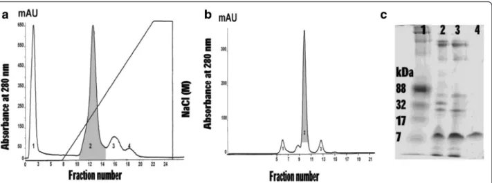
Venom separation by FPLC and SDS-PAGE
Crude venom from Daboia siamensis in lyophilized form was dissolved with 0.02 M phosphate buffer, pH 6.0. The venom solution (approximately 40 mg/ mL) was applied to ion exchange chromatography on HiTrap™ CM FF column. The column was
pre-equilibrated with 0.02 M phosphate buffer, pH 6.0. The proteins were eluted with 0–1 M NaCl linear gradient in 0.02 M phosphate buffer, pH 6.0, at a flow rate 0.5 mL/min. The proteins were collected and measured at absorbance 280 nm under an AKTA pure Fast Pro-tein Liquid Chromatography system (FPLC, GE Healthcare; Sweden). Four peaks were observed and determined for phospholipase A2 activity. Peak 2,
showed PLA2 enzyme activity (gray shaded area in
Fig. 1a), was then further purified by size exclusion chromatography in a pre-equilibrated Superdex 75 10/ 100 GL column. Elution was carried out with 10 mM PBS, pH 7.3, at room temperature. The flow rate was adjusted to 0.5 mL/min., and 1 mL fraction was col-lected in each tube. The proteins were detected by ab-sorbance at 280 nm under a Unicorn 6.3 Software. Four peaks were collected and tested for PLA2activity.
Peak 2 showed a PLA2 activity (gray shaded area in
Fig. 1b) indicating a low molecular weight protein (below 17 kDa) on SDS-PAGE (Fig. 1c).
Each PLA2 fraction was analyzed on SDS containing
polyacrylamide slab gels according to a modified method of Laemmli. The 12.5 % acrylamide in 3.0 M Tris–HCl, pH 8.8, was prepared in the presence of 0.1 % SDS. The venom samples (10 μg of protein) were mixed with a sample buffer and loaded onto a 12.5 % gel under non-reducing conditions. Electrophoresis was carried out at room temperature using 30 mA in the separating process. The precision plus protein standards were used as molecular weight markers. The electrophoresis was stopped when the marker dye reached the bottom of the gel. The electrophoresis gel was stained for 1 h with the Coomassie Brilliant Blue R-250 (0.2 % in methanol/ water/acetic acid, 46.5: 46.5:7) and the excess stain was then removed by de-staining (methanol/acetic acid, 25: 12.5) with several changes of de-staining solution until the background was clear.
Determination of molecular mass
The gel containing dssPLA2 was purified by a
conven-tional method using a clean scalpel to cut off a strip of 15 % Tris-glycine SDS-PAGE. The sample was analyzed by mass spectrometry. Briefly, the excised gel was shred-ded and washed three times with 200 μL of 25 mM NH4HCO3/50 % acetonitrile (ACN). The gels were
dehy-drated with 200 μL of 100 % CAN, then rehydrated by 12.5μg/mL of sequencing grade trypsin (Promega, USA) and incubated at 37 °C for 16 h. The supernatant was transferred to a new tube and added 100 μL of 50 % ACN/0.5 % formic acid. The mixture was finally dried by speedvac and suspended in 10μL of 50 % ACN/0.1 % formic acid. The peptides were analyzed by MS/MS using microTOF-Q II™ ESI-Qq-TOF mass spectrometer
(Bruker, Germany) equipped with an online nanoESI source. MASCOT is a software search engine that iden-tifies proteins based on peptide sequence databases. Mass tolerance of parent and fragmented ions were 1.0 Da and 0.6 Da, respectively. MS/MS ions score ≥38
were considered significant hits.
Phospholipase A2activity
Phospholipase A2activity was performed by Holzer and
Mackessy method [24]. The sample (50 μL) was mixed with 3 mM 4-nitro-3-(octanoyloxy) benzoic acid 1:1 ratio (v/v) and incubated at 37 °C for 20 min. Triton X-100 (2.5 %) was added to the reaction mix and the ab-sorbance was measured at 425 nm by ELISA reader. A standard curve of absorbance as a function of chromo-phore (3-hydroxy-4-nitrobenzoic acid) concentration showed that a change in absorbance of 0.10 AU at 425 nm was equivalent to 25.8 nmoles of chromophore release. The chromophore has an extinction coefficient
of 5039 in this system. This sample was used as PLA2
fraction in the study of migration inhibition, apoptosis, cytotoxic activity, Notch signaling and BRAF V600E mu-tation genes expression on SK-MEL-28 cell line.
Cytotoxic analysis
The MTT assay was chosen for determining the cyto-toxic effect of venoms [25, 26]. Briefly, cells were seeded into 96-well plates (NUNC, Denmark) at concentration of 5 × 10⁴cells/mL and incubated at 37 °C with 5 % CO2
for 24 h. The cells were treated with various concentra-tions of venom (0.25, 0.5, 1, 2 and 4μg/mL) for 24 and 72 h. Cell viability was determined by adding 20 μL of MTT (2.5 mg/mL) for 3 h. An absolute DMSO (150μL) was added to dissolve the formazan crystal. In the exper-iments, absorbance was measured at 540 nm and un-treated cells were used for setting 100 % viability.
Apoptosis analysis
SK-MEL-28 cells were seeded into 6-well plates and incu-bated at 37 °C with 5 % CO2for 24 h. The dssPLA2(0.25
and 0.5μg/mL) were added then incubation continued for the next 24 houra. Apoptotic and necrotic cells were de-tected with AnnexinV apoptosis detection kit (USA), which is based on the observation after initial apoptosis. Cells translocate the membrane phosphatidylserine (PS) from the inner face of the plasma membrane to the cell surface. PS can be detected by staining with a fluorescent isothiocyanate (FITC) conjugate of AnnexinV while propi-dium iodide (PI) binds to the cellular DNA in necrotic cells. The staining process was performed following the manufacturer’s instructions. Cells were acquired and ana-lyzed by FACSCalibur flow cytometry and CellQuest Pro software (Becton Dickinson, San Jose, USA).
Fig. 1A dssPLA2was isolated fromDaboia siamensisvenom. (a) Ion exchange chromatography ofDaboia siamensisvenom on HiTrap™CM FF column.
Protein was eluted with linear gradient 0–1 M NaCl. The peak with gray-shaded area was PLA2activity. (b) Peak 2 from the first step was applied on
Inhibition of cell migration
Wound healing assay was performed for inhibition of cell migration [23]. SK-MEL-28 cells were seeded into 24-well plates (NUNC, Denmark) at 5 × 10⁵ cells/mL concentra-tion and incubated at 37 °C with 5 % CO2for 24 h. After
24 h, the cells were scratched and washed twice with medium without fetal bovine serum. The cells were treated with various concentrations of dssPLA2and
doxo-rubicin; an anticancer drug (2, 1, 0.5, 0.25 and 0 μg/mL) for 24 h. Inhibition of migration was determined by meas-uring the distance of the edge of scratching. The equation of migration inhibition was 100–[((Z-Tn)/Z) × 100]. Z is the negative distance at time 0. Tn is the experiment dis-tance at time 24 h.
Real time-reverse transcription-PCR (qRT-PCR)
qRT-PCR was performed by following manufacturers’ rec-ommendations (RBC Sciences). Total RNA was extracted from the SK-MEL-28 cells that were treated at 0.25μg/mL of dssPLA2for 24 and 72 h. Untreated of SK-MEL-28 cells
were used as a negative control. One microgram of total RNA was employed to generate cDNA using random hex-amer primers. One microliter of 1:10 cDNA dilution was applied to PCR reactions. Sequences of specific primers were Notch1 forward: 5’ -CAGCCTGCACAACCAGA-CAGA-3’; reverse: 5’ -TGAGTTGATGAGGTCCTCCAG-3’; Notch2 forward: 5’ -AAAAATGGGGCCAACCGAGAC-3’; reverse: 5’-TCATCCAGAAGGCGCACA A-3’; Notch3 forward: 5’-AGATTCTCATCCGAAACCGCTCTA-3’; re-verse: 5’-GGGGTCTCCTCCTTGC TATCCTG-3’; BRAF V600E forward: 5’-AGGTGATTTTGGTCTAGCTACA GA-3’; reverse: 5’-TAGTAACTCAGCAGCATCTCAGGG C-3’;β-actin forward: 5’-ACCAACTGGGACGACATG GA CAA-3’; reverse: 5’-GTGGTGGTGAAGCTGTAGCC-3’.
qRT-PCR was performed based on SYBR Green (RBC Biosciences) to quantify the relative expression of BRAF V600E mutation gene and Notch signaling receptors: N1, N2, N3 genes. The mRNA level of theβ-actin gene was de-tected in each cDNA sample to normalize the expression level of gene of interest (GOI). The ratio of GOI and β -actin was compared among samples. The fold change of GOI expression was acquired by setting the values from the untreated cells to one. Calculation of expression level of each gene that was normalized to the expression level ofβ -actin by Delta Delta Ct equation:∆∆Ct =∆Ct (Target gene
treated–Reference gene treated)–∆Ct (Target gene
con-trol–Reference gene control). The mRNA expression gene was calculated by 2-(∆∆CT). CT was a threshold cycle.
Statistical analysis
The statistical significance of the results was analyzed usingt-test (Primer of Biostatistics version 3.02). Differ-ences between the mean values of results were consid-ered statistical significant atp< 0.05.
Results
Purification of phospholipase A2
In the process of purification, Peak 2 from the second step showed the highest PLA2 activity that increased
11.67 folds in the final step. The percent yield of dssPLA2was 27.0 % of crude venom protein. The size of
dssPLA2was confirmed, 16.4 kDa, by using mass
spec-trometry. The summary of the purified method was dis-played in Fig. 1. The purified protein was determined for biological activities.
Cytotoxic analysis
SK-MEL-28 and CCD-1064sk cells were treated with dssPLA2 at concentrations from 0 to 4 μg/mL for 24
and 72 h of incubation. dssPLA2 effects included
dose-and time-dependent cytotoxicity on SK-MEL-28 cells. Viability of cells at 0.25, 0.5, 1, 2 and 4 μg/mL were, respectively, 82.26 % ± 6.73, 72.38 % ± 7.66, 49.19 % ± 5.09, 32.52 % ± 3.3 and 17.82 % ± 4.17 for 24 h whereas were 69.15 % ± 5.38, 32.65 % ± 3.42, 15.95 % ± 2.76, 10.23 % ± 3.35 and 5.3 % ± 1.21 for 72 h when compared with untreated cells. Viability of SK-MEL-28 cells at 72-h incubation was decreased approxi-mately three fold at 1, 2 and 4 μg/mL and 1- and 2-fold at 0.25 and 0.5 μg/mL dssPLA2 concentration
when compared with 24 h-incubation. However, dssPLA2 was not toxic to CCD-1064sk cells at the
same conditions (Fig. 2).
Apoptosis analysis
dssPLA2 at 0.25 and 0.5 μg/mL induced, respectively,
17.16 and 30.60 % apoptosis in SK-MEL-28 cells. How-ever, dssPLA2 also activated necrotic cells at 0.25
(6.53 %) and 0.5 μg/mL (7.05 %), while untreated SK-MEL-28 cells showed signs of necrosis in 1.92 % of the cells (Fig. 3). The results suggest that necrotic cells in-creased when incubation time and concentration of dssPLA2augmented.
Inhibition of cell migration
Wound healing assay was a simple tool to detect inhib-ition of SK-MEL-28 cells migration. The dssPLA2
inhib-ited migration on SK-MEL-28 cells by 31.06 % at 0.25μg/mL, 41.66 % at 0.5μg/mL, 50 % at 1μg/mL and 68.75 % at 2 μg/mL after 24 h of incubation. However, more than 2 μg/mL of dssPLA2did not increase
inhib-ition of migration. Moreover, doxorubicin, an antineo-plastic drug, inhibited less than 50 % of migration at concentrations up to 2μg/mL (Fig. 4).
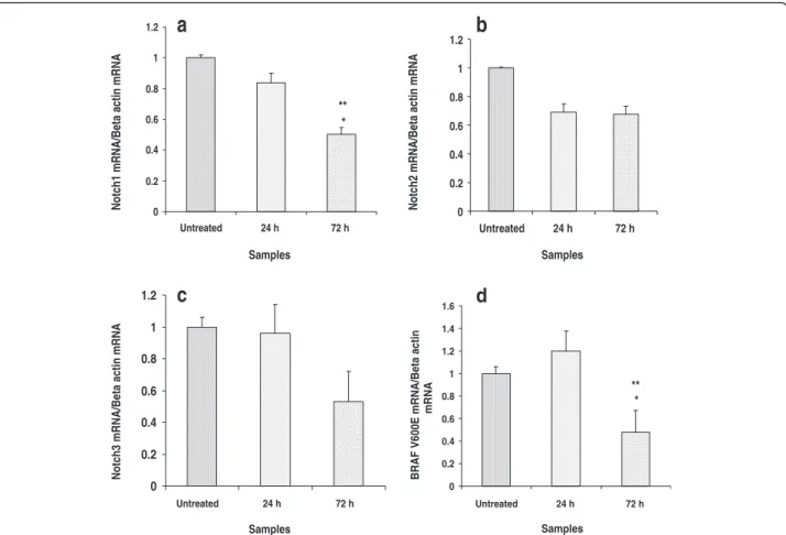
Notch signaling and BRAF V600E mutation genes analysis Effects of dssPLA2to Notch signaling pathway and BRAF
V600E mutation on SK-MEL-28 cells used real-time PCR as a tool for gene expression analysis. SK-MEL-28 cells
were treated with 0.25μg/mL dssPLA2after 24 and 72 h
of incubation. There was no significant change in mRNA expression level of genes on SK-MEL-28 cells between treated and untreated cells for 24 h. N1 and BRAF V600E mutation genes showed significant decrease of mRNA ex-pression level on SK-MEL-28 cells between treated and untreated cells for 72 h (Fig. 5). The dssPLA2did not
sig-nificantly affect N2 and N3 genes.
Discussion
Snake venom PLA2are low molecular weight enzymes that
have different isoforms depending on their geographical
source and species of snakes [27]. In the present study, the action of dssPLA2 damaged SK-MEL-28 cells by causing
cytotoxicity, apoptosis and inhibition of cell migration at 24 h and gene damage after 72 h of incubation. The dssPLA2 showed no toxicity towards normal skin cells
(CCD-1064sk) by MTT assay. These results agree with a previous work that suggested that venom toxins specifically targets cancer cells without affecting normal ones [23]. It indicates a difference of cell membranes between cancer and normal cells, which reveals the variation of phospho-lipid domains in different cell types [14, 18, 28, 29].
Apoptosis is a programmed cell death that occurs as a defense mechanism when cells are irrecoverably dam-aged. Either lost of apoptosis function or the imbalance between cell division and cell death happen in case of cancer. Therefore, the induction of apoptosis pathway is a way to control the proliferation and inhibition of cancer progression. The effect of dssPLA2(0.25 and 0.5 μg/mL)
on apoptosis (17.6 % and 30.60 %) at 24 h of incubation suggests that dssPLA2induced moderate apoptosis on
SK-MEL-28 cells. Apoptosis pathway contains multiple cas-cades that may be investigated by various techniques. AnnexinV-FITC is a tool to detect phosphatidylserine (PS) on the cell surface in initiating apoptosis. Optimization of concentration and incubation period of dssPLA2in order
to provoke apoptosis on SK-MEL-28 cells could be im-proved in further studies. There were a couple of effects of dssPLA2 on SK-MEL-28 cells at 24 h of incubation.
First, dssPLA2induced apoptosis on SK-MEL-28 cells. In
that moment, it had also provoked 31.60, 41.66, 50.0 and 68.75 % of migration inhibition at the following concen-trations: 0.25, 0.5, 1 and 2 μg/mL. dssPLA2 (0.25 and
0.5μg/mL) had induced death on SK-MEL-28 cells (23.19 and 37.65 %) at 24 h. The remaining surviving cells (76.81 Fig. 2Viability of SK-MEL-28 and CCD-1064sk cells treated with dssPLA2
for 24 and 72 h. Data are expressed as means ± SE of triplicates (n= 4). Treatment that significantly differed from untreated control for 24 and 72 h atp< 0.05 are indicated as * and ** respectively. ***p< 0.05 indicates the significant differences between 24- and 72-h treatment at the same concentration
0 10 20 30 40
0.5 0.25 Untreated
dssPLA2 concentrations (µg/ml)
Tot
a
l a
popt
o
s
is
a
nd ne
c
ros
is
(
%
)
Apoptosis Necrosis
Fig. 3Total apoptosis and necrosis of SK-MEL-28 cells were induced by incubation with dssPLA2for 24 h. Results are expressed as
mean ± SE (n= 4)
0 25 50 75 100
2 1 0.5 0.25 Untreated
Concentrations (µg/ml)
M
igrat
io
n i
n
h
ib
it
ion (
%
) dssPLA2Doxorubicin
*
* *
* ** **
*
Fig 4Inhibition of SK-MEL-28 cell migration by dssPLA2from
Daboia siamensisvenom compared with doxorubicin (anticancer
and 62.35 %) resisted apoptosis and necrosis, and could migrate. Therefore, the cells migration might be inhibited due to other effects of dssPLA2. The minimum
concentra-tion of SK-MEL-28 cells (1 × 10⁵cells/mL) could close a scratch within 24 h without inhibitors (Additional file 1). The results indicated that dssPLA2had different actions
in apoptosis and inhibition of migration on SK-MEL-28 cells at 24 h. However, both effects of dssPLA2should be
studied further.
One of the treatments for metastatic melanoma is based on BRAF V600E mutation gene. The BRAF V600E mutation gene has been reported to promote cancer cell survival and proliferation. This is found in most malignant melanomas [6, 30, 31]. Many selective small molecule inhibitors of BRAF V600E have been studied. Similarly to vemurafenib and dabrafenib, BRAF V600E mutation gene inhibitors have a limitation by fre-quent and rapid onset of resistance [4, 8, 9, 32].
In our study, qRT-PCR was used to determine the mRNA expression of BRAF V600E mutation on SK-MEL-28 cells treated by dssPLA2, compared with
un-treated control. In melanoma cancer, the mutation of
BRAF V600 induced the hyperactive BRAF kinase activ-ity and led to promote the uncontrolled cell proliferation [32]. The BRAF V600E mutation gene was a targeting to melanoma cancer treatment. Therefore, we examined the effect of dssPLA2on BRAF V600E mutation gene to
melanoma therapy. Moreover, Notch signaling genes, which are the cell cycle regulation genes, were also in-vestigated in the same experiment. The Notch signaling pathway plays an important role in balance on the cell proliferation, differentiation and apoptosis pathways. Dysfunction in Notch pathway is connected with tumor mutagenesis in various tissues [12, 13, 30]. When those genes were blocked, the characteristic cell death would appear and dssPLA2might be useful in treatment of this
cancer.
The mRNA expression level of N1, N2, N3 and BRAF V600E genes between treated and untreated SK-MEL-28 cells were not significantly changed after 24 h incubation (Fig. 5). This result indicated that dssPLA2had no effect
of N1, N2, N3 and BRAF V600E mutation genes and were not related to induction of apoptosis and inhibition of cell migration. In contrast, the significantly decrease
0 0.2 0.4 0.6 0.8 1 1.2
Untreated 24 h 72 h
Samples
Notch1 mRNA/Beta actin mRNA
** *
a
0 0.2 0.4 0.6 0.8 1 1.2
Untreated 24 h 72 h
Samples
Notch2 mRNA/Beta actin mRNA
b
0 0.2 0.4 0.6 0.8 1 1.2
Untreated 24 h 72 h
Samples
Notch3 mRNA/Beta actin mRNA
c
0 0.2 0.4 0.6 0.8 1 1.2 1.4 1.6
Untreated 24 h 72 h
Samples
BRAF V600E mRNA/Beta actin
mRNA
** *
d
Fig. 5mRNA expression level of Notch1, Notch2, Notch3 and BRAF V600E mutation genes on SK-MEL-28 cells were induced by 0.25μg/mL of
dssPLA2for 24 and 72 h. The values represent relative gene expression compared to untreated cells. Significant differences between untreated
and treated cells are presented as *p< 0.001. **p< 0.001 indicates difference between 24- and 72-h treatment at the same concentration
in the mRNA expression level of N1 and BRAF V600E mutation genes were observed on SK-MEL-28 cells after 72 h of incubation with dssPLA2 compared with
un-treated cells (Fig. 5a and d). This suggested that N1 and BRAF V600E genes may be connected with induction of cytotoxic process by dssPLA2 due to the mortality on
SK-MEL-28 cells. A number of studies have examined the potential of Notch signaling genes by various agents. The results of these studies showed that down regulation of Notch signaling genes are similar to using dssPLA2. For
example, down regulation of Notch1 provoked inhibition of cell growth and apoptosis in cancer cells [10, 11]. In contrast, the increased expression of Notch components was associated with a worse prognosis for cancer and a shorter survival rate [13]. Synthetic substances and natural products have been studied in order to find a melanoma inhibitor concerning this gene. For example, xanthones significant inhibited mRNA expression of BRAF V600E mutation gene at 48 h [6]. Numerous inhibitors–such as vemurafenib, dabrafebib and trametinib – are potential substances of interest since they target BRAF V600E and MAPK signaling. Unfortunately, the disease could re-emerge and escape from therapy. Therefore, the effect of dssPLA2on melanoma should be evaluated in several
as-pects, especially resistance mechanisms focusing on BRAF V600E mutation gene and BRAF protein. dssPLA2is a
po-tential candidate against melanoma that would prevent the recurrence of the disease.
Conclusions
We reported the effects of dssPLA2, a phospholipase A2
from Daboia siamensis venom, activity on SK-MEL-28
cells. This enzyme strongly inhibited migration and cyto-toxicity and showed significant down regulation of N1 and BRAF V600E mutation genes on SK-MEL-28 cells that might lead to cell damage and death. Interestingly, it pre-sented no toxicity towards CCD-1064sk, a normal skin cell.
Ethics approval
This study was approved by the Queen Saovabha Memorial Institute Committee on the Ethics of Human Research (Reference number R2015-04).
Additional file
Additional file 1:Amount of SK-MEL-28 cells that could close a scratch within 24 h of incubation.(PDF 5480 kb)
Abbreviations
CCD-1064sk:Normal skin fibroblast cell line; DMSO: Dimethyl sulfoxide; dssPLA2: Phosphplipase A2isolated fromDaboia siamensisvenom;
FITC: Fluorescent isothiocyanate; FPLC: Fast protein liquid chromatography; MTT: MTT 3-(4,5-dimethylthiazol-2-yl)-2,5-diphenyltetrazolium bromide; N1, N2, N3: Notch 1, Notch 2, Notch 3; PI: Propidium iodide; PLA2: Phospholipase
A2; PS: Phosphatidylserine; qRT-PCR: Quantitative reverse transcription
polymerase chain reaction; SDS-PAGE: Sodium dodecyl sulfate polyacrylamide electrophoresis; SK-MEL-28: Human skin melanoma cells.
Competing interests
The authors declare that there are no competing interests.
Authors’contributions
SK designed the study, handled all experiments, discussed the results and drafted the manuscript. OK isolated the dssPLA2 and its activity. SB performed on apoptotic analysis. SS worked on qRT-PCR. SP and SB helped in tissue culture processes and statistical analysis. All authors read and approved the manuscript.
Acknowledgements
We are thankful to Dr. Lawan Chanhome for the venoms provided. We are also grateful to Prof. Dr. Visith Sitprija and Prof. Dr. Narongsak Chaiyabutr for revising this manuscript.
Author details
1Research and Development, Queen Saovabha Memorial Institute, Thai Red Cross Society, Bangkok 10330, Thailand.2Department of Medicine, Faculty of Medicine, Cellular Immunology Laboratory Allergy and Clinical Immunology Unit, Chulalongkorn University, Bangkok 10330, Thailand.3Institute of Biotechnology and Genetic Engineering, Chulalongkorn University Institute, Building 3, Phayathai Road, Patumwan, Bangkok 10330, Thailand.
Received: 9 October 2015 Accepted: 10 February 2016
References
1. Hardy KM, Kirschmann DA, Seftor EA, Margaryan NV, Postovit LM, Strizzi L. Regulation of the embryonic morphogen Nodal by Notch4 facilitates manifestation of the aggressive melanoma phenotype. Cancer Res. 2010; 70(24):10340–50. doi:10.1158/0008-5472.
2. Einspahr JG, Stratton SP, Bowden GT, Alberts DS. Chemoprevention of human skin cancer. Crit Rev Oncol Hematol. 2002;41(3):269–85.
3. Kumar R, Angelini S, Czene K, Sauroja I, Kahka-Kemppinen M, Pyrhönen S, et al. BRAF mutations in metastatic melanoma: a possible association with clinical outcome. Clin Cancer Res. 2003;9(9):3362–8.
4. Daveri E, Valacchi G, Romagnoli R, Maellaro E, Maioli E. Antiproliferative effect of Rottlerin on SK-MEL-28 melanoma cells. Evid Based Complement Alternat Med. 2015;2015(article ID 545838):9. doi:10.1155/2015/545838. 5. Jang GH, Lee M. BH3-mimetic gossypol-induced autophagic cell death in mutant
BRAF melanoma cells with high expression of p21cip1. Life Sci. 2014;102(1):41
–8.
6. Wang JJ, Zhang W, Sanderson BJS. Altered mRNA expression related to the apoptotic effect of three Xanthones on human melanoma SK-MEL-28 cell line. Biomed Res Int. 2013;2013(article ID 715603):10. doi:10.1155/2013/715603. 7. Wu P, Nielsen TE, Clausen MH. FDA-approved small-molecule kinase
inhibitors. Trends Pharmacol Sci. 2015;36(7):422–39. http://dx.doi.org/10.
1016/j.tips.2015.04.005.
8. Alcalá AM, Flaherty KT. BRAF inhibitors for the treatment of metastatic melanoma: Clinical trials and mechanisms of resistance. Clin Cancer Res. 2012;18(1):33–9.
9. Sullivan RJ, Flaherty KT. Resistance to BRAF-targeted therapy in melanoma. Eur J Cancer. 2013;49(6):1297–304.
10. Wang Z, Zhang Y, Li Y, Banerjee S, Liao J, Sarkar FH. Down-regulation of Notch-1 contributes to cell growth inhibition and apoptosis in pancreatic cancer cells. Mol Cancer Ther. 2006;5(3):483–93.
11. Suwanjunee S, Wongchana W, Palaga T. Inhibition of gamma-secretase affects proliferation of leukemia and hepatoma cell lines through Notch signaling. Anticancer Drugs. 2008;19(5):477–86.
12. Panelos J, Massi D. Emerging role of Notch signaling in epidermal differentiation and skin cancer. Cancer Biol Ther. 2009;8(21):1986–93.
13. Lachej N, Didziapetriene J, Kazbariene B, Kanopiene D, Jonušiene V.
Association between Notch signaling pathway and cancer. Acta Med Litu. 2012;19(4):427–37.
14. Saikia D, Bordoloi NK, Chattopadhyay P, Choklingam S, Ghosh SS, Mukherjee AK. Differential mode of attack on membrane phospholipids by an acidic phospholipase A2(RVVA-PLA2-I) fromDaboia russellivenom. Biochim
15. Soares AM, Giglio JR. Chemical modifications of phospholipase A2from
snake venom: effects on catalytic and pharmacological properties. Toxicon. 2003;42(8):855–68.
16. Zouari-Kessentini R, Luis J, Karray A, Kallech-Ziri O, Srairi-Abid N, Bazaa A, et al. Two purified and characterized phospholipases A2fromCerastes
cerastesvenom, that inhibit cancerous cell adhesion and migration. Toxicon.
2009;53(4):444–53.
17. Cumming BS, Mchowat J, Schnellmann RG. Phospholipase A2s in cell injury
and death. J Pharmacol Exp Ther. 2000;294(3):793–9.
18. Mukherjee AK. Correlation between the phospholipids domains of the target cell membrane and the extent ofNaja kaouthiaPLA2-induced
membrane damage: evidence of distinct catalytic and cytotoxic sites in PLA2molecules. Biochim Biophys Acta. 2007;1770(2):187–95.
19. Jensen LB, Burgess NK, Gonda DD, Spencer E, Wilson-Ashworth HA, Driscoll E, et al. Mechanisms governing the level of susceptibility of erythrocyte membranes to secretory phospholipase A2. Biophys J. 2005;88(4):2692–705.
20. Chen YJ, Lin HC, Chen KC, Lin SR, Cheng TL, Chang LS. Taiwan cobra phospholipase A2suppresses ERK-mediated ADAM17 maturation, thus
reducing secreted TNF-αproduction in human leukemia U937 cells. Toxicon. 2014;86:79–88.
21. Chiou YL, Lin SR, Hu WP, Chang LS. Modulated mechanism of
phosphatidylserine on the catalytic activity ofNaja naja atraphospholipase
A2andNotechis scutatus scutatusnotexin. Toxicon. 2014;92:113–22.
22. Bazaa A, Luis J, Srairi-Abid N, Kallech-Ziri O, Kessentini-Zouari R, Defilles C, et al. MVL-PLA2, a phospholipase A2 fromMacrovipera lebetina
transmediterraneavenom, inhibits tumor cells adhesion and migration.
Matrix Biol. 2009;28(4):188–93.
23. Khunsap S, Pakmanee N, Khow O, Chanhome L, Sitprija V, Suntravat M, et al. Purification of a phospholipase A2fromDaboia russelii siamensisvenom
with anticancer effects. J Venom Res. 2011;2:42–51.
24. Holzer M, Mackessy SP. An aqueous endpoint assay of snake venom phospholipase A2. Toxicon. 1996;34(10):1149–55.
25. Mosmann T. Rapid colorimetric assay for cellular growth and survival: application to proliferation and cytotoxicity assays. J Immunol Methods. 1983;65(1–2):55–63.
26. Sieuwerts AM, Klijn JG, Peters HA, Foekens JA. The MTT tetrazolium salt assay scrutinized: how to use this assay reliably to measure metabolic activity of cell culturesin vitrofor the assessment of growth characteristics, IC50-values and
cell survival. Eur J Clin Chem Clin Biochem. 1995;33(11):813–23.
27. Jan VM, Guillemin I, Robbe-Vincent A, Choumet V. Phospholipase A2
diversity and polymorphism in European viper venoms: paradoxical molecular evolution in Viperinae. Toxicon. 2007;50(8):1140–61.
28. Kojima K. Molecular aspects of the plasma membrane in tumor cells. Nagoya J Med Sci. 1993;56(1–4):1–18.
29. Barros GAC, Pereira AV, Barros LC, Lourenço Jr A, Calvi SA, Santos LD, et al.
In vitroactivity of phospholipase A2 and of peptides fromCrotalus durissus
terrificusvenom against amastigote and promastigote forms ofLeishmania
(L.)infantum chagasi. J Venom Anim Toxins incl Trop Dis. 2015;24:21–48.
doi:10.1186/s40409-015-0049-0.
30. Maddodi N, Bhat KMR, Devi S, Zhang SC, Setaluri V. Oncogenic BRAFV600E
induces expression of neuronal differentiation marker MAP2 in melanoma cells by promoter demethylation and down-regulation of transcription repressor HES1. J Biol Chem. 2010;285(1):242–54.
31. Senft D, Berking C, Graf SA, Kammerbauer C, Ruzicka T, Besch R. Selective induction of cell death in melanoma cell lines through targeting of Mcl-1 and A1. PLoS One. 2012;7(1):e30821. doi:10.1371/journal.pone.0030821. 32. Bucheit AD, Davies MA. Emerging insights into resistance to BRAF inhibitors
in melanoma. Biochem Pharmacol. 2014;87(3):381–89.
• We accept pre-submission inquiries
• Our selector tool helps you to find the most relevant journal
• We provide round the clock customer support
• Convenient online submission
• Thorough peer review
• Inclusion in PubMed and all major indexing services
• Maximum visibility for your research
Submit your manuscript at www.biomedcentral.com/submit