Cells Stimulated with
Pasteurella multocida
Toxin
Rebecca C. Babb1¤, Karen A. Homer1, Jon Robbins2, Alistair J. Lax1*
1King’s College London, Department of Microbiology, Dental Institute, Guy’s Hospital, London, United Kingdom,2King’s College London, Wolfson Centre for Age-Related Disease, London, United Kingdom
Abstract
Many bacterial toxins covalently modify components of eukaryotic signalling pathways in a highly specific manner, and can be used as powerful tools to decipher the function of their molecular target(s). The Pasteurella multocida toxin (PMT) mediates its cellular effects through the activation of members of three of the four heterotrimeric G-protein families, Gq, G12 and Gi. PMT has been shown by others to lead to the deamidation of recombinant Gaiat Gln-205 to inhibit its intrinsic GTPase activity. We have investigated modification of native Gasubunits mediated by PMT in Swiss 3T3 cells using 2-D gel electrophoresis and antibody detection. An acidic change in the isoelectric point was observed for the Gasubunit of the Gq and Gifamilies following PMT treatment of Swiss 3T3 cells, which is consistent with the deamidation of these Gasubunits. Surprisingly, PMT also induced a similar modification of Ga11, a member of the Gqfamily of G-proteins that is not activated by PMT. Furthermore, an alkaline change in the isoelectric point of Ga13was observed following PMT treatment of cells, suggesting differential modification of this Gasubunit by PMT. Gswas not affected by PMT treatment. Prolonged treatment with PMT led to a reduction in membrane-associated Gai, but not Gaq. We also show that PMT inhibits the GTPase activity of Gq.
Citation:Babb RC, Homer KA, Robbins J, Lax AJ (2012) Modification of Heterotrimeric G-Proteins in Swiss 3T3 Cells Stimulated withPasteurella multocida Toxin. PLoS ONE 7(11): e47188. doi:10.1371/journal.pone.0047188
Editor:Esteban Chaves-Olarte, Universidad de Costa Rica, Costa Rica
ReceivedMarch 16, 2012;AcceptedSeptember 12, 2012;PublishedNovember 5, 2012
Copyright:ß2012 Babb et al. This is an open-access article distributed under the terms of the Creative Commons Attribution License, which permits unrestricted use, distribution, and reproduction in any medium, provided the original author and source are credited.
Funding:This work was supported by a studentship grant to RCB from the Biotechnology and Biological Sciences Research Council, UK. The funders had no role in study design, data collection and analysis, decision to publish, or preparation of the manuscript.
Competing Interests:The authors have declared that no competing interests exist. * E-mail: [email protected]
¤ Current address: Centre for Haematology, Division of Experimental Medicine, Faculty of Medicine, Imperial College London, London, United Kingdom
Introduction
Heterotrimeric G-proteins are a family of key signal transduc-tion proteins that intercede between the many G-protein coupled receptors (GPCR) that the cell uses to interrogate its local environment and downstream signalling pathways that ultimately regulate fundamental cellular choices [1]. G-proteins are divided into 4 classes (Gq, G12, Giand Gs) according to their constituent
alpha subunit, which is a guanine nucleotide binding protein that can exist in an inactive GDP-bound or an active GTP-bound form [2]. Activation of a GPCR causes a conformational change in its cognate Gasubunit that triggers GDP to be exchanged for GTP. The activated state persists until GTP is hydrolysed to GDP by the intrinsic GTPase activity of the Ga subunit. G-proteins are also subject to reversible tyrosine phosphorylation and lipid modifica-tions during their activation cycle, but the regulatory role of these events is not fully understood [3]. Each G-protein class activates a characteristic set of downstream targets. The Gsand Gifamilies
activate or inhibit adenylate cyclase, respectively [4]. The Gq
family activates phospholipase C (PLC) [5], while the G12family is
particularly linked to activation of the Rho GTPase [6]. Intracellularly-acting bacterial protein toxins enzymatically modify a limited and precise set of cellular proteins to modulate their function. The Pasteurella multocida toxin (PMT) activates multiple signalling pathways in cultured cells leading characteris-tically to a strong mitogenic response [7]. PMT has been shown to
activate members of the Gq, G12 and Gifamilies [8–13]. PMT
catalyses the deamidation of recombinant Giat Gln-205 to inhibit
its intrinsic GTPase activity [14]. We describe here the effects of PMT on all four classes of heterotrimeric G-proteins in Swiss 3T3 cells using two-dimensional (2-D) gel electrophoresis and other techniques.
Materials and Methods
Reagents
Cell culture reagents were obtained from Invitrogen. (c-32P) GTP was obtained from PerkinElmer LAS. Anti-Gaq/11(sc-392),
anti-Ga11(sc-394), anti-Gas(sc-387), anti-Ga13(sc-410) and
anti-Gai-2 (internal: sc-7276) antibodies were from Santa Cruz
Biotechnology. Anti-Gaq(371752), anti-Gai-1(371720), anti-Ga i-1-2 (371723) and anti-Gai-1-3 (371729: which is known to cross
react with Gai-1 and Gai-2) antibodies were purchased from
Calbiochem-Novabiochem. Phospho-FAK (Tyr397) was from New England Biolabs Ltd. All reagents used for 2-D gel electrophoresis were from GE HealthCare, unless otherwise stated. Recombinant PMT was purified essentially as described [15]. A recombinant His-tagged Gaq subunit (371765) was purchased from
Calbio-chem-Novabiochem. Recombinant His6-tagged human Gai-1was
USA) [16]. All other chemical reagents were of analytical grade and were obtained from Sigma-Aldrich, unless otherwise stated.
Cell culture
Swiss 3T3 cells, originally developed by Todaro and Green [17], and kindly provided by Theresa Higgins (Cancer Research UK, London, UK) were cultured as described [9]. Cells were grown to confluence and used when quiescent, before the addition of PMT or bombesin (Calbiochem-Novabiochem). The tyrosine kinase inhibitors Su6656 and St638 (Calbiochem-Novabiochem) were prepared in DMSO, diluted in DMEM containing 0.1% DMSO and added to cell cultures to give a final concentration of 100mM 1 h prior to treatment with PMT.
Preparation of Swiss 3T3 membranes and cytoplasmic fractions
Swiss 3T3 cells were grown in 145 mm dishes, rinsed twice with ice cold PBS and scraped into 2 ml of PBS containing proteinase inhibitors (CompleteTM, Roche Diagnostics). Cells from 10 dishes were pooled, collected by centrifugation (200g, 10 min, 4uC), and washed cell pastes were frozen at 270uC until required. The frozen cell pastes (,5 mg) were thawed on ice and suspended in 5 ml of membrane buffer (10 mM Tris-HCl, 10 mM MgCl2,
0.1 mM EDTA, pH 7.4, containing proteinase inhibitors). The cells were ruptured by 25 passes through a 23-gauge needle, and the resulting homogenate was centrifuged at 800gfor 10 min to remove unbroken cells and nuclei. The supernatants were transferred to fresh tubes and centrifuged at 50,000 g for 10 min. The supernatant containing cytoplasmic proteins was transferred to a fresh tube, snap frozen in liquid nitrogen and stored at270uC. The pellet was washed and suspended in 10 ml of membrane buffer. After a second centrifugation step the membrane pellet was suspended in membrane buffer to a protein concentration of 1 mg/ml and stored at270uC.
SDS PAGE and urea gel electrophoresis
Membrane proteins were resolved by SDS PAGE on 12.8% acrylamide/0.06% bis acrylamide gels, or on these same gels containing 6M urea to separate the closely migrating Ga11and
Gaqsubunits as described [18]. Proteins were transferred to PVDF
membranes and immunoblotted as described below.
2-D gel electrophoresis
Swiss 3T3 membrane proteins were resolved by 2-D gel electrophoresis, as described [19]. The immunodetection of Ga subunits was performed by incubating the membrane overnight at 4uC with primary antibody at a dilution of 1:1000, followed by incubation with horseradish peroxidase-coupled secondary anti-body at a dilution of 1:10000 (SouthernBiotech) for 1 h at room temperature. The membrane was incubated with ECLTM chemiluminescent substrate (GE HealthCare) and signals were detected using an automatic X-Ray film processor (Jungwon Precision Industries Co.).
Calcium microfluorimetry
Intracellular calcium was recorded as given previously [20]. Briefly, Swiss 3T3 cells were plated onto 19 mm glass cover slips and incubated in 5mM Indo –AM (1 hour, 37uC, in the dark, Calbiochem). Cover slips were placed in a custom built chamber allowing gravity fed superfusion (10–12 ml/min) of a modified Krebs solution. Bombesin was applied by switching a multiway tap to a solution containing it and was removed by switching back to a bombesin free solution. The waste was removed by a peristaltic
pump. Recordings were performed at room temperature by subtraction of background light and recording the emitted light from individual cells at 405 and 488 nm. The emission ratio (R) was converted to a calcium concentration after calibration (see reference 20] in which [Ca]i (nM) = 1028(R-0.86)/(12-R) and autofluorescence was less that 4%. .
Trypsin protection assay
The trypsin protection assay was adapted from Evanko et al.
[21]. Briefly, membrane fractions (100mg) were incubated with PMT, bombesin, GTPcS or GTP at the required concentrations at 37uC for times indicated. Membrane fractions were centrifuged at 18,0006g for 10 min at 4uC and the pellet was resuspended in
12.8ml of solubilisation buffer (20 mM Tris-HCl, pH 7.5, 100 mM NaCl, 2 mM MgCl2, 0.1 mM EDTA, 1 mM
dithio-threitol, 10% glycerol, 1% C12E10 (polyoxyethylene 10-lauryl
ether), 0.1 mM phenylmethylsulfonyl fluoride), vortexed, incubat-ed on ice for 20 min and centrifugincubat-ed at 18,0006g for 10 min at 4uC. The supernatant was then transferred to a new microfuge tube, treated with 4ml of trypsin mixture (100mM GDP, 1.5 mg/ ml trypsin in solubilisation buffer) for 30 min at 30uC. The trypsin activity was neutralised with 3ml of soybean trypsin inhibitor (3 mg/ml). Trypsin-resistant fragments were resolved by SDS-PAGE, and detected by immunoblotting using antiserum against Gaq/11. The induction of trypsin protection by GTPcS and GTP
alone or in the presence of bombesin or PMT were quantified relative to untrypsinised Gq using scanning densitometry
(Gene-Tools, Syngene). Data were analysed using factorial analysis of variance (ANOVA) by Dr Ron Wilson (King’s College London). Unactivated Gasubunits (GDP-bound) are highly susceptible to tryptic digestion; however tryptic cleavage is inhibited when G-proteins are activated (GTP-bound) as most cleavage sites are conformationally protected, and a product resulting from a small N-terminal cleavage can be visualised [22].
Measurement of high-affinity GTPase activity
Determination of GTPase activity was essentially as described [23]. High-affinity GTPase activity was determined by subtraction of Pirelease in membranes incubated with 50mM of GTP (low-affinity GTPase activity) from that with 0.5mM GTP (total GTPase activity).
Results
PMT stimulates an acidic modification of Gaq and Gai
family proteins
Gaq/11antiserum detected both Gaqand Ga11subunits at an
apparent molecular mass of 42 kDa in membranes prepared from quiescent Swiss 3T3 cells. Separating these subunits on a urea gel showed that Gaq(which aberrantly runs slower in this system than
G11[24]) was more abundantly expressed than Ga11in these cells
(Fig. 1A). A similar relative abundance has been shown in rat neurons [25]. Four distinct Gaq/11molecular isoforms, designated
q-II, q-III, q-V and q-VI, were resolved by 2-D gel electrophoresis followed by immunoblotting with anti-Gq/11 antibody in
mem-branes derived from untreated cells (Fig. 1B). These plus two additional isoforms, q-I and q-IV, were detected by 2D PAGE and Western blot analysis of membrane fractions derived from cells treated with PMT (150 pM) for 4 h (Fig. 1C; Table 1).
Antiserum directed only against Gaqdetected two isoforms with
pI values corresponding to q-II and q-III (Fig. 1D; Table 1) in untreated cells. The Gaqantiserum detected an additional isoform
isoforms in untreated cells with pI values corresponding to the isoforms q-V and q-VI (Fig. 1F; Table 1) and one additional isoform with a pI value corresponding to q-IV in PMT-treated cells (Fig. 1G; Table 1).
We excluded the possibility that the Ga11antibody could react
with Gaqby testing the ability of the Gaqand Ga11antibodies to
react with a recombinant Gaqsubunit. The Gaqbut not the Ga11
antiserum could detect the Gaq subunit (Fig. 1H). The
experi-mentally determined pI values for the Gaq/11isoforms (Table 1)
are similar to the predicted pI values of 5.48 and 5.70 for murine Gaqand Ga11, respectively [26,27].
The expression of the Gai-1, Gai-2and Gai-3subclasses, which
have the widest tissue expression pattern of this family [28], was analysed in Swiss 3T3 cells using specific antisera. The Gai-1-2
(directed against Gai-1and Gai-2) and Gai-1-3 antisera (directed
against Gai-1, Gai-2and Gai-3) each detected an abundant protein
band at 40 kDa in membranes from Swiss 3T3 cells. The antiserum specific for only Gai-1detected a weak band (Fig. 2A),
although this antiserum could be shown to react strongly with a recombinant Gai-1 subunit (Fig. 2B), demonstrating a low
abundance of Gai-1in Swiss 3T3 cells.
Gai-1isoforms were present at low abundance in membranes
prepared from either untreated or PMT-treated Swiss 3T3 cells as determined by 2-D gel electrophoresis followed by immunoblot-ting (Fig. 2C, D; Table 2). The Gai-1-2antiserum detected two Gai
isoforms in untreated and PMT-treated cells, designated I and i-II (Fig. 2E, F; Table 2), with a reproducible change in the relative abundance of the isoforms after PMT treatment. The Gai-1-3
antiserum detected 3 Gaiisoforms in untreated cells, two of which
appeared to correspond to i-I and i-II; the third isoform was designated i-IV (Fig. 2G; Table 2). The Gai-1-3 antiserum also
detected these and one additional isoform, i-III in PMT-treated cells (Fig. 2H; Table 2).
The predicted pI values of murine Gai-1, Gai-2and Gai-3are
5.69, 5.28 and 5.50, respectively [29]. It seems probable that isoforms i-I and i-II detected by the Gai-1-2antiserum belong to
the Gai-2subclass, as isoforms of the Gai-1subclass are expected to
have a more basic pI, and Gai-1was not detected in Swiss 3T3
cells. Isoforms i-III and i-IV are therefore likely to belong to the Gai-3 subclass. Orthet al.resolved Gai-1and Gai-2from mouse
embryonic fibroblast cells by 2-D gel electrophoresis at an unspecified pI value and showed that PMT treatment of these cells caused an acidic pI shift consistent with deamidated recombinant Gai-2[14]. Our results suggest that PMT catalyses
the acidic covalent modification of Gai-2and Gai-3.
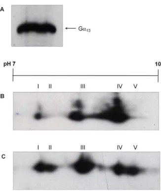
PMT induces an alkaline modification of Ga13
The two members of the Ga12 family, Ga12 and Ga13, are
ubiquitously expressed [30]. Ga13 was detected in Swiss 3T3
membranes using antiserum against Ga13(Fig. 3A). Three Ga13
isoforms, 13-I, 13-III and 13-IV, were identified in membranes from Swiss 3T3 cells (Fig. 3B). Two additional isoforms, 13-II and 13-V, were detected in membranes derived from PMT-treated cells (Fig. 3C; Table 3). The additional Ga13isoforms seem to be
the result of an alkaline pH shift, in contrast to the effect of PMT on Gaq/11and Gaiisoforms. Under our experimental conditions,
Ga12could not be resolved by 2-D gel electrophoresis.
PMT does not induce any modification of Gas
The alpha subunits of the ubiquitously expressed Gsfamily can
be expressed as four distinct forms as a result of alternative mRNA splicing [31]. Swiss 3T3 cells were shown to express both large (55 kDa) and small (52 kDa) forms of Gas, with Gas-large being
more abundantly expressed than Gas-small (Fig. S1A). Six
isoforms of Gas-large (s-I to s-VI) and two isoforms of Gas-small
(s-VII and s-VIII) were resolved in membranes derived from Swiss 3T3 cells by 2-D gel electrophoresis, followed by immunoblotting with the Gas/olf antiserum (Fig. S1B; Table S1). The Gas-large
isoforms were detected at a more acidic pI than the Gas-small
isoforms, which concurs with previous findings [19]. PMT showed Figure 1. PMT induces the covalent modification of Gaqand
Ga11.(A) Membrane proteins from Swiss 3T3 cells were separated by urea gel electrophoresis and Western blotted with anti-Gaq/11antibody.
The locations of Gaqand Ga11are indicated by arrows. Membrane
proteins from Swiss 3T3 cells (B,D,F) left untreated or (C,E,G) treated with 150 pM PMT for 4 h were separated by 2-D gel electrophoresis and Western blotted with (B,C)anti-Gaq/11, (D,E)anti-Gaqor (F,G) anti-Ga11
antibody. (H) Recombinant Gaq subunit (lane 1) and membrane
proteins from Swiss 3T3 cells (lane 2) were separated by SDS PAGE and Western blotted with anti-Gaq (left panel) or -Ga11(right panel)
antibody. Samples from at least 3 independent experiments were resolved with similar results.
no discernable effect on the pI or molecular mass of the Gas
subunits (Fig. S1C; Table S1).
PMT stimulates the stable covalent modification of G-proteins
It was important to establish whether the additional isoforms detected in PMT-treated cells arose as a consequence of normal activation induced by PMT or if they were directly PMT-modified. Cells were challenged with the neuropeptide bombesin, which acts through a Gq-coupled receptor to stimulate
phospho-lipase C (PLC) activation culminating in the release of Ca2+
from intracellular stores [32]. Bombesin at a concentration of 30 nM effectively stimulated Ca2+
release from cells (Fig. 4A), but no additional Gaq/11 isoforms were detected by 2D PAGE and
Western blot analysis of a membrane fraction derived from cells exposed to bombesin (Fig. 4B, C). This suggested that Gaq
-coupled receptor activation did not stimulate the stable covalent modification of Gaq.
We have previously demonstrated that PMT induced the phosphorylation of Gaqon Tyr369 [9]. We stimulated membrane
fractions with bombesin in the presence of sodium vanadate, a potent tyrosine phosphatase inhibitor, in order to prevent the dephosphorylation of Gaq/11. Bombesin activation of Gaq/11in
the membrane fractions was confirmed by the trypsin protection assay. Bombesin significantly enhanced GTPcS binding to Gaq/11
(p = 0.002), by up to 50% in some cases, the most likely explanation being that its action accelerated the rate of nucleotide exchange (Fig. 4D). The additional isoforms detected in mem-branes stimulated with bombesin appeared to be identical to those found in PMT-treated cells. However, the additional Gaq/11and
Gai isoforms were also found in membranes derived from
unstimulated cells, that had been treated with sodium vanadate alone (Fig. 4E, F and H). These findings suggest that PMT modification of Gaq/11and Gaiproduces a similar pI shift as the
tyrosine phosphorylation of these Gasubunits.
The appearance of the additional isoforms observed in PMT-treated cells could not be blocked by the competitive kinase inhibitors Su6656 or St638, although these inhibitors were effective at blocking pervanadate-induced phosphorylation of focal adhesion kinase (FAK) (Fig. S2). We have previously shown that a mutant PMT (PMTC1165S) can stimulate the tyrosine phosphor-ylation of Gq, although it does not activate Gq downstream
signalling [9]. Treatment of Swiss 3T3 cells with PMTC1165Sdid not result in the covalent modification of Gaq or Gai(Fig. S3).
Moreover, tyrosine phosphorylation is a transient reversible modification that cannot be readily detected unless tyrosine phosphatases are inhibited.. The PMT-induced modification of
Ga subunits was detected in the absence of sodium vanadate, indicating that the PMT-induced modification was covalent and stable.
Prolonged treatment of cells with PMT has differential effects on G-proteins
PMT treatment decreased the abundance of some of the pre-existing Gaq/11 and Gai isoforms in membrane fractions. To
explore if PMT caused G-protein removal from membranes, Swiss 3T3 cells were treated with PMT at a concentration of 1 nM for 16 h. This treatment did not cause loss of Gaq/11 from the
membrane (Fig. 5A), but resulted in the complete loss of the most basic isoforms of Gaq and Ga11, q-III and q-VI, respectively
(Fig. 5B, C, and D), while isoforms q-II and q-IV did not undergo an evident change in abundance. We speculate that the loss of detection of Gaq/11 isoforms q-III and q-VI is a result of the
covalent modification of these isoforms to q-I and q-IV, respectively, induced by PMT.
In contrast, prolonged treatment of Swiss 3T3 cells with PMT generally resulted in the almost complete loss of Gai from
membranes (Fig. 5E, F). It is unlikely that the failure to detect the Gaiisoforms reflects a modification that interferes with the Gai-1-3
antigen recognition site, which is at the C-terminus of Gai, as the
loss of Gaifrom membranes could also be demonstrated with an
antiserum against an internal epitope of Gai-2(Fig. 5F).
Cytoplas-mic extracts of cells that had received prolonged treatment with PMT were probed with anti-Gai-1-3antibody, but no increase in
Gai subunits could be detected in these fractions (Fig. 5G). It
appears that the sequential loss of Gaifrom membranes proceeds
by covalent modification of Gaiisoforms i-II and i-IV to produce
isoforms i-I and i-III, respectively, followed over time by the loss of isoforms i-I and i-III from the membranes (Fig. 5H–J). In some cases only partial loss of Gaiisoforms was observed over this time
period (data not shown).
PMT inhibits the GTPase activity of Gq
PMT did not significantly enhance GTPcS binding to Gaq/11in
contrast to bombesin (data not shown). Due to its enzymatic nature, PMT required a longer incubation time to promote GTP binding to Gaqcompared to bombesin [9]. Therefore, it is likely
that during the course of the incubation, Gaq was gradually
saturated by GTPcS, thereby preventing the detection of PMT-enhanced GTPcS binding to Gaqabove background levels. When
GTP was used instead of GTPcS, PMT significantly enhanced GTP binding to Gaqas measured by trypsin protection (p = 0.03),
by up to 30% (Fig. 6A), in contrast to bombesin (Fig. 6B). This Table 1.Analysis of pI values of Gaqfamily isoforms after treatment with PMT.
Control (pI) PMT-treated (pI)
Isoform Gaq/11 Gaq Ga11 Gaq/11 Gaq Ga11
q-I - - - 5.3960.04 5.4260.02
-q-II 5.4560.04 5.4960.02 - 5.4960.01 5.5160.02
-q-III 5.6160.05 5.5960.05 - 5.6060.08 5.6060.03
-q-IV - - - 5.6460.01 - 5.6460.02
q-V 5.7660.09 - 5.7560.01 5.7660.09 - 5.7360.01
q-VI 5.8960.1 - 5.8560.01 5.8360.02 - 5.8260.05
finding suggested that PMT might inhibit the GTPase activity of Gaq, to prevent the hydrolysis of GTP to GDP.
Bombesin stimulated the steady-state GTPase activity in Swiss 3T3 membrane preparations by up to 30%, whereas pre-treatment of cells with PMT at 150 pM for 4 h reduced the basal and bombesin-stimulated GTPase activity in membrane prepara-tions (Fig. 6C). To further decrease the basal steady-state GTPase level, cells were pre-treated with cholera toxin, which ADP-ribosylates Gasto inhibit its GTPase activity. Cholera toxin caused
an increase in the molecular weight of both the long and short forms of Gas, due to the addition of ADP ribose (Fig. 6D).
Pre-treatment of cells with both cholera toxin and PMT further decreased the basal GTPase activity in membrane preparations, compared to cells pre-treated with PMT alone. Bombesin stimulated the steady state GTPase activity by up to 50% in cells treated with cholera toxin, whereas the additional pre-treatment of cells with PMT reduced the bombesin-stimulated Figure 2. PMT induces the covalent modification of Gai.(A) Membrane proteins from Swiss 3T3 cells were separated by SDS PAGE and Western blotted with anti-Gai-1, anti-Gai-1-2or anti-Gai-1-3antibody, as indicated. (B) A recombinant Gai-1subunit was analysed by SDS PAGE and
Western blotted with anti-Gai-1, -Gai-1-2or -Gai-1-3antibody, as indicated. Membrane proteins from Swiss 3T3 cells (C,E,G) left untreated or (D,F,H)
treated with 150 pM PMT for 4 h were separated by 2-D gel electrophoresis and Western blotted with (C,D) anti-Gai-1, (E,F)anti-Gai-1-2or(G,H)
anti-Gai-1-3antibody. Samples from at least 3 independent experiments were resolved with similar results.
GTPase activity in membrane preparations, indicating that PMT inhibits the GTPase activity of Gaqbut not Gs(Fig. 6C).
Discussion
PMT executes its cellular effects through the activation of the heterotrimeric G-proteins, Gq, G12and Gi[8–13]. This has been
shown to occur in recombinant Giby PMT-induced deamidation
of Gln-205 to glutamic acid, which inhibits its intrinsic GTPase activity [14]. The work we report here complements these studies by investigating covalent modifications of G-proteins in Swiss 3T3 cells treated with PMT. PMT treatment consistently led to the appearance of new isoforms at a lower pI for both Gaqand Ga11.
PMT also stimulated the covalent modification of members of the Gifamily. The Ga12family proteins, unlike the other G-protein
families, have predicted pI values within the alkaline pH range (.pH 8) and such proteins are difficult to resolve by 2-D gel electrophoresis [33]. Ga13, but not Ga12, subunits displayed a
reproducible pattern and PMT treatment led to new Ga13
isoforms at slightly higher pI values. We found no evidence that PMT stimulates the covalent modification of Gas, although the
glutamine residue targeted by PMT is conserved in all G-proteins. Stimulation of Gq-coupled receptors by bombesin only resulted
in the detection of the additional Gaq/11 isoforms observed in
PMT-treated cells when vanadate was present. The addition of sodium vanadate per se led to a similar pattern of isoforms to those observed in PMT-treated cells. However it is likely that these different treatments lead to different modifications. The modifi-cation of Gaq/11 and Gai stimulated by PMT was detected
without sodium vanadate, and is thus indicative of a stable covalent modification such as deamidation, whereas tyrosine phosphorylation is a transient covalent modification. We previ-ously showed that a src family kinase mediates the phosphorylation of Gq in response to PMT [9]. However, pre-treatment of cells
with a specific src kinase family inhibitor, SU6656, or a broad spectrum kinase inhibitor, St638, did not prevent PMT from stimulating the covalent modification of Gaqand Gai, despite each
kinase inhibitor being effective at blocking FAK phosphorylation. It is possible that the kinase inhibitors failed to completely block PMT-stimulated phosphorylation of G-proteins, due to their competitive nature and the enzymatic nature of PMT. However, this would suggest that deamidation by PMT results in the stable phosphorylation of these Gasubunits that is not reversed by the action of phosphatases, which is unlikely.
Deamidation and tyrosine phosphorylation of a Ga subunit would have a similar effect on the isoelectric point. The PMT-induced deamidation of in-vitro translated Gaqand recombinant
Gai-2was reported to cause an acidic pI shift of 0.05 and 0.07,
respectively [14]. This compares with the acidic pI shift of approximately ,0.15 for both Gaq and Gai that we have
observed. There are various possible interpretations of this apparent discrepancy. First, pI shifts are known to be variable and depend on the overall pI of a protein and its local context [34], and thus Gai expressed in E. coli may behave differently
because of the absence of post-translational modifications. Alternatively, the PMT-induced modification in cells may differ from that observed following expression inE. coli.
PMT is reported not to activate G11, as PMT could not induce
the activation of PLC in Gq-deficient cells [12], and further
analysis using Gaq/Ga11chimeras also confirmed that PMT did Table 2.Analysis of pI values of Gaifamily isoforms after
treatment with PMT.
Control (pI) PMT-treated (pI)
Isoform Gai-1 Gai-1,2 Gai-1,2,3 Gai-1 Gai-1,2 Gai-1,2,3
i-I - 5.1160.01 5.0960.03 - 5.1060.01 5.0760.02
i-II - 5.2260.03 5.1860.03 - 5.1760.01 5.1760.02
i-III - - - 5.3460.02
i-IV - - 5.5960.03 - - 5.4560.01
The samples were as described in the legend to Figure 2 and the results are expressed as the mean6standard error of the mean (n = 3).
doi:10.1371/journal.pone.0047188.t002
Figure 3. PMT induces the covalent modification of Ga13.(A) Membrane proteins from Swiss 3T3 cells were separated by SDS PAGE and Western blotted with anti-Ga13antibody. The location of Ga13is
indicated. Membrane proteins from Swiss 3T3 cells left (B) untreated or (C) treated with 150 pM PMT for 4 h were separated by 2-D gel electrophoresis and Western blotted with anti-Ga13antibody. Samples
from at least 3 independent experiments were resolved with similar results.
doi:10.1371/journal.pone.0047188.g003
Table 3.Analysis of pI values of Ga13isoforms after treatment with PMT.
Control (pI) PMT-treated (pI)
Isoform Ga13 Ga13
13-I 8.1560.02 8.1560.07
13-II - 8.2460
13-III 8.5460.02 8.5460.02
13-IV 8.9060.05 8.9060.01
13-V - 9.0460.01
The samples were as described in the legend to Figure 4 and the results are expressed as the mean6standard error of the mean (n = 3).
not lead to G11-linked stimulation of PLC [35].We were therefore
surprised that PMT stimulated the covalent modification of Ga11.
Gaq and Ga11 each contain Gln-209 that is functionally
equivalent to Gln-205 in Gai-2 and it would be unlikely that
Ga11could be deamidated and yet not activated by PMT, as the
loss of the functional Gln would affect the GTPase activity of the G-protein. While this manuscript was in preparation, Kamitaniet al. published evidence that an antibody against deamidated Ga subunits recognised Ga11 in PMT-treated mouse embryonic
fibroblasts that were deficient in Gaq/11but transfected to express
Ga11[36]. This result provides further evidence that G11is also a
substrate for PMT. In their experiments there was a small stimulation of PLC in cells expressing Ga11. All the other papers
addressing this issue have used the same source of Gq/11-deficient
MEF cells, whereas our work uses Swiss 3T3 cells. Further
investigation of these puzzling and partially contradictory results is required.
PMT treatment of cells led to new Ga13 isoforms at slightly
higher (0.09–0.15) pI values. The PMT catalytic triad has high structural similarity to eukaryotic transglutaminases [14], and it is possible that PMT can also function as a transglutaminase, in a similar manner to the cytotoxic necrotizing factor (CNF) which was originally considered to be a deamidase, but was later found to cause transglutamination in cells [37]. Transglutaminases catalyse the acyl transfer between thec-carboxyamide of a peptide bound glutamine (acyl donor) to a primary amine (acyl acceptor). When water functions as an acyl acceptor the result is glutamine deamidation [38]. The choice between deamidation and transglu-tamination is influenced by the environment of the targeted glutamine residue [39,40]. As transglutamination would impart a Figure 4. Sodium vanadate treatment mimics PMT effects on Gaqand Gai.(A) Indo-1 AM labelled Swiss 3T3 cells were treated with 30 nM bombesin for 10 s (marked with solid bar beneath trace) and intracellular Ca2+release was measured. Membrane proteins from Swiss 3T3 cells that
were either (B) untreated or (C) treated with 30 nM bombesin for 1 min were separated by 2-D gel electrophoresis and Western blotted with anti-Gaq/11antibody. (D) Swiss 3T3 membrane proteins were incubated in the presence or absence of 30 nM bombesin for 20 min with or without
0.05 nM GTPcS. The proteins were then analysed for trypsin protection as described under Materials and Methods, and activated Gaq/11 was
separated by SDS PAGE and Western blotted with anti-Gaq/11 antibody. Quantification of activated Gaq/11 (lower panel) was determined by
densitometric scanning and these data were analysed using factorial analysis of variance (ANOVA). The induction of activation shown is relative to the density of the band without GTPcS or bombesin. Bombesin significantly enhanced GTPcS binding to Gaq/11(* p = 0.002). Membrane proteins from
Swiss 3T3 cells were incubated with (E) 1 mM sodium vanadate for 20 min at 37uC or (F) 1 mM sodium vanadate and 30 nM bombesin for 20 min at 37uC, proteins were separated by 2-D gel electrophoresis and Western blotted with anti-Gaq/11antibody. Membrane proteins from Swiss 3T3 cells
were incubated (G) without or (H) with 1 mM sodium vanadate for 20 min at 37uC, proteins were separated by 2-D gel electrophoresis and Western blotted with anti-Gai-1-3antibody. Samples from at least 3 independent membrane preparations were resolved with similar results.
positive charge to produce an alkaline shift, it is possible that PMT preferentially transglutaminates Ga13in cells.
The removal of G-proteins from the membrane is a regulatory phenomenon that can follow prolonged G-protein activation [41]. The ADP-ribosylation of Gs by cholera toxin leads to its
downregulation, although ADP-ribosylation of Gai by pertussis
toxin does not result in its degradation [42]. We observed that prolonged treatment of cells with PMT caused the loss of Gai, but
not Gaq, from membranes prepared from Swiss 3T3 cells.
Furthermore Gaicould not be detected in the cytoplasm following
prolonged PMT treatment. Orth et al. had suggested that overnight treatment of Swiss 3T3 cells with 1 nM PMT uncoupled Gaifrom its receptor, as the Gi-linked agonist lysophosphatidic
acid could not stimulate GTPcS binding to Gaiin membranes
derived from these cells [10]. The loss of Gifrom the membrane
that we observed over this time period would provide a more likely explanation for their observation. Furthermore, the site of the PMT-induced modification, Gln-205, is not thought to be linked to receptor interaction. A similar differential degradation has been observed with Rho proteins following modification by CNF [43]. We found that PMT could promote the binding of GTP to Gaq/11, whereas bombesin could not, which suggested that the
action of PMT inhibits the GTPase activity of Gaq/11. PMT
significantly inhibited the bombesin-mediated stimulation of steady-state GTPase activity in Swiss 3T3 membrane prepara-tions. These results complement the demonstration that PMT
inhibits the GTPase activity of E. coli-expressed Gai [10,14].
Furthermore, pre-treatment of cells with cholera toxin and PMT resulted in a greater inhibition of GTPase activity, supporting the view that PMT does not affect Gs.
In conclusion, our results demonstrate that treatment of Swiss 3T3 cells with PMT induces the irreversible modification of G-proteins belonging to the Giand Gqfamilies resulting in an acidic
pI shift, which is consistent with the observation that PMT catalyses deamidation of recombinantly expressed Gicausing a
similar shift in pI. We found that PMT inhibits the intrinsic GTPase activity of Gq, which complements the finding that
PMT-stimulated deamidation of Gai-2inhibits its GTPase activity. We
showed that stimulation of cells with PMT results in the degradation of Gi which provides an explanation for the
observation that PMT-treatment blocks Gi activation by a
receptor agonist. The unexpected modification of Ga11 requires
further investigation. We demonstrated that PMT treatment causes an alkaline pI shift in Ga13and speculate that PMT might
preferentially transglutaminate Ga13. Working with cells enables
the PMT/G-protein interaction to be investigated in a more natural context than when working with recombinantly expressed proteins. However, the further interpretation of results is impeded by the near impossibility of purifying these low abundance proteins in a modified form from cell lines, and thus both vitro and in-vivo studies are required to unravel the complexity of the toxin/G-protein interactions.
Figure 5. Prolonged exposure of Swiss 3T3 cells to PMT causes the loss of Gaibut not Gqfrom cell membranes.(A) Membrane proteins from Swiss 3T3 cells left untreated (lane 1) or treated with PMT at 1 nM for 16 h (lane 2) were separated by SDS PAGE and Western blotted with anti-Gaq/11antibody. Membrane proteins from Swiss 3T3 cells (B) left untreated, or treated with 1 nM PMT for (C) 4 h or (D) 16 h were separated by 2-D
gel electrophoresis and Western blotted with anti-Gaq/11antibody. Samples from at least 3 independent experiments were resolved with similar
results. Membrane proteins from Swiss 3T3 cells left untreated (lane 1) or treated with 1 nM PMT for 16 h (lane 2) were separated by SDS PAGE and Western blotted with (E) anti-Gai-1-3antibody or (F) an antibody recognising an internal epitope of Gai-2. (G) Cytoplasmic proteins from Swiss 3T3
cells left untreated (lane 1) or treated with 1 nM PMT for 16 h (lane 2) were separated by SDS PAGE and Western blotted with anti-Gai-1-3antibody.
Membrane proteins from Swiss 3T3 cells (H) left untreated, treated with 1 nM PMT for (I) 4 h or (J) 16 h were separated by 2-D PAGE and Western blotted with anti-Gai-1-3antibody. Samples from 3 independent experiments were resolved with similar results.
Supporting Information
Figure S1 PMT does not induce the covalent modifica-tion of Gas. (A) Membrane proteins from Swiss 3T3 cells were separated by SDS PAGE and Western blotted with anti-Gas
antibody. Membrane proteins from Swiss 3T3 cells left (B) untreated or (C) treated with 150 pM PMT for 4 h were separated by 2-D gel electrophoresis and Western blotted with anti-Gas
antibody. Samples from at least 3 independent experiments were resolved with similar results.
(TIF)
Figure S2 Kinase inhibitors do not block PMT induced modification of Gaq/11or Gai.Swiss 3T3 cells were either not treated (Lane 1) or pre-treated (Lane 2) for 1 h with (A) SU6656 or (B) St638, then stimulated with 0.5 nM pervanadate for 5 min. The cells were lysed in SDS-buffer and proteins were resolved by SDS PAGE followed by Western blotting with an anti-phospho-FAK antibody. Three independent experiments gave similar results. Swiss 3T3 cells were (C, D, G, H) not treated or
pre-treated with either (E,I) SU6656 or (F,J) St638 and then either treated with (D,E,F,H,I,J) 150 pM PMT or (C,G) not treated with PMT. Samples were resolved from 3 independent experi-ments with similar results. Membrane proteins were separated by 2-D gel electrophoresis and Western blotted with (C–F) anti-Gaq/ 11antibody or (G–J) anti-Gai-1-3antibody. Samples were resolved
from 2 independent experiments with similar results. (TIF)
Figure S3 Mutant PMT does not induces the covalent modifications of Gaqor Gai.Membrane proteins from Swiss 3T3 cells (A, C) left untreated or (B, D) treated with 150 pM PMTC1165S for 4 h, separated by 2-D gel electrophoresis and
Western blotted with either (A, B) anti-Gaq/11or (C, D) anti-Ga i-1-3 antibodies. Samples from 3 independent experiments were
resolved with similar results. (TIF)
Table S1 Analysis of pI values of Gs family isoforms
after treatment with PMT.The samples were as described in Figure 6. PMT inhibits the GTPase activity of Gq.(A) Membrane proteins were incubated in the presence or absence of 150 pM PMT for 1 h
with 0.5 nM GTP and tested in a trypsin protection assay as described in Materials and Methods. Proteins were separated by SDS PAGE and Western blotted with anti-Gaq/11antibody. Quantification of activated Gaq/11(lower panel) was determined by densitometric scanning and the data were
analysed using factorial analysis of variance (ANOVA). The induction of activation shown is relative to the density of the band without GTP or PMT. PMT significantly enhanced GTP binding to Gq(* p = 0.03). (B) Membrane proteins were incubated in the presence or absence of 30 nM bombesin
20 min with 0.5 nM GTP and tested in a trypsin protection assay as described in Materials and Methods. Proteins were separated by SDS PAGE and Western blotted with anti-Gaq/11antibody. Samples from at least 3 independent membrane preparations were resolved with similar results. (C)
Membranes derived from Swiss 3T3 cells that had either been treated or untreated with 150 pM PMT for 4 h or 100 ng cholera toxin, or both, were treated with or without 30 nM bombesin for 20 min in the presence of [c-32P] GTP. All the experimental conditions were repeated three times, and all data are presented as mean6standard deviation (SEM). The results for the groups were compared using single-factor analysis of variance (one-way ANOVA), followed by Newnan-Keuls test used to determine differences between groups. Significant changes are indicated by an asterisk (*P,0.05,
***P,0.001). (D) Membranes derived from Swiss 3T3 cells that had either been untreated (lane 1) or treated with 150 pM PMT for 4 h (lane 2), or 100 ng cholera toxin for 16 h (lane 3), or both PMT and cholera toxin (lane 4) were resolved by SDS PAGE followed by Western blotting with an anti-Gasantibody. Samples from at least 3 independent membrane preparations were resolved with similar results.
the legend to Figure. S1 and the results are expressed as the mean 6standard error of the mean.
(DOC)
Acknowledgments
We thank Susmitha Rao (King’s College London Dental Institute) for technical support with 2-D gel electrophoresis, and Dr Ron Wilson (King’s College London Dental Institute) for help with statistical analysis.
Author Contributions
Conceived and designed the experiments: RB KH JR AL. Performed the experiments: RB KH JR. Analyzed the data: RB KH JR AL. Contributed reagents/materials/analysis tools: RB KH JR. Wrote the paper: RB KH JR AL.
References
1. Sprang SR (1997) G protein mechanisms: insights from structural analysis. Annu Rev Biochem 66: 639–678.
2. Oldham WM, Hamm HE (2008) Heterotrimeric G protein activation by G-protein-coupled receptors. Nat Rev Mol Cell Biol 9: 60–71.
3. Chen CA, Manning DR (2001) Regulation of G proteins by covalent modification. Oncogene 20: 1643–1652.
4. Sunahara RK, Dessauer CW, Gilman AG (1996) Complexity and diversity of mammalian adenylyl cyclases. Annu Rev Pharmacol Toxicol 36: 461–480. 5. Hubbard KB, Hepler JR (2006) Cell signalling diversity of the Gqafamily of
heterotrimeric G proteins. Cell Signal 18: 135–150.
6. Suzuki N, Hajicek N, Kozasa T (2009) Regulation and physiological functions of G12/13-mediated signaling pathways. Neurosignals 17: 55–70.
7. Rozengurt E, Higgins T, Chanter N, Lax AJ, Staddon JM (1990)Pasteurella multocidatoxin: potent mitogen for cultured fibroblasts. Proc Natl Acad Sci U S A 87: 123–127.
8. Murphy AC, Rozengurt E (1992)Pasteurella multocidatoxin selectively facilitates phosphatidylinositol 4,5-bisphosphate hydrolysis by bombesin, vasopressin, and endothelin. Requirement for a functional G protein. J Biol Chem 267: 25296– 25303.
9. Baldwin MR, Pullinger GD, Lax AJ (2003)Pasteurella multocidatoxin facilitates inositol phosphate formation by bombesin through tyrosine phosphorylation of Gaq. J Biol Chem 278: 32719–32725.
10. Orth JHC, Fester I, Preuss I, Agnoletto L, Wilson BA, et al. (2008) Activation of Gaiand subsequent uncoupling of receptor-Gaisignaling byPasteurella multocida toxin. J Biol Chem 283: 23288–23294.
11. Orth JHC, Lang S, Taniguchi M, Aktories K (2005)Pasteurella multocida toxin-induced activation of RhoA is mediated via two families of Gaproteins, Gaqand Ga12/13. J Biol Chem 280: 36701–36707.
12. Zywietz A, Gohla A, Schmelz M, Schultz G, Offermanns S (2001) Pleiotropic effects of Pasteurella multocida toxin are mediated by Gqdependent and -independent mechanisms - involvement of Gqbut not G11. J Biol Chem 276: 3840–3845.
13. Mullan PB, Lax AJ (1996)Pasteurella multocidatoxin is a mitogen for bone cells in primary culture. Infect Immun 64: 959–965.
14. Orth JHC, Preuss I, Fester I, Schlosser A, Wilson BA, et al. (2009)Pasteurella multocidatoxin activation of heterotrimeric G proteins by deamidation. Proc Natl Acad Sci U S A 106: 7179–7184.
15. Ward PN, Miles AJ, Sumner IG, Thomas LH, Lax AJ (1998) Activity of the mitogenic Pasteurella multocidatoxin requires an essential C-terminal residue. Infect Immun 66: 5636–5642.
16. Kimple RJ, De Vries L, Tronche`re H, Behe CI, Morris RA, et al. (2001) RGS12 and RGS14 GoLoco motifs are Gaiinteraction sites with guanine nucleotide dissociation inhibitor activity. J Biol Chem 276: 29275–29281.
17. Todaro GJ, Green H (1963) Quantitative studies of the growth of mouse embryo cells in culture and their development into established lines. J Cell Biol 17: 299– 313.
18. Svoboda P, Milligan G (1994) Agonist-induced transfer of theasubunits of the guanine-nucleotide-binding regulatory proteins Gqand G11and of muscarinic m1 acetylcholine receptors from plasma membranes to a light-vesicular membrane fraction. Eur J Biochem 224: 455–462.
19. Matousˇek P, Novotny´ J, Svoboda P (2004) Resolution of Gsaand Gqa/G11a proteins in membrane domains by two-dimensional electrophoresis: the effect of long-term agonist stimulation. Physiol Res 53: 295–303.
20. Hayat S, Wigley CB, Robbins J (2003) Intracellular calcium handling in rat olfactory ensheathing cells and its role in axonal regeneration. Mol Cell Neurosci 22: 259–270.
21. Evanko DS, Thiyagarajan MM, Wedegaertner PB (2000) Interaction with Gbc is required for membrane targeting and palmitoylation of Gas, and Gaq. J Biol Chem 275: 1327–1336.
22. Fung BK-K, Nash CR (1983) Characterization of transducin from bovine retinal rod outer segments. II. Evidence for distinct binding sites and conformational
changes revealed by limited proteolysis with trypsin. J Biol Chem 258: 10503– 10510.
23. Cassel D, Selinger Z (1976) Catecholamine-stimulated GTPase activity in turkey erythrocyte membranes. Biochim Biophys Acta 452: 538–551.
24. Mullaney I, Mitchell FM, McCallum JF, Buckley NJ, Milligan G (1993) The human muscarinic M1 acetylcholine receptor, when expressed in CHO cells, activates and downregulates both Gqaand G11aequally and non-selectively. FEBS Letts 324: 241–245.
25. Caulfield MP, Jones S, Vallis Y, Buckley NJ, Kim G-D, et al. (1994) Muscarinic M-current inhibition via Gaq/11anda-adrenoceptor inhibition of Ca2+current via Gaoin rat sympathetic neurones. J Physiol (Lond) 477: 415–422. 26. Wettschureck N (2009) G protein alpha q. UCSD-Nature Molecule Pages
(doi:10.1038/mp.a000978.01).
27. Kurrasch DM, Huang J, Wilkie TM (2004) G protein alpha 11. UCSD-Nature Molecule Pages (doi:10.1038/mp.a000970.01).
28. Jones DT, Reed RR (1987) Molecular cloning of five GTP-binding protein cDNA species from rat olfactory neuroepithelium. J Biol Chem 262: 14241– 14249.
29. Bajpayee NS, Jiang M (2010) G protein alpha i1. UCSD-Nature Molecule Pages (doi:10.1038/mp.a000974.01).
30. Strathmann MP, Simon MI (1991) Ga12 and Ga13 subunits define a fourth class of G proteinasubunits. Proc Natl Acad Sci U S A 88: 5582–5586. 31. Kozasa T, Itoh H, Tsukamoto T, Kaziro Y (1988) Isolation and characterization
of the human Gsagene. Proc Natl Acad Sci U S A 85: 2081–2085. 32. Takuwa N, Takuwa Y, Bollag WE, Rasmussen H (1987) The effects of bombesin
on polyphosphoinositide and calcium metabolism in Swiss 3T3 cells. J Biol Chem 262: 182–188.
33. Go¨rg A, Obermaier C, Boguth G, Harder A, Scheibe B, et al. (2000) The current state of two-dimensional electrophoresis with immobilized pH gradients. Electrophoresis 21: 1037–1053.
34. Zhu K, Zhao J, Lubman DM, Miller FR, Barder TJ (2005) Protein pIshifts due to posttranslational modifications in the separation and characterization of proteins. Anal Chem 77: 2745–2755.
35. Orth JHC, Lang S, Aktories K (2004) Action ofPasteurella multocidatoxin depends on the helical domain of Gaq. J Biol Chem 279: 34150–34155.
36. Kamitani S, Ao S, Toshima H, Tachibana T, Hashimoto M, et al. (2011) Enzymatic actions ofPasteurella multocidatoxin detected by monoclonal antibodies recognizing the deamidatedasubunit of the heterotrimeric GTPase Gq. FEBS J 278: 2702–2712.
37. Schmidt G, Selzer J, Lerm M, Aktories K (1998) The Rho-deamidating cytotoxic necrotizing factor 1 fromEscherichia colipossesses transglutaminase activity: cysteine 866 and histidine 881 are essential for enzyme activity. J Biol Chem 273: 13669–13674.
38. Folk JE, Chung SI (1973) Molecular and catalytic properties of transglutamin-ases. Adv Enzymol Relat Areas Mol Biol 38: 109–191.
39. Stamnaes J, Fleckenstein B, Sollid LM (2008) The propensity for deamidation and transamidation of peptides by transglutaminase 2 is dependent on substrate affinity and reaction conditions. Biochim Biophys Acta 1784: 1804–1811. 40. Esposito C, Caputo I (2005) Mammalian transglutaminases: identification of
substrates as a key to physiological function and physiopathological relevance. FEBS J 272: 615–631.
41. Milligan G (1993) Agonist regulation of cellular G protein levels and distribution: mechanisms and functional implications. Trends Pharmacol Sci 14: 413–418. 42. Milligan G, Unson CG, Wakelam MJO (1989) Cholera toxin treatment
produces down-regulation of the alpha-subunit of the stimulatory guanine-nucleotide-binding protein (Gs). Biochem J 262: 643–649.