Volume 2013, Article ID 590329,6pages http://dx.doi.org/10.1155/2013/590329
Research Article
Antigenic Peptides Capable of Inducing Specific
Antibodies for Detection of the Major Alterations Found in
Type 2B Von Willebrand Disease
Marina de Oliveira Paro,
1Cyntia Silva Ferreira,
1Fernanda Silva Vieira,
1Marcos Aurélio de Santana,
1William Castro-Borges,
1Maria Sueli Silva Namen-Lopes,
2Sophie Yvette Leclercq,
3Cibele Velloso-Rodrigues,
4and Milton Hércules Guerra de Andrade
11Departamento de Ciˆencias Biol´ogicas/DECBI, Instituto de Ciˆencias Exatas e Biol´ogicas/ICEB,
Universidade Federal de Ouro Preto, N´ucleo de Pesquisas em Ciˆencias Biol´ogicas/NUPEB, Campus Universit´ario Morro do Cruzeiro, 35400-000 Ouro Preto, MG, Brazil
2Fundac¸˜ao Centro de Hematologia e Hemoterapia, Hemominas, Servic¸o de Pesquisa, Alameda Ezequiel Dias, 321,
Bairro Santa Efigˆenia, 30130-110 Belo Horizonte, MG, Brazil
3Fundac¸˜ao Ezequiel Dias, FUNED, Rua Conde Pereira Carneiro, 80, Bairro Gameleira, 30510-010 Belo Horizonte, MG, Brazil 4Departamento B´asico, Instituto de Ciˆencias Biol´ogicas, Universidade Federal de Juiz de Fora, Campus de Governador Valadares,
´
Area da Sa´ude, Avenida Doutor Raimundo Monteiro de Resende, 330, Centro, 35010-177 Governador Valadares, MG, Brazil
Correspondence should be addressed to Milton H´ercules Guerra de Andrade; [email protected] Received 11 March 2013; Revised 19 June 2013; Accepted 20 June 2013
Academic Editor: Jean-Marie Zajac
Copyright © 2013 Marina de Oliveira Paro et al. This is an open access article distributed under the Creative Commons Attribution License, which permits unrestricted use, distribution, and reproduction in any medium, provided the original work is properly cited. Von Willebrand disease (VWD) is an inherited hemorrhagic disorder promoted by either quantitative or qualitative defects of the von Willebrand factor (VWF). The disease represents the most common human coagulopathy afflicting 1.3% of the population. Qualitative defects are subdivided into four subtypes and classified according to the molecular dysfunction of the VWF. The differential diagnosis of the VWD is a difficult task, relying on a panel of tests aimed to assess the plasma levels and function of the VWF. Here, we propose biochemical approaches for the identification of structural variants of the VWF. A bioinformatic analysis was conducted to design seven peptides among which three were representatives of specific amino acid sequences belonging to normal VWF and four encompassed sequences found in the most common VWD subtype 2B. These peptides were used to immunize mice, after which, peptide-specific immunoglobulins were purified. This resulted in four Ig preparations capable of detecting alterations in the subtype 2B VWD plus additional three antibody fractions targeting the normal VWF. The panel of antibodies could serve many applications among them (1) assessment of VWF: antigen interaction, (2) VWF multimer analysis, and (3) production of monoclonal antibodies against VWF for therapeutic purposes as in thrombotic thrombocytopenic purpura.
1. Introduction
Von Willebrand disease (VWD) is an inherited hemorrhagic disturbance related to quantitative and/or qualitative defects
of the von Willebrand factor (VWF) [1,2]. VWD prevalence
varies between 0.8 and 2.0%, depending on the investigated population, being considered the most common coagulopa-thy afflicting humans [3].
of clinical outcomes. Through different laboratory criteria it is possible to identify three primary types of the disease [5]. Alterations on the plasma levels of VWF are associated with VWD types 1 and 3, whereas structural and functional defects
of VWF result in VWD type 2 [3,6–11]. VWD types 1 and
3 reflect, respectively, partial and complete deficiency of the VWF. VWD type 2 is also classified into four subtypes. In subtype 2A, there is enhanced platelet adhesion caused by the selective deficiency of high molecular mass multimers of VWF (HMW). In subtype 2B VWD, it is observed increased affinity of VWF by platelet glycoprotein Ib (GpIb) associated with loss of HMW-VWF and mild thrombocytopenia. In contrast, in subtype 2M, a pronounced reduction in platelet adhesion is described, even considering the relatively normal size of the VWF multimers. Variants of the VWF in the subtype 2N display reduced affinity by coagulation factor VIII (FVIII). Altogether these six VWD types correlate with diverse clinical outcomes each requiring adequate
therapeu-tic interventions [4,12]. Whilst some patients with type 2B
VWD can be treated with desmopressin, patients who do not show a satisfactory response to this drug should receive
VWF/FVIII-containing products [13,14].
Considering the current difficulties associated with diag-nosis of the qualitative defects of the VWF, the development of novel biochemical approaches that would allow detection of such molecular alterations is of great biotechnological interest. The possibility of a precise and direct diagnosis of qualitative defects in the VWF would certainly permit appli-cation of a better oriented medical approach to afflicted patients. In this context, it is worth emphasizing that anti-bodies capable of detecting structural variants of the VWF are not commercially available. The design of peptides for synthesis and further generation of antibodies to be employed in the identification of structural alterations in the VWF might therefore represent a convenient way forward.
It has previously been shown that amino acid substitu-tions R1306W, R1308C, V1316M, and R1341Q account for 90%
prevalence of the subtype 2B VWD [6,8]. These mutations
in the A1 domain of VWF are responsible for a “gain-of-function” defect allowing for an increased affinity of large multimers to platelets in the circulation [15]. In the present investigation we have generated a panel of antipeptide-specific antibodies useful at detecting the aforementioned structural variants of the VWF.
2. Materials and Methods
2.1. Ethics Statement. All experiments involving mice were
conducted according to approved guidelines for animal use and care defined by the Local Ethics Committee on Animal Experimentation (CEUA/UFOP). Healthy individuals were informed previously of the investigatory nature of this study, and after giving their consent, plasma samples were obtained under the procedures approved by the Local Ethics Commit-tee from Hemominas Foundation, MG, Brazil.
2.2. Selection of VWF Peptides for Synthesis. As stated
previ-ously the VWF amino acid substitutions R1306W, R1308C,
V1316M, and R1341Q are collectively found in the majority of subtype 2B VWD patients [6]. In order to design signature
peptides that would represent such mutations,Homo
sapi-ens VWF sequences were retrieved from the International
Society on Thrombosis and Haemostasis database,
avail-able athttp://www.vwf.group.shef.ac.uk/. Predicted peptide
sequences for synthesis obeyed the following criteria: (1) peptide size should be within the 8 to 10 mer range; (2) pep-tides should contain one aromatic residue for estimation of peptide yield after synthesis and HPLC purification (in the absence of an aromatic residue, this was added at the peptide C-terminal); (3) the location of the peptide sequences in the crystal structure of the VWF (available at http://www.pdbj.org/), revealed by the Swiss-Pdb Viewer 3.7, should demonstrate their exposure to the solvent, meaning that highly hydrophobic peptide sequences were not consid-ered in this study.
The selected peptide sequences were then aligned toMus
musculusVWF sequence using ClustalW2 (http://www.ebi
.ac.uk/Tools/msa/clustalw2/) to reveal species specific amino acid differences as predictors for successful production of anti-VWF peptide antibodies in mice.
2.3. Synthesis, Characterization, and Production of
Poly-clonal Antibodies against VWF Peptides. Peptides selected
for synthesis were representatives of the normal and altered versions of the VWF found in the major subtypes of type 2 VWD (Table 1). Peptides were synthesized using the solid-phase protocol essentially as described by Merrifield [16]. Briefly, the support matrix consisted of Rink Amide Resin HL (Merck, Germany) at 0.78 mmol/g for an expected
maximum yield of 40𝜇M of peptides per synthesis, using
Fmoc-derivatized amino acids. Synthetic peptides were then purified via reversed-phase chromatography using a C18 column (Shim-pack CLD-ODS Shimadzu, Japan) on a Shi-madzu HPLC system. Selected peptide peaks were recovered for analysis through direct injection onto an electrospray-operating mass spectrometer (LCMS-IT-ToF, Shimadzu, Japan) in positive ionization mode and capillary voltage set to 4,500 V. Mass spectrometric data were acquired over 10 ms,
after which m/z values obtained were compared with the
expected molecular masses for the synthesized peptides. Synthetic peptides representatives of both normal and altered VWF were individually coupled to the highly immu-nogen carrier protein keyhole limpet hemocyanin (KLH, Sigma) at 1 : 1 ratio (1 mg peptide: 1 mg KLH) using glutaral-dehyde as the crosslinking agent [17]. Polyclonal anti-KLH
peptide antibodies were raised in maleSwissmice aged ten
weeks. Immunization regimen consisted of three
intraperi-toneal administrations of 50𝜇g of the KLH-peptide conjugate
prepared in 10% aluminium hydroxide as adjuvant, each at a 15 days interval. Blood was withdrawn after 45 days post-immunization.
Table 1: Proposed synthetic peptides for detection of the major qualitative alterations found in VWD type 2B. Major qualitative
alterations found in VWD type 2B
Designed 8 mer peptides for synthesis Theoretical [M + H]+
Observed [M + H]+
Respective peptide from
M. musculusVWF R1306W MEWLRISY 1097.54 1113.56∗
MERLHIS
R1308C MERLCISY 1014.47 1014.50
Respective peptide found inH. sapiensVWF: MERLRISY 1067.56 1082.57∗ V1316M SQKWVRMA 1005.52 1020.54∗
SQKRIRVA Respective peptide found inH. sapiensVWF: SQKWVRVA 973.55 972.57
[−1H+]
R1341Q RPSELQRY 1048.55 1048.57
RPSELRR Respective peptide found inH. sapiensVWF: RPSELRRY 1076.59 1076.62
∗Peptide mass containing an oxidized methionine.
described previously [18]. The antisera from mice immunized with KLH coupled to altered VWF peptides were loaded individually in affinity columns bearing the corresponding normal versions of the VWF peptide. This procedure aimed the subtraction of antibodies targeting the normal VWF.
Approximately 100𝜇L of the unbound fraction was collected
for analysis during the first chromatographic step, whilst the remaining were immediately submitted to further six rounds of chromatography to guarantee complete removal of antinormal VWF antibodies. For each chromatographic step bound fractions were eluted in 5 mL 0.1 M glycine pH 2.6 and collected in tubes containing 5 mL of 0.4 M Tris-HCl pH 8.0 for pH neutralization. Alkaline phosphatase labeled anti-mouse IgG, at 1 : 2000 dilution, was used to detect the presence of IgGs found in both nonretained and retained fractions from each chromatographic step, using the western blotting technique [19].
The next approach involved confirmation that antibodies targeting the normal VWF have been thoroughly subtracted after the six passages in affinity columns containing immo-bilized altered VWF peptides. For this purpose, 1 mL of
pooled plasma from human healthy donors (𝑛 = 5) was first
precipitated in 20% ethanol to enrich for normal VWF.
Approximately 10𝜇g aliquots of the precipitated plasma were
resuspended in protein loading buffer and loaded in three different lanes for separation on a 7% SDS-PAGE. The gel was transferred to a PVDF (polyvinylidene fluoride) membrane
and each lane western blotted with either a 100𝜇L fraction
obtained from the crude antisera or the two 100𝜇L
non-retained fractions recovered from the first and sixth chro-matographic steps.
The combined eluates from each chromatographic step containing immunoglobulins targeting a given altered VWF peptide were lyophilized, resuspended in 1 mL of phosphate buffer pH 7.4, and individually loaded onto the Sepharose-4B affinity column bearing the respective altered VWF peptide. Bound IgGs targeting the altered VWF peptide were eluted exactly as described above.
2.5. Production of Carrier Albumins Bearing Altered VWF Synthetic Peptides and Their Recognition by Anti-VWF Specific
Antibodies. Aiming to produce a model sample for detection
of altered VWF through the western blotting technique, puri-fied albumin was conjugated to two representative synthetic
peptides (SQKWVRMA and RPSELQRY,Table 1) [20] at a
1 : 1 ratio, respectively. Briefly, to each 1 mg of the respective
peptides it was added 17𝜇L of N-Ndiisopropylcarbodiimide,
diluted 1 : 25 in dimethylformamide (DMF), and 20𝜇L of
1 M N-hydroxysuccinimide (also diluted in DMF). Reaction proceeded during 5 min at room temperature, after which
50𝜇L of 0.4 M sodium acetate (in DMF) was added. After
fur-ther 2 min incubation, at room temperature, 1 mL of bovine serum albumin (BSA, Sigma), at 1 mg/mL, prepared in 0.05 M ammonium bicarbonate pH 7.5, was combined. Coupling reactions occurred during 1 h followed by dialysis of each preparation in 0.1 M ammonium acetate over 24 h. Peptide-derivatized albumins were lyophilized and resuspended in
100𝜇L of saline.
Five 𝜇g aliquots of peptide-conjugated albumin were
loaded into three lanes and separated through 12% SDS-PAGE, followed by Coomassie staining. A replica gel was pro-duced and transferred to a PVDF membrane. Antipeptide-specific IgG purified through affinity chromatography, at
approximately 0.15𝜇g/𝜇L, was used as the primary
anti-body, at a final dilution of 1 : 500. The reaction was allowed to proceed for 3 h at room temperature. Development of the reactive band was achieved using alkaline phosphatase labeled anti-mouse IgG (Sigma), at 1 : 2000 dilution for 2 h, followed by addition of the alkaline phosphatase sub-strates nitroblue tetrazolium/5-bromo-4-chloro-3-indolyl-phosphate (NBT/BCIP, Sigma) as per the manufacturer’s instructions.
3. Results and Discussion
250
1 2 3
(kDa) Approx. Mr
(a)
250
4 5 6
(kDa) Approx. Mr
(b)
50
25
66
66
8
7 9 10
8 9 10
(kDa) Approx. Mr (kDa)
Approx. Mr
(c)
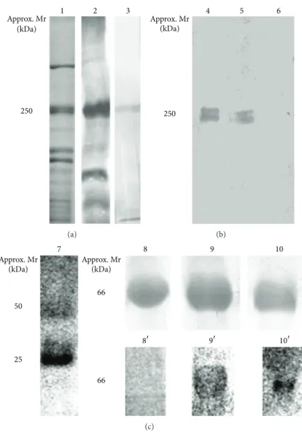
Figure 1: SDS-PAGE and western blotting approaches featuring the major steps involved in the production of a panel of anti-(KLH-peptide) antibodies for detection of VWD subtype 2B. (a)Lane 1, 10% SDS-PAGE profile of pooled human plasma after enrichment for normal VWF using ethanol precipitation;Lane 2, western blotting for detection of normal VWF, at approximately 250 kDa, using the commercially available antibody against VWF/FVIII—note the presence of additional protein bands being recognized, particularly at lower mass;Lane 3, a representative Western blotting reaction obtained with the generated panel of anti-(KLH-peptide) antibodies, targeting both normal and altered VWF. (b) Detection of normal VWf by western blotting following the subtractive affinity chromatography;Lane 4, detection of VWF prior to depletion of antibodies against the normal factor;Lanes 5and6, detection of VWF using the nonretained fractions from the first and sixth chromatographic steps, respectively—note that 6 column passages proved sufficient for complete removal of anti-(KLH-peptide) antibodies. (c)Lane 7, 10% SDS-PAGE profile representative of the eluates obtained after affinity purification of antibodies targeting specifically altered VWf peptides—note the presence of IgG heavy and light chains at approximately 50 and 25 kDa, respectively;Lanes 8,9, and10, 10% SDS-PAGE profile of nonderivatized BSA, BSA-(RPSELRR) and BSA-(SQKRIRVA), respectively;Lanes 8,9, and10, corresponding western blotting reactions obtained using control sera, anti-(KLH-RPSELRR) and anti-(KLH-SQKRIRVA), respectively.
out immunoglobulins targeting the normal VWF from the antisera raised with the altered VWF peptides. Given that the
correspondingMus musculusVWF peptides are MERLHIS,
SQKRIRVA, and RPSELRR, we anticipated that the observed amino acid differences between human and mice VWF would justify the peptides capabilities for generating specific antibodies in mice (Table 1).
After immunization, the obtained antisera were tested for their ability to specifically recognize the VWF enriched
from plasma samples of human healthy donors.Figure 1(a),
lane 3 is a representative reactivity obtained for all antisera raised with either the altered or normal versions of VWF peptides. A unique band at approximately 250 kDa was observed coinciding exactly with a major band, at the same
mass, observed inFigure 1(a), lane 2. This was probed with
(Figure 1(a), lane 2) revealing that the antisera generated herein are more suitable for specific recognition of the human VWF.
The result obtained inFigure 1(a), lane 3 is a proof that
the antisera produced using altered VWF peptides also con-tain IgGs targeting the normal VWF. Aiming to remove those immunoglobulins from the preparations, a subtraction experiment was conducted. Firstly, each individual antiserum obtained by immunization with a given altered VWF peptide was submitted to affinity chromatography on a Sepharose 4B column, containing the respective immobilized normal VWF peptide. Through collection of both non-retained and retained fractions during 6 chromatographic rounds, it was observed complete removal of IgGs targeting normal
VWF. This can be visualized in Figure 1(b) (lanes 4, 5,
and 6), which represents the enriched plasma probed with the antiserum prior to affinity chromatography (lane 4) or probed with the retained fractions from the first (lane 5) and sixth (lane 6) column passages. From this result we expected the immunoglobulins targeting specifically the altered VWF peptides to be found in the combined non-retained fractions. Isolation of anti-altered VWF specific immunoglobulins was achieved through new rounds of affinity chromatogra-phies employing Sepharose 4B bearing immobilized altered VWF peptides. Bound immunoglobulins were eluted from each individual column and the presence of specific IgGs (heavy and light chains at approx. 50 and 25 kDa, resp.) con-firmed by western blotting, using alkaline phosphatase labeled anti-mouse IgG (Figure 1(c), lane 7).
Given the experienced difficulty in obtaining a plasma sample from a VWD patient, for whom a qualitative alter-ation of the subtype 2B has been undoubtedly confirmed, we have engineered a model protein as a manner to test the diagnostic usefulness of our produced antisera. In this approach, nonconjugated BSA, BSA-SQKWVRMA plus BSA-RPSELQRY were separated using 1D 12% SDS-PAGE and the gel lanes western blotted with their respective
pep-tide-specific antisera. As shown inFigure 1(c), nonconjugated
albumin, which served as a control, was not detected by any of the anti-altered VWF peptide specific antiserum (Figure 1(c),
represented by lane 8/8). In contrast, the two derivatized
albumins were recognized by the respective antisera gener-ated against the two aforementioned altered VWF peptides
(Figure 1(c), lanes 9/9and 10/10).
Finally, it is worth mentioning that the protocol used to produce the chimera albumins guarantees minimal coupling of the peptides. This should have resulted in a more realistic model samples being produced. Such information is of rel-evance considering that high sensitivity for detecting quali-tative alterations in the VWF in human plasma is obviously desirable.
4. Conclusions
In this study, a combination of bioinformatic analysis, peptide synthesis, and affinity chromatography allowed the genera-tion of antipeptide specific antibodies capable of recognizing the most common qualitative defects associated with type
2B VWD. These should provide speed and innovation when potentially applied to the diagnosis of human VWD.
Abbreviations
VWF: Von Willebrand factor VWD: Von Willebrand disease.
Conflict of Interests
Authors do not declare conflict of interests.
Acknowledgments
This work was supported in part by the Conselho Nacional de Desenvolvimento Cient´ıfico e Tecnol´ogico
(MCT/CNPq/CT-SA ´UDE no. 57/2010–Clinical Genetics) and Fundac¸˜ao de
Amparo a Pesquisa do Estado de Minas Gerais (FAPEMIG-CDS-APQ-03515-10 and CBB-APQ-03096-09).
References
[1] U. Budde and R. Schneppenheim, “von Willebrand factor and von Willebrand disease,”Reviews in Clinical and Experimental Hematology, vol. 5, no. 4, pp. 335–368, 2001.
[2] A. H. James, “Von Willebrand disease in women: awareness and diagnosis,”Thrombosis Research, vol. 124, no. 1, pp. S7–S10, 2009. [3] E. J. Werner, E. H. Broxson, E. L. Tucker, D. S. Giroux, J. Shults, and T. C. Abshire, “Prevalence of von Willebrand disease in children: a multiethnic study,”Journal of Pediatrics, vol. 123, no. 6, pp. 893–898, 1993.
[4] D. Lillicrap, “Von Willebrand disease-Phenotype versus geno-type: deficiency versus disease,”Thrombosis Research, vol. 120, no. 1, pp. S11–S16, 2007.
[5] S. Keeney and A. M. Cumming, “The molecular biology of von willebrand disease,”Clinical and Laboratory Haematology, vol. 23, no. 4, pp. 209–230, 2001.
[6] G. Castaman, A. B. Federici, F. Rodeghiero, and P. M. Man-nucci, “Von Willebrand’s disease in the year 2003: towards the complete identification of gene defects for correct diagnosis and treatment,”Haematologica, vol. 88, no. 1, pp. 94–108, 2003. [7] G. Castaman, J. C. J. Eikenboom, R. M. Bertina, and F.
Rodegh-iero, “Inconsistency of association between type 1 von Wille-brand disease phenotype and genotype in families identified in an epidemiological investigation,”Thrombosis and Haemostasis, vol. 82, no. 3, pp. 1065–1070, 1999.
[8] D. Ginsburg, J. E. Sadler, T. Abe et al., “Von Willebrand disease: a database of point mutations, insertions, and deletions,” Throm-bosis and Haemostasis, vol. 69, no. 2, pp. 177–184, 1993. [9] F. Rodeghiero, G. Castaman, and A. Tosetto, “Von Willebrand
factor antigen is less sensitive than ristocetin cofactor for the diagnosis of type I von Willebrand disease—results based on an epidemiological investigation,”Thrombosis and Haemostasis, vol. 64, no. 3, pp. 349–352, 1990.
[11] J. E. Sadler, T. Matsushita, Z. Y. Dong, E. A. Tuley, and L. A. Westfield, “Molecular mechanism and classification of von Wil-lebrand disease,”Thrombosis and Haemostasis, vol. 74, no. 1, pp. 161–166, 1995.
[12] J. E. Sadler, U. Budde, J. C. J. Eikenboom et al., “Update on the pathophysiology and classification of von Willebrand disease: a report of the Subcommittee on von Willebrand factor,”Journal of Thrombosis and Haemostasis, vol. 4, no. 10, pp. 2103–2114, 2006.
[13] M. S. Enayat, A. M. Guilliatt, W. Lester, J. T. Wilde, M. D. Will-iams, and F. G. H. Hill, “Distinguishing between type 2B and pseudo-von Willebrand disease and its clinical importance,”
British Journal of Haematology, vol. 133, no. 6, pp. 664–666, 2006.
[14] F. Rodeghiero, G. Castaman, and A. Tosetto, “Optimizing treat-ment of von Willebrand disease by using phenotypic and molecular data,”Hematology, vol. 2009, pp. 113–123, 2009. [15] D. Meyer, E. Fressinaud, L. Hilbert, A.-S. Ribba, J.-M. Lavergne,
and C. Mazurier, “Type 2 von Willebrand disease causing defec-tive von Willebrand factor-dependent platelet function,”Best Practice and Research, vol. 14, no. 2, pp. 349–364, 2001. [16] R. B. Merrifield, “Solid-phase peptide synthesis,”Advances in
Enzymology and Related Areas of Molecular Biology, vol. 32, pp. 221–296, 1969.
[17] D. Drenckhahn, T. J¨ons, and F. Schmitz, “Chapter 2 production of polyclonal antibodies against proteins and peptides,”Methods in Cell Biology, vol. 37, pp. 7–56, 1993.
[18] I. Matsumoto, Y. Mizuno, and N. Seno, “Activation of sepharose with epichiorohydrin and subsequent immobilization of ligand for affinity adsorbent,”Journal of Biochemistry, vol. 85, no. 4, pp. 1091–1098, 1979.
[19] H. Towbin, T. Staehelin, and J. Gordon, “Electrophoretic trans-fer of proteins from polyacrylamide gels to nitrocellulose sheets: procedure and some applications,”Proceedings of the National Academy of Sciences of the United States of America, vol. 76, no. 9, pp. 4350–4354, 1979.
Submit your manuscripts at
http://www.hindawi.com
Hindawi Publishing Corporation
http://www.hindawi.com Volume 2014
Anatomy
Research International
Peptides
Hindawi Publishing Corporation
http://www.hindawi.com Volume 2014
Hindawi Publishing Corporation http://www.hindawi.com
International Journal of
Volume 2014
Zoology
Hindawi Publishing Corporation
http://www.hindawi.com Volume 2014
Molecular Biology International
Genomics
International Journal ofHindawi Publishing Corporation
http://www.hindawi.com Volume 2014
The Scientific
World Journal
Hindawi Publishing Corporationhttp://www.hindawi.com Volume 2014
Hindawi Publishing Corporation
http://www.hindawi.com Volume 2014
Bioinformatics
Advances inMarine Biology
Journal ofHindawi Publishing Corporation
http://www.hindawi.com Volume 2014 Hindawi Publishing Corporation
http://www.hindawi.com Volume 2014
Signal Transduction
Journal ofHindawi Publishing Corporation
http://www.hindawi.com Volume 2014
BioMed
Research International
Evolutionary Biology International Journal of
Hindawi Publishing Corporation
http://www.hindawi.com Volume 2014
Hindawi Publishing Corporation
http://www.hindawi.com Volume 2014
Biochemistry Research International
Archaea
Hindawi Publishing Corporation
http://www.hindawi.com Volume 2014
Hindawi Publishing Corporation
http://www.hindawi.com Volume 2014
Genetics
Research International
Hindawi Publishing Corporation
http://www.hindawi.com Volume 2014
Advances in
Virology
Hindawi Publishing Corporation http://www.hindawi.com
Nucleic Acids
Journal ofVolume 2014
Stem Cells
International
Hindawi Publishing Corporation
http://www.hindawi.com Volume 2014
Hindawi Publishing Corporation
http://www.hindawi.com Volume 2014
Enzyme
Research
Hindawi Publishing Corporation
http://www.hindawi.com Volume 2014
International Journal of