Ye LiI, Haifeng HuII, Jingchen LiuIII, Qingsan ZhuIV, Rui GuV
Effects of aquaporin 4 and inward rectifier
potassium channel 4.1 on medullospinal edema after
methylprednisolone treatment to suppress acute spinal
cord injury in rats
1Acta Cir Bras. 2018;33(2):175-184
Abstract
Purpose: To investigate the effects of aquaporin 4 (AQP4) and inward rectifier potassium channel 4.1 (Kir4.1) on medullospinal edema after treatment with methylprednisolone (MP) to suppress acute spinal cord injury (ASCI) in rats.
Methods: Sprague Dawley rats were randomly divided into control, sham, ASCI, and MP-treated ASCI groups. After the induction of ASCI, we injected 30 mg/kg MP via the tail vein at various time points. The Tarlov scoring method was applied to evaluate neurological symptoms, and the wet–dry weights method was applied to measure the water content of the spinal cord.
Results: The motor function score of the ASCI group was significantly lower than that of the sham group, and the spinal water content was significantly increased. In addition, the levels of AQP4 and Kir4.1 were significantly increased, as was their degree of coexpression. Compared with that in the ASCI group, the motor function score and the water content were significantly increased in the MP group; in addition, the expression and coexpression of AQP4 and Kir4.1 were significantly reduced.
Conclusion: Methylprednisolone inhibited medullospinal edema in rats with acute spinal cord injury, possibly by reducing the coexpression of aquaporin 4 and Kir4.1 in medullospinal tissues.
Key words: Methylprednisolone. Spinal Cord Injuries. Aquaporin 4. Potassium Channels. Rats.
DOI: http://dx.doi.org/10.1590/s0102-865020180020000009
IAssociate Professor, Department of Orthopaedics, China-Japan Union Hospital, Jilin University, Changchun, China. Conception, design, intellectual and scientific content of the study; acquisition of data; manuscript writing; critical revision.
IIAttending Doctor, Department of Orthopaedics, China-Japan Union Hospital, Jilin University, Changchun, China. Acquisition of data, manuscript writing.
IIIProfessor, Department of Orthopaedics, China-Japan Union Hospital, Jilin University, Changchun, China. Scientific content of the study, acquisition of data, manuscript writing.
IVProfessor, Department of Orthopaedics, China-Japan Union Hospital, Jilin University, Changchun, China. Acquisition of data.
with the degree of brain edema, and Kir4.1
was transported by AQP4-coupled mediating
water6. However, the combined effects of Kir4.1 and AQP4 on medullospinal edema secondary to spinal cord injury have not been reported.
Methylprednisolone (MP) is the most
widely studied and recognized glucocorticoid
drug for the treatment of spinal cord injury; it
is also the only Food and Drug
Administration-approved drug for the clinical treatment of ASCI in the United States7. In response to spinal
cord injury, MP mediates antioxidative, anti-inflammatory, and immunosuppressive effects;
stabilizes cell and lysosomal membranes;
reduces medullospinal tissue deficiency at the injured site; improves blood flow to the
injured spinal cord segment; reduces edema;
increases the activity of Na+/K+-dependent ATP enzyme; increases the resting potentials and excitabilities of spinal cord motor fibers; and promotes the generation and conduction
of spinal cord impulses8. In this study, we observed the impact of MP on medullospinal
edema in rats with ASCI, and investigated the
Kir4.1- and AQP4-related mechanisms of MP-mediated suppression of medullospinal edema in rats with ASCI.
■
Methods
This study was carried out in strict
accordance with the recommendations in
the Guide for the Care and Use of Laboratory
Animals of the National Institutes of Health.
The animal use protocol has been reviewed
and approved by the Institutional Animal Care and Use Committee (IACUC) of Jilin University.
Healthy, male Sprague Dawley (SD) rats (250–300 g) were provided by the
Experimental Animal Center of Jilin University; rat breeding and experimental manipulations
were performed in accordance with the
management and protection requirements for experimental animals. The rats were
bred by special individuals, and maintained with 6 rats to a cage, with free access to
■
Introduction
Acute spinal cord injury (ASCI) is a trauma-induced spinal cord disorder; current
research mainly focuses on preventing secondary spinal cord injuries, promoting regeneration of the spinal cord, and replacing the injured medullospinal tissues1. Spinal cord injuries are usually divided into primary and secondary injuries; the former refers to direct
and irreversible tissue injury to the spinal cord,
which may be followed by secondary spinal cord injury. Injury results in the release of a large
number of self-destructive mediators that lead to hypoxia–ischemia, edema, degeneration,
and necrosis of the spinal cord2. Therefore,
protecting the uninjured portions of the spinal
cord and suppressing medullospinal edema are the key early treatments for ASCI. However, the mechanisms of ASCI-induced medullospinal
edema are still unclear. Aquaporins (AQPs) are closely related to tissue edema and AQP4
is the most studied isoform in the central nervous system, as it is widely distributed in the brain and spinal cord; it is closely related to trauma-induced brain and medullospinal edema. Oklinski et al.3 found that AQP4 is
mainly expressed in the astrocytes of the rat spinal cord, as well as in the gray matter and white matter of the spinal cord; it is most highly expressed on the membranes of perivascular astrocytes in the gray matter. AQP4 participates in the transportation of water molecules in the brain, as well as the regulation of electrolytes and osmotic pressure, in the physiological state; under pathological conditions, it is involved in tissue edema caused by bodily injury4.
Overexpression of AQP4 in the gray matter
of the spinal cord indicates that AQP4 might
play an important role in regulating the water balance of the spinal cord. Inward rectifier potassium channel 4.1 (Kir4.1) participates in not only the formation of action potentials, but also water transportation, in brain tissues5. The
food and drinking water on a 12-h light/dark
cycle, at 22°C±3°C. We utilized MP
(Sigma-Aldrich, USA); a BCA protein concentration detection kit, polyvinylidene fluoride (PVDF)
membranes, 100 ug of protein were loaded per lane, and protein molecular weight standards
(Invitrogen, USA); anti-AQP4 (PA1220, BOSTER, China) and anti--Kir4.1 (A02619, BOSTER, China) monoclonal antibodies, anti-β-actin primary antibody (1:1000 dilution, WL0002, wanleibio, China), and horseradish peroxidase conjugated anti-rabbit secondary antibody (0.2 µg/ml, WLA023, wanleibio, China), a vertical
electrophoresis and membrane-transferring
system (Hoefer, USA); and a quantitative polymerase chain reaction (PCR) instrument (Exicycler 96, BIONEER).
Experimental grouping and the establishment of an ASCI animal model
SD rats were randomly divided into control, sham, ASCI, and MP groups, with
10 rats in each group. After weighing them, we anesthetized the rats by intraperitoneal injection of 10% chloral hydrate (3 mg/kg); we then fixed them in the prone position on the operating table, removed their back fur,
and disinfected their skin. The T6 spinous process was set as the center, and a single,
approximately 5-cm incision was made along the middle of the back to expose the T5 to T7
spinous processes and lamina; the T6 spinous process and lamina were removed to reveal
approximately 0.5 cm of dural sac. The width of
a spinal cord clip was adjusted to 1 mm and its 2 blades were placed along the bilateral sides of the dural sac of the rats in the ASCI and MP
groups, then quickly adjusted to 2 mm and clipped to the spinal cord for approximately
1 min; spasm and swing of the rats’ lower limbs and tail indicated that the spinal cord had been damaged, then the spinal cord clip was removed, The bilateral deep fascia,
subcutaneous tissues, and skin were sutured.
The control group did not undergo surgery, and
the sham-group rats were incised to expose the dural sac without the use of a spinal cord clip to
damage the spinal cord. After the surgery, the
rats from each group were fed separately, and
assisted with urination and defecation in the early post-operative stages. The rats in the MP group were slowly injected with 30 mg/kg MP solution via the tail vein within 1 h of the ASCI
surgery; the total, 23-h dose was calculated
according to 5.4 mg/kg/h, and injected once every 8 h; the injection time points were post-operative days 3 and 7.
Tarlov scoring
A modified Tarlov scoring method was applied to score the motor functions of the
rats in each group at 8 h, 24 h, 3 d, and 7 d post-surgery, to determine the success of the
modeling. The scoring criteria were as follows: 0, complete paralysis without reaction to
lower limb acupuncture; 1, complete paralysis
with reaction to lower limb acupuncture, but
inability to move limbs; 2, limb movement, but inability to stand or stand stably (<5 s); 3, ability to stand, but not walk; 4, ability to walk a few unstable steps; 5, ability to walk slowly but
inflexibly with some defects; 6, ability to walk normally. The rats with motor nerve function
scores from 0 to 2 were deemed to successfully
model ASCI, and were selected for subsequent experiments.
Water content detection in medullospinal tissues
Five rats in each group were randomly
selected, and anesthetized with 10% chloral hydrate (3 mg/kg); the incision of the original
approach was reopened, the paraspinal
muscles were peeled to expose the T5 to T7
lamina, then the T5 to T7 spinal cord segments
were removed. The medullospinal tissues of
according to the following formula: (wet weight
− dry weight)/wet weight × 100%.
Hematoxylin & Eosin staining of medullospinal
tissues
The T5 to T7 spinal cord segments were collected from the remaining 5 rats in each
group. Part of the medullospinal tissues were fixed in neutral formalin, embedded in paraffin, and sectioned; Sections of 5 µm were stained with hematoxylin & Eosin (H&E) to observe
the pathological changes in the medullospinal
tissues, and the rest of the sections were used for immunofluorescence detection. The remaining medullospinal tissues were used for western blotting.
Immunofluorescence detection
Each section was dried in a 65°C oven
for 30 min, then dewaxed in grade I and II xylene solutions. After soaking in a gradient of ethanol solutions, the sections were rinsed with PBS and subjected to a 10-min antigen
retrieval. Bovine serum albumin blocking
solution (1%) was added to the sections for
10 min, followed by rinsing with PBS, and
overnight incubation with the PBS-diluted primary antibodies against Kir4.1 (1:50) and AQP4 (1:100) in a wet box at 4°C. The diluted
fluorescent secondary antibodies were then added to the sections and allowed to stand at room temperature for 60 min. After washing, DAPI was added to the sections for nuclear staining. We then added half a drop of anti-fluorescent quencher to the sections and mounted them with coverslips. The sections
were then observed and photographed under
a fluorescence microscope.
Western blot analysis
We placed 100 mg of medullospinal
tissue into 1 ml of protein lysate solution and quickly homogenized to prepare tissue homogenates. We then extracted total protein,
and determined the concentration by BAC. We
boiled 100 μl of the samples in 2× SDS gel loading buffer, then performed 1-h gel electrophoresis
to separate the proteins. The membrane was
washed in TBS buffer (200 mM Tris [pH 7.5], 200 mM NaCl) and blocked with 10% powdered
milk in TBS for 1 h. The membrane was washed
twice in TBS. The primary antibody was added in milk-TBS buffer for 2 h at room temperature
(or at 4°C overnight). The membrane was then
washed three times in TBS TT buffer (TBS with 2% Triton X and 0.5% Tween). The goat anti-rabbit secondary antibody was then added
and incubated at room temperature for 6 h.
After developing the film, we performed a semi-quantitative analysis on the proteins, with the control group as 100%, to analyze the expression of the target proteins.
Statistical analysis
SPSS18.0 statistical software was used for statistical analyses. The normally distributed measurement data were expressed as x±s,
the intergroup averages were compared by
ANOVA, and multiple comparisons were made
among the groups by the least significant difference t-test; comparisons where P<0.05
were considered statistically significant.
■
Results
The impact of MP on post-ASCI motor nerve
functions
Compared with the sham group, the
ASCI group had significantly reduced motor nerve function scores at each time point (P
< 0.05); compared with the ASCI group, the
MP group had significantly increased motor function scores on post-operative days 3 and 7 (P
< 0.05) (Figure 1). We did not detect significant
differences at any time point between the sham and control groups, indicating that the incision alone did not damage motor function
Figure 1 - Impacts of MP on Tarlov scores at
different post-ASCI time points in SD rats (x±SD,
n = 10). Compared with Group sham, *P < 0.05; compared with Group ASCI, #P< 0.05.
Effects of MP on tissue water content in rats with ASCI
Seven days after injury, the water content in the ASCI group was significantly higher than that in the sham group (76.91±8.64 vs. 60.85±5.37, P < 0.05); however, compared with that in the ASCI group, the water content
in the MP group was significantly reduced (76.91±8.64 vs. 64.91±6.88, P < 0.05). The comparison between the sham and control
groups showed no significant difference in
the water content, indicating that the surgery
did not result in medullospinal edema in rats (60.85±5.37 vs. 60.65±4.27, P > 0.05).
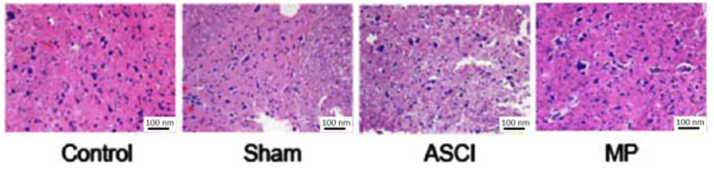
MP alters spinal cord morphology post-ASCI
The rats in the control and sham groups had normal spinal cord morphologies; however,
the medullospinal tissues of the rats in the ASCI group exhibited extensive hemorrhaging in the central tube and the central gray matter,
as well as severe edema in the perivascular
tissues and neurons. In the ASCI group, the
number of neurons was reduced, the cell gaps were widened, the nuclei were condensed,
partial medullospinal necrosis had occurred, partial spinal white matter degeneration had
occurred, and cysts and vacuoles had formed. Our results showed that in the MP group, the
boundaries between spinal gray matter and white matter were clearer than ASCI group.
The range of bleeding and phenomenon of
nuclear condensation were reduced. The edema in perivascular tissues and neurons was
also reduced, the edema volume was improved (Figure 2).
Figure 2 - Impacts of MP on post-ASCI spinal cord morphologies (HE ×200).
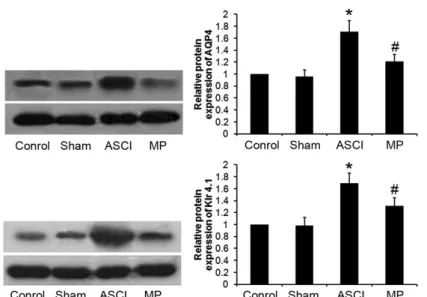
Effect of MP on the expression of AQP4 and Kir4.1 in post-ASCI medullospinal tissues
Compared with that in the sham group,
the expression of AQP4 and Kir4.1 in the ASCI group was significantly increased (P < 0.05).
However, in the MP group, compared with
that in the ASCI group, the expression of AQP4 and Kir4.1 was significantly downregulated
(P < 0.05) (Figure 3); there was no significant
difference in the expression of AQP4 or Kir4.1
Figure 3 - Impacts of MP on expressions of AQP4 and Kir4.1 in post-ASCI medullispinal tissues (x±SD, n = 10).
Compared with Group sham, *P < 0.05; compared with Group ASCI, #P < 0.05.
Detection of AQP4 and Kir4.1 coexpression in post-ASCI medullospinal tissues by immunofluorescence
AQP4 and Kir4.1, which were
coexpressed in post-ASCI medullospinal tissues, were mostly distributed in the spinal gray matter and less abundant in the spinal white matter; they were abundantly expressed
on the membranes of the spinal pia mater, the
central tube, and perivascular astrocytes of the spinal cord, but absent in neurons. In the ASCI group, compared with that in the sham
group, the coexpression of AQP4 and Kir4 was significantly increased, while it was significantly
reduced in the MP group compared with that in
the ASCI group; the coexpression of AQP4 and Kir4.1 was not significantly different between
the sham and control groups (Figure 4).
■
Discussion
ASCI is a common cause of motor
dysfunction in modern society, where it is
primarily caused by sports, falls, violence,
or traffic accidents9. With the continuous
developments of modern society, the probability of ASCI is increased yearly; most
patients are young or middle-aged, and suffer
from paraplegia, loss of the ability to work,
motor dysfunction, or even death, which results in heavy burdens on society and the patients’
families. ASCI includes primary and secondary spinal cord injury processes. The former refers to the direct damage caused by spinal canal
destruction and dislocation, which compress
the spinal cord and cause fractures; usually,
this kind of external force-mediated injury is
irreversible. Secondary injury follows primary injury, and is caused by a variety of factors. This kind of injury can occur within several minutes or a few days, and includes medullospinal edema, spinal cord hemorrhage, changes to free
radicals in local tissues, microtubule circulation
disorder, or reperfusion injury, which can harm
the spinal cord by inducing devastating lesions in the tissues around the foci, followed by gradual neurological dysfunction10. The degree of secondary spinal cord injury is greater than that of primary injury, but most cases are reversible or preventable.
Medullospinal edema is one of the key pathological processes of secondary spinal cord injury; it has an important impact on the
recovery of spinal cord function, and affects
the prognosis and treatment of spinal cord injury. The degree of post-ASCI edema is closely
related to motor functions; medullospinal edema starts from the central portion of the spinal gray matter, then gradually spreads to the surrounding tissues and the spinal white matter, causing injury aggravation owing
to compression of the spinal cord and an abnormal microtubule environment11. The
occurrence and development of medullospinal
edema induces ischemia and hypoxia, or even death in severe conditions, in neurons, which eventually leads to neuronal dysfunction; therefore, identifying strategies to reduce
the occurrence of post-ASCI edema is a
non-negligible issue. Currently, the lack of effective
treatments for ASCI is a worldwide problem; secondary ASCI typically causes catastrophic
damage to patients, so effective treatments
could preserve and improve peri-lesion
nerve tissue function, thereby extending and improving the lives of patients with ASCI12. Therefore, measures that reduce the loss of
neuronal functions would have research and therapeutic value.
MP is the most effective drug currently
recognized for the treatment of ASCI, but its
specific mechanisms of action have not been identified. Its main mechanism is to inhibit the generation of free oxygen radicals, thus resisting the peroxidation of blood lipids; meanwhile, it could also improve microtubule circulation, inhibit inflammation, reduce intracellular calcium influx, and maintain the excitability of
neurons13. In this study, intervention with MP
improved the neurological function scores of
rats with ASCI, and reduced the water content and degree of edema in their medullospinal
tissues.
AQPs are a family of water-specific membrane proteins that exist on cell
membranes, where they form pores and
control the exchange of extracellular and
intracellular water. Currently, a total of 13 AQPs (AQP0–AQP12) have been found in mammals;
AQP4 is the most widely expressed and
distributed in the nervous system, especially
in the brain and the medullospinal tissues14.
vivo, and maintain the water balance in vivo.
AQP4 has been reported to participate in the formation of tissue edema caused by post-injury hemorrhage, inflammation, or tumors15.
Manley found that the expression of AQP4 in edematous brain tissue was higher than that in normal brain tissue, and that the deficiency
of AQP4 reduced edema in the brain and capillaries16. Solenov et al.17 found that edema
was significantly reduced in rat medullospinal tissues that lacked AQP4. Nesic also reported that AQP4 is expressed in the spinal gray matter of normal rats, and that this expression tended to increase after ASCI; furthermore, this trend was spatiotemporally related to the
water content in the spinal cord, and could last for several days18. Therefore, the expression of
AQP4 positively correlated with medullospinal edema, and the downregulation of AQP4 could
reduce its severity.
Kir has strong effects on K+ influx and can shift extracellular K+ into cells. Recent studies found that Kir participated in water transportation, and was closely related to edema; moreover, the expression of Kir4.1 was increased in edematous tissues19. Zhang20 found
that the expression of Kir4.1 in rats with focal cerebral ischemia and reperfusion positively
correlated with the degree of cerebral edema. However, Kir4.1 was transported via the
AQP4-coupled mediating water, not independently20. The water balance regulatory roles of AQP4 were also closely related to Kir4.121. Nagelhus22
found strict colocalization between AQP4 and
Kir4.1, and Amiry23 confirmed that
AQP4-mediated water molecule transportation was related to the siphoning of K+. Therefore, the coexpression of AQP4 and Kir4.1 indicated interaction between them at the molecular level, and their coexpression was associated with normal water transportation in brain tissues.
We showed that the expression of AQP4 and Kir4.1 in the medullospinal tissues was significantly increased in the ASCI group
compared with that in the sham group; they
exhibited the phenomenon of co-expression, which was also significantly upregulated in
injured rats. In the MP group, compared with
that in the ASCI group, however, the expression of AQP4 and Kir4.1 in the medullospinal tissues was significantly decreased, as was their coexpression. These results suggested that the increased coexpression and expression
levels of AQP4 and Kir4.1 might be involved
in the formation of medullospinal edema and the induction of pathological changes in medullospinal tissues, as part of secondary ASCI; the inhibitory effects of MP on medullospinal edema might be associated with its reduction of the coexpression and expression levels of AQP4 and Kir4.1. Recent study demonstrated that MP administration following SCI reduced AQP4 expression and exacerbates edema24. The Cabrera-Aldana’s24 study showed that
SCI increased AQP4 expression in the spinal cord white matter and that MP diminished
such increase to baseline levels. Moreover,
MP increased the extravasation of plasma components after SCI and enhanced tissue
swelling and edema. Our results showed that MP inhibited medullospinal edema in rats with
ASCI, possibly by reducing the coexpression of AQP4 and Kir4.1 in medullospinal tissues. The
inconsistency between the two studies might
be attributable to the difference in SCI models
and types of edema.
■
Conclusions
Methylprednisolone inhibits
medullospinal edema in rats with acute spinal cord injury, which might be related to its roles
in reducing the coexpression and expression
levels of AQP4 and Kir4.1 in the medullospinal
tissues. This study could provide a theoretical and experimental basis for the application of glucocorticoids in treating ASCI, as well as
provide new ideas for developing AQP4- and
■
References
1. Varma AK, Das A, Wallace G 4th, Barry J, Vertegel AA, Ray SK, Banik NL. Spinal cord injury: a review of current therapy, future treatments, and basic science frontiers. Neurochem Res. 2013 May;38(5):895-905. doi: 10.1007/s11064-013-0991-6.
2. Peterson SL, Anderson AJ. Complement and spinal cord injury: traditional and non-traditional aspects of complement cascade function in the injured spinal cord microenvironment. Exp Neurol. 2014 Aug;258:35-47. doi: 10.1016/j. expneurol.2014.04.028.
3. Oklinski MK, Lim JS, Choi HJ,
Oklinska P, Skowronski MT, Kwon TH.
Immunolocalization of Water Channel Proteins AQP1 and AQP4 in Rat Spinal Cord. J Histochem Cytochem. 2014 Aug;62(8):598-611. doi: 10.1369/0022155414537495.
4. Wang BF, Cui ZW, Zhong ZH, Sun YH, Sun
QF, Yang GY, Bian LG. Curcumin attenuates
brain edema in mice with intracerebral
hemorrhage through inhibition of AQP4 and AQP9 expression. Acta Pharmacol Sin. 2015 Aug;36(8):939-48. doi: 10.1038/
aps.2015.47.
5. Jiang X, Huang Y, Lin W, Gao D, Fei Z. Protective effects of hydrogen sulfide in a rat model of traumatic brain injury via activation of mitochondrial adenosine triphosphate-sensitive potassium channels and reduction of oxidative stress. J Surg Res. 2013 Oct;184(2):e27-e35. doi: 10.1016/j.
jss.2013.03.067.
6. Yan JH, Khatibi NH, Han HB, Hu Q, Chen CH, Li L, Yang XM, Zhou CM. p53-induced uncoupling expression of aquaporin-4 and inwardly rectifying K+ 4.1 channels in cytotoxic edema after subarachnoid hemorrhage. CNS Neurosci Ther. 2012 Apr;18(4):334-42. doi: 10.1111/j.1755-5949.2012.00299.x.
7. Karamouzian S, Akhtarshomar S, Saied
A, Gholamhoseinian A. Effects of methylprednisolone on neuroprotective effects of delay hypothermia on spinal cord injury in rat. Asian Spine J. 2015 Feb;9(1):1-6. doi: 10.4184/asj.2015.9.1.1.
8. Wu Y, Collier L, Pan J, Qin W, Bauman
WA, Cardozo CP. Testosterone reduced methylprednisolone-induced muscle
atrophy in spinal cord-injured rats. Spinal
Cord. 2012 Jan;50(1):57-62. doi: 10.1038/ sc.2011.91.
9. Jia X, Kowalski RG, Sciubba DM, Geocadin RG. Critical care of traumatic spinal cord injury. J Intensive Care Med. 2013 Jan-Feb;28(1):12-23. doi: 10.1177/0885066611403270.
10. Grant RA, Quon JL, Abbed KM. Management of acute traumatic spinal cord injury. Curr Treat Options Neurol. 2015 Feb;17(2):334. doi: 10.1007/s11940-014-0334-1.
11. Wang YF, Fan ZK, Cao Y, Yu DS, Zhang YQ,
Wang YS. 2-Methoxyestradiol inhibits the up-regulation of AQP4 and AQP1 expression after spinal cord injury. Brain Res. 2011 Sep;1370:220-6. doi: 10.1016/j.
brainres.2014.12.045.
12. Gensel JC, Zhang B. Macrophage activation and its role in repair and pathology after spinal cord injury. Brain Res. 2015 Sep;1619:1-11. doi: 10.1016/j.brainres.2014.12.045.
13. Nash HH, Borke RC, Anders JJ. Ensheathing
cells and methylprednisolone promote
axonal regeneration and functional recovery in the lesioned adult rat spinal cord. J Neurosci. 2002 Aug;22(16):7111-20. doi:
20026746.
14. Filippidis AS, Kalani MY, Rekate HL. Hydrocephalus and aquaporins: the role of aquaporin-4. Acta Neurochir Suppl. 2012;113:55-8. doi: 10.1007/978-3-7091-0923-6_12.
15. Verkman AS, Mitra AK. Structure and function of aquaporin water channels. Am J Physiol Renal Physiol. 2000 Jan;278(1):F13-28. PMID: 10644652.
16. Manley GT, Fujimura M, Ma T, Noshita N, Filiz F, Bollen AW, Chan P, Verkman AS. Aquaporin-4 deletion in mice reduces brain edema after acute water intoxication and ischemic stroke. Nat Med. 2000 Feb;6(2):159-63. doi: 10.1038/72256. 17. Solenov EI, Vetrivel L, Oshio K, Manley
GT, Verkman AS. Optical measurement of
swelling and water transport in spinal cord
slices from aquaporin null mice. J Neurosci Methods. 2002 Jan;113(1):85-90. PMID:
11741725.
2006 Dec;143(3):779-92. doi: 10.1016/j. neuroscience.2006.08.079.
19. Dibaj P, Kaiser M, Hirrlinger J, Kirchhoff F, Neusch C. Kir4.1 channels regulate swelling of astroglial processes in experimental medullispinal edema. J Neurochem. 2007 Dec;103(6):2620-8. doi: 10.1111/j.1471-4159.2007.04979.x.
20. Zhao M, Bousquet E, Valamanesh F, Farman N, Jeanny JC, Jaisser F, Behar-Cohen FF. Differential regulations of AQP4
and Kir4.1 by triamcinolone acetonide and
dexamethasone in the healthy and inflamed retina. Invest Ophthalmol Vis Sci. 2011 Aug;52(9):6340-7. doi:
10.1167/iovs.11-7675.
21. Tham DK, Moukhles H. Regulation of Kir4.1 and AQP4 expression and stability at the
basolateral domain of epithelial MDCK cells
by the extracellular matrix. Am J Physiol Renal Physiol. 2011 Aug;301(2):F396-409. doi: 10.1152/ajprenal.00315.2010.
22. Nagelhus EA, Horio Y, Inanobe A, Fujita A, Haug FM, Nielsen S, Kurachi Y, Ottersen
OP. Immunogold evidence suggests
that coupling of K+ siphoning and water transport in rat retinal Müller cells is
mediated by a coenrichment of Kir4.1 and
AQP4 in specific membrane domains. Glia. 1999 Mar;26(1):47-54. PMID: 10088671.
23. Amiry-Moghaddam M, Otsuka T, Hurn PD,
Traystman RJ, Haug FM, Froehner SC, Adams ME, Neely JD, Agre P, Ottersen OP, Bhardwaj
A. An alpha-syntrophin-dependent pool of AQP4 in astroglial end-feet confers
bidirectional water flow between blood and brain. Proc Natl Acad Sci U S A. 2003 Feb;100(4):2106-11. doi: 10.1073/ pnas.0437946100.
24. Cabreraaldana EE, Ruelas F, Aranda C, Rinconheredia R, Martínezcruz A, Reyessánchez A, Guizar-Sahagún G, Tovar-y-Romo LB. Methylprednisolone administration following spinal cord injury reduces aquaporin 4 expression and exacerbates edema. Mediators Inflamm. 2017 May;2017(6):1-7. doi: 10.1155/2017/4792932.
Correspondence:
Rui Gu
Department of Orthopaedics
China-Japan Union Hospital, Jilin University
Changchun 130033 China
Phone: +86 431 84995117
cnlydoc@126.com
Received: Oct 16, 2017 Review: Dec 18, 2017 Accepted: Jan 19, 2018
Conflict of interest: none Financial source: none
1Research performed at Department of Central
Laboratory, China-Japan Union Hospital, Jilin