Prognostic factors predicting a
fatal outcome in hiv-negative
children with neurotuberculosis
Murilo Gimenes Rodrigues1, Jaime Lin1, Marcelo Rodrigues Masruha1, Luiz Celso Pereira Vilanova1, Thais Soares Cianciarullo Minett2
ABSTRACT
Objective: To identify prognostic factors predicting a fatal outcome in HIV-negative children with neurotuberculosis based on clinical, epidemiological, and laboratory findings. Method: The clinical records of all in-patients diagnosed with neurotuberculosis from 1982 to 2005 were evaluated retrospectively. The following prognostic parameters were examined: gender, age, close contact with a tuberculosis-infected individual, vaccination for bacillus Calmette-Guérin, purified protein derivative (PPD) of tuberculin results, concomitant miliary tuberculosis, seizures, CSF results, and hydrocephalus. Results: One hundred forty-one patients diagnosed with neurotuberculosis were included. Seventeen percent of the cases resulted in death. The factors that were correlated with a negative outcome included lack of contact with a tuberculosis-infected individual, negative PPD reaction, coma, and longer hospitalisation time. A multiple logistic regression analysis was performed to identify which of these factors most often resulted in death. Conclusion: Coma at diagnosis, lack of tuberculosis contact, and a non-reactive PPD were the most important predictors of fatality in patients with neurotuberculosis.
Key words: tuberculosis, meningitis, central nervous system, prognosis, child.
Fatores prognósticos de letalidade da neurotuberculose em crianças HIV-negativas
RESUMO
Objetivo: Identificar elementos prognósticos para a letalidade da neurotuberculose na criança, a partir das manifestações clínicas, dados epidemiológicos e laboratoriais. Método: Registros de pacientes internados durante o período de 1982 a 2005 foram retrospectivamente avaliados. Os elementos prognósticos considerados foram: sexo, idade, história de contato íntimo com indivíduo com tuberculose, vacinação com o bacilo de Calmette-Guérin (BCG), teste tuberculínico (PPD), concomitância de tuberculose miliar, convulsões, resultados da análise do LCR e presença de hidrocefalia. Resultados: 141 pacientes com diagnóstico de neurotuberculose foram incluídos. Dezessete por cento dos pacientes foram a óbito. Os fatores associados ao óbito foram história negativa de contágio, ausência de reatividade ao teste de PPD, coma e tempo de internação prolongado. Análise por regressão logística múltipla foi usada para investigar as relações entre os elementos prognósticos e o desfecho óbito. Conclusão: Os fatores prognósticos na previsão de óbito nos pacientes com neurotuberculose foram a presença de coma no momento do diagnóstico, a ausência de história de contágio e a ausência de reação ao PPD.
Palavras-chave: tuberculose, meningite, sistema nervoso central, prognóstico, crianças.
Correspondence Murilo Gimenes Rodrigues Setor de Neurologia Infantil Depto. de Neurologia e Neurocirurgia Universidade Federal de São Paulo Rua Botucatu 720
04023-900 São Paulo SP - Brasil E-mail: [email protected]
Received 31 January 2010 Received in final form 20 March 2010 Accepted 26 March 2010
1Department of Neurology and Neurosurgery, Federal University of São Paulo, São Paulo SP, Brazil; 2Department of Preventive
Medicine, Federal University of São Paulo, São Paulo SP, Brazil.
Tuberculosis remains one of the most severe infectious diseases and more than eight million new cases are diagnosed
over-population1. In addition, the human immunodeiciency
virus (HIV) has caused a resurgence of tuberculosis2,3.
According to the World Health Organisation, 22 countries are responsible for more than 80% of tubercu-losis cases. Among these countries, Brazil occupies the 14th position, reporting more than 60 cases per 100,000
inhabitants, 50 million infected people, 92,000 new cas-es, and 8,400 deaths annually4.
Although pulmonary tuberculosis is more frequent, neurotuberculosis is considered a more severe form be-cause even when treated, it often leads to death or neu-rological sequelae5,6.
Important prognostic indicators of neurotuberculo-sis include age (especially very young or old individuals), clinical stage at diagnosis, presence of neurological dei-cits, presence of other forms of tuberculosis, presence of other diseases, malnutrition, seizures, and resistance to tuberculosis treatment5.
However, the clinical manifestations of tuberculosis difer in children and adults. In children, a closed caseous lesion is commonly the primary infection. Children also present extrapulmonary cases more frequently and exhib-it a higher incidence of miliary dissemination7.
his study aims to identify prognostic factors that may predict a fatal outcome in HIV-negative children who suf-fer from tuberculosis based on clinical, epidemiological, and laboratory indings.
METHOD
The study was performed at the Nossa Senhora da Glória Children’s Hospital in Vitória ES, Brazil. he clin-ical records of all in-patients diagnosed with neurotuber-culosis from 1982 to 2005 were evaluated retrospectively. We used the diagnostic criteria for neurotuberculosis pro-posed and validated by Ahuja et al.8 and Doerr et al.9.
Patients with a deinitive or probable diagnosis of neu-rotuberculosis were included5. A deinitive diagnosis was
deined by either the presence of alcohol acid-resistant bacillus in the cerebrospinal luid (CSF), a positive cul-ture for Mycobacterium tuberculosis, or a conclusive post-mortem examination. A probable diagnosis was deined by neurologically abnormal signs and symptoms accom-panied by two or more of the following conditions: [A] CSF with pleocytosis and predominance of lymphocytes (characteristic of the neutrophilic dominance of early tu-berculosis) without evidence of other infection; [B] pre-vious or concomitant contact (either intradomiciliary or otherwise signiicant) with a tuberculosis-infected indi-vidual, or a positive culture for Mycobacterium tubercu-losis from any part of the body other than the central ner-vous system; [C] radiological evidence of pulmonary tu-berculosis; [D] a reactive tuberculin test without a vaccine scar; and [E] a cranial computed tomography (CT) scan
compatible with tuberculous meningoencephalitis (i.e., a CT showing two or more of the following features: exu-dates in the basal cisterns or sylvian sulcus, hydrocepha-lus, tuberculomas, infarcts, or gyral enhancement). Since 1986, when ELISA for HIV becomes available to us, all patients were tested for HIV infection and positive cas-es were excluded.
The following prognostic parameters were exam-ined: gender, age, previous or concomitant contact with a tuberculosis-infected individual (either intradomicili-ary or other prolonged contact), vaccination for bacillus Calmette-Guérin (BCG) (this immunisation is required in Brazil and can be conirmed by the presence of a vac-cine scar on the right deltoid or by cross-referencing with a vaccine register), presence of tuberculin puriied protein derivative (PPD), concomitant miliary tuberculosis, sei-zures, and hydrocephalus. A CSF analysis was also per-formed, and the results were statistically analyzed.
To classify the severity of the disease in our patients, we used the clinical stages deined by the British Medical Research Council (BMRC): stage I - patients with gener-al symptoms (fever, anorexia, headache, and vomiting); stage II - patients with meningeal irritation accompanied by a diminished level of consciousness; and stage III - pa-tients with coma, decortication, or descerebration5.
RT23 PPD tuberculin was used and applied following the Mantoux technique via an intradermic route in the middle third of the anterior forearm. he dose was 0.1 ml, which is equivalent to 2 TU (tuberculin units). he larg-er transvlarg-erse diametlarg-er of the indurate palpable area was measured with a millimetre ruler 72 to 96 hours after tu-berculin application. he result was recorded in millime-tres: up to 4 mm (non-reactive); 5 to 9 mm (weak reac-tive); 10 mm or more (strong reactive)10.
A Chi-square (c2) or Fisher statistical test was used
to compare categorical prognostic factors between sur-viving and deceased patients. A Student’s t-test (t) was used to compare the mean values of the continuous mea-surements. A multiple logistic regression analysis was used to investigate the relationship between fatality and the prognostic factors that were found to be signiicant in the univariate analysis. Missing data were excluded, and outliers were not removed from the model. he odds ra-tios (OR) with 95% conidence intervals (CI) are report-ed. All tests were two tailed; a p-value<0.05 was consid-ered to indicate a statistically signiicant diference. All statistical analyses were conducted on a personal com-puter with the SPSS for Windows (version 11.5.1) soft-ware package.
RESULTS
One hundred forty-one cases of children with neuro-tuberculosis from 1982 to 2005 were reviewed. A dein-itive diagnosis was established in twenty-three patients (16%); the remaining cases (84%) were children with a probable diagnosis.
he incidence age varied from 3 to 156 months (medi-an 24 months). Among the 141 cases, 87 (62%) were boys, 68 (48%) had been previously immunised with the BCG vaccine, 109 (77%) reported either an intradomiciliary or other signiicant contact with a tuberculosis-infected pa-tient, and 27 (19%) presented concomitant miliary tuber-culosis. he mean length of hospitalisation varied from 2 to 270 days (median 60 days). he main clinical indings are listed in Table 1.
CSF analysis
The Koch bacillus was identified in the CSF of 23 (16%) of the children. Of those, 9 (39%) presented a pos-itive CSF culture, 9 (39%) presented a pospos-itive direct ex-amination, and 5 (22%) presented positive results using both methods.
he mean±SD of the CSF protein, glucose, and cell count was 143.2 (17-1034) mg/100 ml, 34.9 (0-100) mg/100 ml, and 325.1 (10-5120) cells/mm3, respectively.
A hundred-fourteen (81%) patients presented CSF pro-tein readings of above 40 mg/100 ml and 109 (77%) pre-sented CSF glucose readings lower than 50 mg/100 ml.
Neuroimaging
Computed tomography scans were performed on 62 (44%) of the patients. Among these scans, 54 (87%) dis-closed abnormalities. Hydrocephalus was the most preva-lent abnormality (79% of the scans), followed by infarct ar-eas (18%) and tuberculomas (16%). Interestingly, isolated tuberculomas were found in 11 (17.7%) of the scans, and
isolated infarct areas were found in 2 (3%). One patient pre-sented with concomitant tuberculomas and infarct areas.
PPD test
he PPD test was performed during the irst week of hospitalisation. Of the 141 patients, 69 (49%) of the pa-tients were non-reactive, 14 (10%) were weakly reactive, and 58 (41%) were strongly reactive.
Among the children with previous immunisation, the PPD test reactivity did not vary signiicantly: 70 (50%) were non-reactive, 18 (13%) were weakly reactive, and 53 (37%) were strongly reactive (c2(2)=6.0; p=0.050).
Table 1. Frequency of the signs and symptoms of neurotuberculosis patients at the time of admission.
Signs and symptoms %
Fever 96
Nuchal rigidity 79
Vomiting 63
Spasticity 52
Drowsiness 50
Anorexia 49
Seizures 46
Apathy 45
Cough 38
Headache 36
Weight loss 33
Irritability 32
Cranial nerve palsies 30
Stupor 19
Slow photomotor relex 18
Diarrhoea 16
Coma 13
Constipation 9
Table 2. Comparison of prognostic factor frequencies in surviving and deceased neurotuberculosis patients. Factors include previous BCG immunisation, positive tuberculosis contact, miliary tuberculosis, PPD reactivity, CT abnormalities, coma, epileptic seizures, and CSF polymorphonuclear predominance.
Prognostic factors n Survivors (%) Deceased (%) c2 (1) p
BCG immunisation 141 46 58 1.18 0.277
Contact 141 80 58 5.38 0.020*
Miliary tuberculosis 141 20 12 – 0.569+
PPD reactivity 128 54 29 4.61 0.032*
CT abnormalities 62 85 100 – 0.590+
Coma 141 6 47 28.40 <0.001*
Seizures 141 43 62 3.13 0.077
PMN predominance 140 27 29 0.06 0.807
Lethality
Twenty-four (17%) patients died. Tables 2 and 3 com-pare the prognostic parameters of surviving and deceased patients.
he factors associated with a negative outcome were a negative history of contact with a tuberculosis-infect-ed individual, a negative PPD reaction, coma, or a longer hospitalisation time.
A multiple logistic regression analysis was performed to identify which of the preceding factors most often re-sulted in fatalities (Table 4). We found that coma at the time of hospitalisation and a negative tuberculosis con-tact history were the factors most strongly correlated with fatality.
he diference in the proportion of deceased patients between the deinite (13%) and probable (18%) cases was not signiicant (Fisher exact test, p=0.765).
DISCUSSION
We found that coma at the time of admission, a nega-tive tuberculosis contact history, and a non-reacnega-tive PPD were most commonly associated with fatalities in neuro-tuberculosis patients.
Our series had a low percentage of fatalities (17%), a result that is similar to data from Mahadevan et al. (16%)11
and Lee et al. (10%)7. In our study, most of the patients
were in clinical stages I or II as deined by the BMRC scale, which may explain the low percentage of lethal cases. Despite these results, the high lethality of neurotu-berculosis is what makes it more severe than other forms of tuberculosis12. Some authors have tried to identify the
prognostic factors of neurotuberculosis13-15. he level of
consciousness at the time of admission or at the time that a speciic therapy is established is the main prognostic factor that predicts disease lethality and sequelae16,17. In
our study, more than 60% of the 24 patients who died were in coma when hospitalised.
Among our patients, 77% had a history of close con-tact with an individual who was a conirmed tuberculo-sis suferer. In other studies, the history of close contact with tuberculosis varies widely from 20 to 75%18-20.
Ab-sence of close contact with a tuberculosis-infected indi-vidual, however, tends to be strongly correlated with fatal-ities. In our study, 15% of the patients had a negative his-tory of close contact and 30% of these patients eventually died. he absence of close contact may be related to high-er fatalities because of a delay in establishing an appro-priate treatment protocol. More than half of our patients began treatment during the irst few days of hospitalisa-tion, most of whom reported a positive epidemiological history that caused us to suspect tuberculosis. Another explanation is that these patients could have some kind of immunological deicient mechanisms, being more sus-ceptible to develop neurotuberculosis, even with a small inoculus.
Molavi et al.19 and Hosoglu et al.21, over a decade later,
addressed the importance of contact history as an impor-tant and useful epidemiological tool for the early diagno-sis of tuberculodiagno-sis. heir reports, however, failed to cor-relate this data with the patient’s prognosis. While a his-tory of close contact did reduce the time between diag-nosis and treatment, in our experience this history is
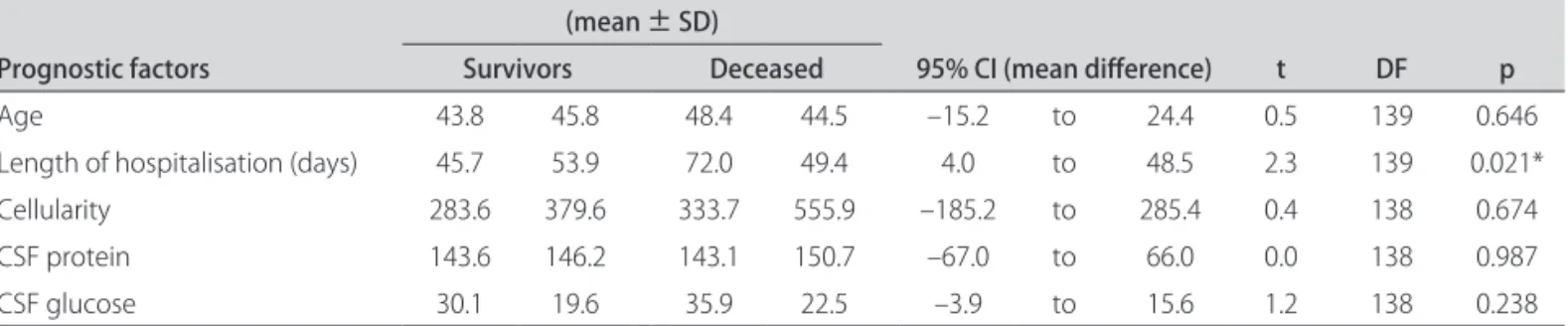
of-Table 3. Comparison of the prognostic factor mean values among surviving and deceased neurotuberculosis patients.
Prognostic factors
(mean ± SD)
95% Ci (mean diference) t DF p Survivors Deceased
Age 43.8 45.8 48.4 44.5 –15.2 to 24.4 0.5 139 0.646
Length of hospitalisation (days) 45.7 53.9 72.0 49.4 4.0 to 48.5 2.3 139 0.021*
Cellularity 283.6 379.6 333.7 555.9 –185.2 to 285.4 0.4 138 0.674
CSF protein 143.6 146.2 143.1 150.7 –67.0 to 66.0 0.0 138 0.987
CSF glucose 30.1 19.6 35.9 22.5 –3.9 to 15.6 1.2 138 0.238
*Signiicant; SD: standard deviation; CI: conidence interval; DF: degrees of freedom; CSF: cerebrospinal luid.
Table 4. Likelihood of death based on results from the multiple logistic regression analysis using the prognostic factors as independent variables.
Prognostic factors B SE Wald c2 OR 95% Ci (OR) p
Coma –2.11 0.53 15.65 0.12 0.04 to 0.35 <0.001*
Hospitalisation time –0.01 0.01 3.45 0.99 0.97 to 1 0.063
Absence of contact 1.24 0.58 4.6 3.47 1.11 to 10.82 0.032*
Absence of PPD reactivity 0.81 0.5 2.59 2.25 0.84 to 6.05 0.108
ten diicult to obtain from the patients or their relatives because of the continuing stigma surrounding tubercu-losis and due to ignorance about the disease.
In our study, a non-reactive PPD was found in 63% of the patients and this was shown to have a negative im-pact on patient outcomes. A non-reactive PPD would not be unusual if the disease were in an advanced stage, when patients present severe cellular immunity impairment22.
his may also be explained by a previous deiciency of im-mune mechanisms. he correlation between a negative prognosis and PPD insensitivity may be a direct conse-quence of a typical meningeal reaction failure23,24. Succi et
al.25 found that, among 111 children with
neurotubercu-losis, those with a consistently negative PPD results had more extensive neurological involvement.
CT scans were performed on only 44% of the patients because many of them were treated between 1982 and 1990, a decade when this resource was largely unavail-able in our centre. However, every patient in our study who died presented an abnormal CT examination that suggested hydrocephalus, infarct areas, or tuberculomas. Tan et al.26 examined a group of 18 patients with
neuro-tuberculosis and found that the presence of hydroceph-alus was the only factor associated with a negative prog-nosis; however, this prognosis was also correlated with a more advanced disease stage.
he high incidence of hydrocephalus in our series re-lects the typical physiopathological presentation of neu-rotuberculosis, which includes a blockage of CSF drain-age-especially in the basal cisterns27. However, we found
only a weak correlation between hydrocephalus and fatali-ties, perhaps because our patients presented only mild hy-drocephalus. Although hydrocephalus is a common fea-ture of neurotuberculosis and is associated with a nega-tive prognosis, early treatment can prevent the progres-sion of ventricular dilatation28. If these patients had been
diagnosed earlier, the prognosis could potentially have been better29.
The presence of seizures has also been correlated with eventual fatality. Paganini et al.30 reported that
sei-zures were an important predictor of fatal outcomes in 40 children with tuberculosis. In this report, seizures were manifested late in the disease and were usually associated with more advanced stages of tuberculosis24. In our study,
however, seizures were not related to death. Importantly, our study only evaluated symptoms after hospitalisation. In contrast with Paganini et al.30, who regarded seizure
as a late manifestation of tuberculosis, our study found that, after hospitalisation, seizures could be present in any stage of the disease.
he presence of CSF alterations also failed to predict a negative prognosis. Succi et al.25 assessed the clinical
evo-lution of patients with diferent intensities of pleocytosis, concluding that CSF alterations increased the likelihood of fatality but noting that this correlation was not statis-tically signiicant. In contrast, Singh et al.31 found that
pa-tients who presented protein concentrations above 200 mg/dL, glucose lower than 20 mg/dL, and chlorides low-er than 650 mg/dL exhibited a highlow-er incidence of fatali-ty. However, Singh et al. only included adult patients and failed to analyse their data for statistical signiicance.
In our study, the concomitance of miliary tuberculo-sis was also not associated with fatality. Succi et al.25
con-cluded that the presence of radiological alterations, which indicate the presence of miliary tuberculosis, did not in-crease the likelihood of death. Our study used the same radiological criteria to determine the presence of miliary tuberculosis. It is possible that those patients who sur-vived long enough to present an image that was detect-able and indicated miliary dissemination had a stronger immune response. hus, these patients exhibited a sur-vival rate similar to those patients without miliary dis-semination. In contrast, Paganini et al.30 reported that
ex-tra-meningeal foci of tuberculous meningitis were pre-dictors of fatality. hey hypothesised that the detection of extra-meningeal tuberculosis in neurotuberculosis pa-tients may be an important tool for early diagnosis and may lead to a better prognosis.
Rodrigues et al.32 argued that BCG immunisation may
protect children against the most severe forms of tuber-culosis and result in a better prognosis. In our study, BCG immunisation did not afect the prognosis of neurotu-berculosis. However, because all of the patients enrolled in our study already presented a severe form of the dis-ease, we were unable to evaluate whether BCG immuni-sation afects the prognosis of tuberculosis by protecting against the most severe forms. Interestingly, more than half of our patients had been immunised with BCG but still developed neurotuberculosis, calling into question the ability of the BCG vaccine to protect against severe forms of tuberculosis.
Our retrospective study was limited by the fact that neuroimaging was not performed with all patients. Ad-ditionally, our diagnoses of tuberculosis were based on clinical criteria and we failed to secure microbiological conirmation.
REFERENCES
Schachter EN. Tuberculosis: a global problem at our doorstep. Chest 2004; 1.
126:1724-1725.
Corbett EL, Watt CJ, Walker N, et al. The growing burden of tuberculosis: 2.
global trends and interactions with the HIV epidemic. Arch Intern Med 2003;163:1009-1021.
Snider DE Jr., Roper WL. The new tuberculosis. N Engl J Med 1992;326: 3.
703-705.
WHO. Global tuberculosis control: epidemiology, strategy, inancing - WHO 4.
report 2009. 2009.
Hosoglu S, Geyik MF, Balik I, et al. Predictors of outcome in patients with tu-5.
berculous meningitis. Int J Tuberc Lung Dis 2002;6:64-70.
Girgis NI, Sultan Y, Farid Z, et al. Tuberculosis meningitis, Abbassia Fe-6.
ver Hospital-Naval Medical Research Unit No. 3-Cairo, Egypt, from 1976 to 1996. Am J Trop Med Hyg 1998;58:28-34.
Lee LV. Neurotuberculosis among Filipino children: an 11 years experience 7.
at the Philippine Children’s Medical Center. Brain Dev 2000;22:469-474. Ahuja GK, Mohan KK, Prasad K, Behari M. Diagnostic criteria for tuberculous 8.
meningitis and their validation. Tuber Lung Dis 1994;75:149-152. Doerr CA, Starke JR, Ong LT. Clinical and public health aspects of tubercu-9.
lous meningitis in children. J Pediatr 1995;127:27-33.
Bailey WC, Thompson DH, Greenberg HB. A technique for standardizing 10.
the jet injector and Mantoux tuberculin skin tests. Public Health Rep 1974; 89:465-467.
Mahadevan B, Mahadevan S, Serane VT. Prognostic factors in childhood tu-11.
berculous meningitis. J Trop Pediatr 2002;48:362-365.
Gusmao Filho FA, Marques HH, Marques-Dias MJ, Ramos SR. [Central ner-12.
vous system tuberculosis in children: 1. Clinical and laboratorial presenta-tion]. Arq Neuropsiquiatr 2001;59:71-76.
Misra UK, Kalita J, Roy AK, Mandal SK, Srivastava M. Role of clinical, radio-13.
logical, and neurophysiological changes in predicting the outcome of tu-berculous meningitis: a multivariable analysis. J Neurol Neurosurg Psychi-atry 2000;68:300-303.
Ramachandran P, Duraipandian M, Nagarajan M, Prabhakar R, Ramakrish-14.
nan CV, Tripathy SP. Three chemotherapy studies of tuberculous meningi-tis in children. Tubercle 1986;67:17-29.
Saitoh A, Pong A, Waecker NJ, Jr., Leake JA, Nespeca MP, Bradley JS. Pre-15.
diction of neurologic sequelae in childhood tuberculous meningitis: a re-view of 20 cases and proposal of a novel scoring system. Pediatr Infect Dis J 2005;24:207-212.
Farinha NJ, Razali KA, Holzel H, Morgan G, Novelli VM. Tuberculosis of the 16.
central nervous system in children: a 20-year survey. J Infect 2000;41:61-68. Nunes C, Cunha S, Gomes I, Lucena R, Moraes D, Melo A. [Prognostic fac-17.
tors of tuberculous meningoencephalitis lethality]. Arq Neuropsiquiatr 1998;56:772-777.
Yeo IK, Tannenbaum T, Scott AN, et al. Contact investigation and geno-18.
typing to identify tuberculosis transmission to children. Pediatr Infect Dis J 2006;25: 1037-1043.
Molavi A, LeFrock JL. Tuberculous meningitis. Med Clin North Am 1985;69: 19.
315-331.
Bateman DE, Newman PK, Foster JB. A retrospective survey of proven cases 20.
of tuberculous meningitis in the Northern Region, 1970-1980. J R Coll Phy-sicians Lond 1983;17:106-110.
Hosoglu S, Ayaz C, Geyik MF, Kokoglu OF, Ceviz A. Tuberculous meningitis 21.
in adults: an eleven-year review. Int J Tuberc Lung Dis 1998;2:553-557. Boras Z, Juretic A, Rudolf M, Uzarevic B, Trescec A. Cellular and humoral im-22.
munity to puriied protein derivative (PPD) in PPD skin reactive and non-reactive patients with pulmonary tuberculosis: comparative analysis of an-tigen-specific lymphocyte proliferation and IgG antibodies. Croat Med J 2002;43:301-305.
Waecker NJ Jr., Connor JD. Central nervous system tuberculosis in children: 23.
a review of 30 cases. Pediatr Infect Dis J 1990;9:539-543.
Illingworth RS. Miliary and meningeal tuberculosis; diiculties in diagnosis. 24.
Lancet 1956;271:646-649.
Succi RCM. Meningoencefalite tuberculosa na infância: estudo de 358 ca-25.
sos. Aspectos clínicos, laboratoriais e fatores prognósticos. Tese. Universi-dade de São Paulo. São Paulo, 1990.
Tan EK, Chee MW, Chan LL, Lee YL. Culture positive tuberculous meningi-26.
tis: clinical indicators of poor prognosis. Clin Neurol Neurosurg 1999;101: 157-160.
Altunbasak S, Alhan E, Baytok V, Aksaray N, Yuksel B, Onenli N. Tuberculous 27.
meningitis in children. Acta Paediatr Jpn 1994;36:480-484.
Schoeman JF, Van Zyl LE, Laubscher JA, Donald PR. Serial CT scanning in 28.
childhood tuberculous meningitis: prognostic features in 198 cases. J Child Neurol 1995;10:320-329.
Gusmao Filho FA, Marques-Dias MJ, Marques HH, Ramos SR. [Central ner-29.
vous system tuberculosis in children: 2. Treatment and outcome]. Arq Neu-ropsiquiatr 2001;59:77-82.
Paganini H, Gonzalez F, Santander C, Casimir L, Berberian G, Rosanova MT. 30.
Tuberculous meningitis in children: clinical features and outcome in 40 cases. Scand J Infect Dis 2000;32:41-45.
Singh NK, Singh P, Tripathi K, Srivastava PK, Singh DS. Prognostic factors 31.
and sequelae of tuberculous meningitis in adults. J Indian Med Assoc 1985; 83:50-53.
Rodrigues LC, Diwan VK, Wheeler JG. Protective efect of BCG against tuber-32.