(1) Universidade Federal de Alfenas, UNIFAL, UNIFENAS and UNINCOR (postdoctorate ICB/USP), MG, Brazil. (2) Departamento de Microbiologia do Instituto de Ciências Biomédicas da Universidade de São Paulo, SP, Brazil. (3) Departamento de Análises Clínicas da Universidade de São Paulo, SP, Brazil
(4) UNINCOR, Três Corações, MG, Brazil.
(5) Departamento de Análises Clínicas da Universidade de São Paulo, SP, Brazil. (6) Universidade Federal de Alfenas, UNIFAL, Alfenas, MG, Brazil.
Correspondence to: Dr. Jorge Kleber Chavasco, Departamento de Microbiologia, Instituto de Ciências Biomédicas II, Universidade de São Paulo, Av. Prof. Lineu Prestes 1374, 05508-900 São Paulo, SP, Brazil. Tel: 55.11.3091-7294; Fax: 55.11.3091-7354; e-mail: [email protected]
MOLECULAR IDENTIFICATION OF Candida dubliniensis ISOLATED FROM ORAL LESIONS OF
HIV-POSITIVE AND HIV-NEGATIVE PATIENTS IN SÃO PAULO, BRAZIL
Jorge Kleber CHAVASCO(1), Claudete Rodrigues PAULA(2), Mario Hiroyuki HIRATA(3), Natanael Atilas ALEVA(4), Carlos Eduardo de MELO(5), Walderez GAMBALE(2), Luciana da Silva RUIZ(2) & Marília Caixeta FRANCO(6)
SUMMARY
Candida dubliniensis is a new, recently described species of yeast. This emerging oral pathogen shares many phenotypic and biochemical characteristics with C. albicans, making it hard to differentiate between them, although they are genotypically distinct. In this study, PCR (Polymerase Chain Reaction) was used to investigate the presence of C. dubliniensis in samples in a culture collection, which had been isolated from HIV-positive and HIV-negative patients with oral erythematous candidiasis. From a total of 37 samples previously identified as C. albicans by the classical method, two samples of C. dubliniensis (5.4%) were found through the use of PCR. This study underscores the presence of C. dubliniensis, whose geographical and epidemiological distribution should be more fully investigated.
KEYWORDS: HIV-positive; HIV-negative; Candida dubliniensis; PCR.
INTRODUCTION
An increased incidence of fungal infections has been well documented throughout the last decade. The most important factor contributing to this phenomenon has been the increased number of immunocompromised individuals. As a result, many species previously unassociated with human diseases have become important pathogens, some examples being Penicillium marneffei, Emmonsia pasteurina and Candida dubliniensis6,30.
Candida dubliniensis was first identified as a new species in 1995 in Dublin, Ireland39.Since then, infections by this yeast have been
widely reported in a large number of HIV-positive and AIDS patients23,
being isolated mainly from the oral cavity9. Moreover, C. dubliniensis
has been implicated as a causative agent for oral candidoses and HIV-negative individuals, both in healthy individuals and diabetics42,43.
This species shares many phenotypic and biochemical characteristics with C. albicans39, making it difficult to differentiate
between the two species, since C. dubliniensis expresses the serotype A of C. albicans, and is able to form germ tubes and abundant numbers of chlamydoconidia3,4,39,41. Moreover, C. dubliniensis is characterized
by a high resistance to fluconazole, and the susceptible isolates are able to develop resistance to this drug in vitro26,40. This high degree of
similarity between the two species has contributed to the identification of some isolates of C. dubliniensis as C. albicans31.This species has
most likely been present in the community for long time, although identified as C. albicans36.
Therefore, various phenotypic methods for the identification of C. dubliniensis and its differentiation from C. albicans have been reported. These tests include: the formation of chlamydospores39; the pattern of
carbohydrate assimilation33, β-D-glucosidase activity35; the color of
colonies after seeding in different mediums such as CHROMagar Candida, Staib agar37, Niger agar17, Tobacco agar13 and others; as well
as growth in Sabouraud agar at temperatures between 42 and 45 °C31.
However, individual variations among the strains have been reported for these phenotypic characteristics3,39, raising the necessity to study
its genotypic characteristics. Analyses of the DNA of different samples of C. dubliniensis have demonstrated that this species presents conserved sequences of DNA elements, these being important in the identification of isolates for a differential diagnosis of candidiasis between C. dubliniensis and C. albicans11.
Currently, there exists a wide variety of molecular techniques able to identify C. dubliniensis, which include: DNA tests using analyses with restriction endonucleases, methods based in pulsed field electrophoresis, DNA tests using probes, as well as PCR-based methods16,19,28,42.
routine laboratories; it is therefore necessary to know the phenotypic and genotypic characteristics of the isolates to obtain a final characterization. Studies on the incidence of this yeast, carried out by reference laboratories, are necessary for a better understanding of the epidemiology of this new species, especially in South America, where its frequency is not well known5.
In this study, PCR (polymerase chain reaction) was used to identify the presence of C. dubliniensis in samples isolated from HIV-positive and HIV-negative patients with oral erythematous candidiasis, in the city of São Paulo, Brazil.
MATERIAL AND METHODS
Yeast isolates: This study involved 39 isolates of yeasts from HIV-positive and HIV-negative patients with erythematous oral candidiasis, which had originally been identified by the classical method15. All the
patients released the HIV tests by the ELISA method. These samples were kept, for nine months, at the culture collection of the Laboratory of Pathogenic Yeasts, Department of Microbiology, Biomedical Science Institute, University of São Paulo (ICB II/USP). Parallel to this, analyses were made of standard samples of C. albicans (LSHT 330) and C. dubliniensis (ATCC 777).
DNA analysis: The DNA analyses were carried out on the isolated samples using PCR, according to MAGEE et al.18, PFALLER et al.29
andSMITH et al.38.
DNA extraction: Each sample was inoculated into 5 mL of YPD medium and incubated for 18 hours at 37 °C. After incubation, 1.5 mL of the culture was transferred to an Eppendorf tube and centrifuged at 10,000 rpm for five minutes, under refrigeration. The supernatant was discarded, the pellet was resuspended by vortexing after addition of 1 mL sorbitol (Merck), and the resulting suspension was centrifuged at 10,000 rpm for two minutes. The pellet was resuspended in 1 mL of lyticase buffer (50,000 U - Sigma) with 350 µg/mL of that enzyme. The Eppendorf tube was then incubated at 30 °C in a humidified incubator for 30 minutes and subsequently centrifuged at 10,000 rpm for one minute. The supernatant was discarded and 0.5 mL of the digestion solution was added to the tube containing the pellet. The tube was then incubated at 70 °C for 30 minutes in a humidified incubator. The Eppendorf tube was then left at room temperature for 10 minutes, followed by the addition of 50 µL of 5M potassium acetate solution. After 60 minutes at 0 °C, the Eppendorf tube was centrifuged at 10,000 rpm for 15 minutes. The supernatant was transferred to another Eppendorf tube containing 1 mL of 95% ethanol (Merck) for the precipitation of DNA. Then the tube was centrifuged at 10,000 rpm for five minutes and the sediment was washed twice with 0.5 mL of 70% ethanol (Merck). The precipitated and washed DNA was centrifuged at 10,000 rpm for five minutes. Then, the sediment was resuspended in 100 µL of TE (Tris + EDTA).
Estimation of the quantity of DNA: The agarose gel (Sigma) was prepared at 1% in TBE buffer and placed on an acrylic plate for an 8-toothed comb, and covered by TBE run buffer on a horizontal electrophoresis plate. Each well was loaded with 10 µL of a mixture of the extracted DNA with bromophenol blue stain (V/V). The run was carried out at 90 V for 30 minutes, until the material reached the
opposite end of the gel. The gel was stained with ethidium bromide in a concentration of 1 µL of a 20 mg/mL stock solution in 100 µL of distilled water, for 15 minutes. The total-DNA band was observed in a UV transilluminator.
PCR reaction: According to MANNARELLI & KURTZMAN, 199819. The primers were obtained from Life Technologies do Brasil.
Two pairs of the primers were used: one for C. dubliniensis (sense: CDU2 – 5’AGT TAC TCT TTC GGG GGT GGC CT 3’; anti-sense: NL4CAL – 5’ AAG ATC ATT ATG CCA ACA TCC TAG GTA AA 3’) and another for C. albicans (sense: CAL5 – 5’ TGT TGC TCT CTC GGG GGC GGC CG 3’; anti-sense: NL4CAL – 5’ AAG ATC ATT ATG CCA ACA TCC TAG GTA AA 3’). The mix was prepared in an Eppendorf tube with 10x MgCl2 (2 mM), 0.2 mM of dNTP, 0.4 µM of each primer, 1 U of Taq and 2 µL of the sample, resulting in a final volume of 50.0 µL. Amplification was carried out in a PTC-200 thermal cycler (Peltier Thermal Cycler, MJ Research) as follows: 98 °C for three minutes, 95 °C for one minute, 52 °C for 1.5 minutes, 72 °C for 10 minutes, for total of 35 cycles. After the DNA of the sample was amplified, it was submitted to electrophoresis on a horizontal plate (Horizon 58-Life Technologies) in 1% agarose gel in TBE buffer at 100V for 35 minutes. The gel was then stained with ethidium bromide (Sigma) and the DNA bands were observed in a UV transilluminator fitted with a video camera linked to a computer (Multiimage Light Cabinet by Alpha Innotech Corporation) and photographed.
RESULTS
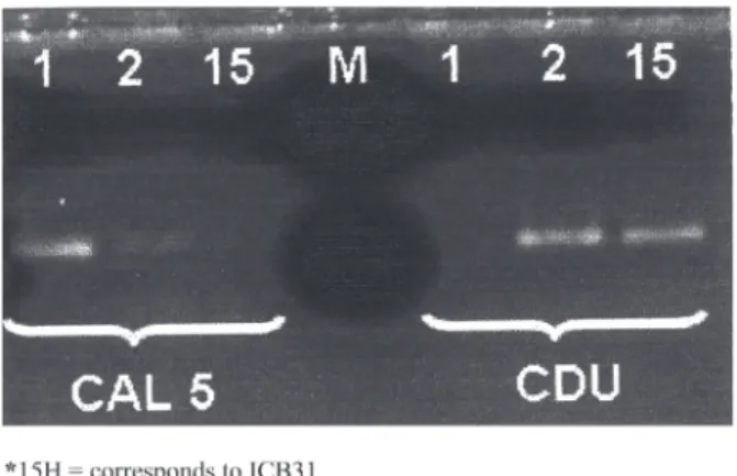
The results obtained in the amplification of the fragments using the primers CAL5 and NL4CAL (C. albicans), and CDU2 and NL4CAL (C. dubliniensis) are shown in Table 1.
The samples numbered from 3 to 24 are from HIV-negative patients, and those numbered from 25 to 39 are from HIV-positive patients. The samples numbered 3, 4, 5, 6, 7, 8, 9, 10, 11, 12, 13, 14, 15, 16, 17, 18, 19, 21, 22, 23, 24, 25, 26, 27, 28, 29, 30, 32, 33, 34, 35, 36, 37, 38 and 39 produced fragments with the primers CAL5 and NL4CAL and were identified as C. albicans. In relation to the primers CDU2 and NL4CAL, only the samples 20 (25 P) and 31 (15 H) produced fragments, being therefore identified as C. dubliniensis (Fig. 1).
Table 1
Results for the samples of yeast used in the PCR reaction with the primers CAL5 and NL4CAL (C. albicans), and CDU2 and NL4CAL (C. dubliniensis)
ICB/USP Results of the amplification Identification by Identification
reference Sample of the fragments the classical method** using PCR
number Primers CAL5 Primers CDU2
and NL4CAL and NL4CAL
01* LSHT 330 + - C. albicans C. albicans
02* ATCC 777 - + C. dubliniensis C. dubliniensis
03 28 P + - C. albicans C. albicans
04 13 P + - C. albicans C. albicans
05 20 P + - C. albicans C. albicans
06 08 P + - C. albicans C. albicans
07 18 P + - C. albicans C. albicans
08 04 P + - C. albicans C. albicans
09 35 P + - C. albicans C. albicans
10 02 P + - C. albicans C. albicans
11 23 P + - C. albicans C. albicans
12 22 P + - C. albicans C. albicans
13 06 P + - C. albicans C. albicans
14 17 P + - C. albicans C. albicans
15 37 P + - C. albicans C. albicans
16 27 P + - C. albicans C. albicans
17 34 P + - C. albicans C. albicans
18 24 P + - C. albicans C. albicans
19 39 P + - C. albicans C. albicans
20 25 P - + C. albicans C. dubliniensis
21 32 P + - C. albicans C. albicans
22 44 P + - C. albicans C. albicans
23 11 P + - C. albicans C. albicans
24 40 P + - C. albicans C. albicans
25 07 H + - C. albicans C. albicans
26 06 H + - C. albicans C. albicans
27 23 PH + - C. albicans C. albicans
28 17 H + - C. albicans C. albicans
29 23 LH + - C. albicans C. albicans
30 26 H + - C. albicans C. albicans
31 15 H - + C. albicans C. dubliniensis
32 01 H + - C. albicans C. albicans
33 14 H + - C. albicans C. albicans
34 09 H + - C. albicans C. albicans
35 13 H + - C. albicans C. albicans
36 03 H + - C. albicans C. albicans
37 21 PH + - C. albicans C. albicans
38 25 H + - C. albicans C. albicans
39 11 H + - C. albicans C. albicans
The size of the fragment amplified by primers CAL5 and NL4CAL, and by primers CDU2 and NA4CAL, being 175 bp, corresponds to those observed by MANNARELLI & KURTZMAN (1998), thus allowing for the identification of the two respective species.
DISCUSSION
C. dubliniensis is a yeast species recently described as an opportunistic pathogen associated with oral candidiasis, particularly in HIV-positive individuals and AIDS patients41.
This species is phenotypically similar to C. albicans, which has resulted in problems in the identification of clinical samples8, as well
as in the reidentification of isolates kept in culture collections and initially identified as C. albicans.
In a retrospective study carried out on a collection of yeast, COLEMAN et al.3 demonstrated that 2% of the isolates originally
identified as C. albicans were actually C. dubliniensis. ODDS et al.27
reidentified2589 cultures in a culture collection initially identified as C. albicans, finding that 2.1% were actually C. dubliniensis. JABRA-RIZK et al.10 foundthat 1.2% of 1251 isolates originally identified as
C. albicans were actually C. dubliniensis. COLOMBO et al.5
investigated the presence of C. dubliniensis among 548 isolates kept in a collection and previously identified as C. albicans, finding that 11 of the isolates were actually C. dubliniensis.
In the present study, it was found that two of 37 samples previously identified as C. albicans were actually C. dubliniensis, for percentage of 5.4%.
In Brazil, C. dubliniensis was isolated for the first time in two AIDS patients in the state of São Paulo. One patient was a 3-year-old child with oropharyngeal candidiasis34 and the other was an adult24.
ALVES et al.1 reported the first three cases of C. dubliniensis isolation
from AIDS patients in the state of Rio Grande do Sul.
According to MARIANO et al.20 in South America the prevalence
of C. dubliniensis isolates appears to be less than that encountered in North America.
The incidence of C. dubliniensis in HIV-positive and AIDS patients observed in Brazil is less than that encountered in Europe and the United States. MILAN et al.25 carried out the first multicenter
prospective study of the oral incidence of C. dubliniensis in Brazilian HIV-positive and AIDS patients. Their study was conducted over a period of two years, at six medical centers around Brazil that provided treatment for HIV-positive patients. Of a total of 155 samples isolated, 2.8% were identified as C. dubliniensis. In a study done in Ireland, it was found that the incidence of C. dubliniensis ranged from 18 to 32% in HIV-infected individuals3,42,while studies conducted in the United
States have reported rates ranging from11.1 to 17.5%14,21.
The isolation of C. dubliniensis in HIV-negative patients has also been reported2,7,12,22,32,43. Recent studies have shown that this species is
more prevalent in HIV-positive individuals, and it is encountered as a commensal organism that can cause various forms of candidiasis12.
We point out, that in the present study,of the two samples identified as
C. dubliniensis, one was isolated from an HIV-negative patient who only suffered from erythematous candidiasis.
In this study, the genotypic differentiation between C. albicans and C. dubliniensis was carried out by means of PCR, which proved to be a useful and practical method yielding an accurate identification, thereby showing that PCR can be an effective tool for elucidating the epidemiology of C. dubliniensis, and for establishing its clinical significance.
RESUMO
Identificação molecular de amostras de Candida dubliniensis
isoladas de lesões orais de pacientes HIV positivos e negativos em São Paulo, Brasil
Candida dubliniensis é uma nova espécie recentemente descrita. Este patógeno oral emergente compartilha muitas características fenotípicas e bioquímicas com C. albicans dificultando assim a diferenciação entre elas. As mesmas, porém, mostram-se genotipicamente distintas. Este trabalho tem como objetivo identificar, pela técnica de PCR (Polymerase Chain Reaction), a possível presença de C. dubliniensis dentre amostras isoladas de candidose oral eritematosa, provenientes de pacientes HIV positivos e HIV negativos. Em um total de 37 amostras identificadas anteriormente, por método clássico, como C. albicans encontramos duas amostras de C. dubliniensis (5,4%) utilizando a técnica do PCR. Esta técnica mostrou-se útil, prática e com identificação taxonômica mais acurada.
ACKNOWLEDGMENTS
Our thanks go to FAPESP, UNIFAL-MG, USP for financial support, and to Dr. John Norman for his revision of the English manuscript.
REFERENCES
1. ALVES, S.H.; MILAN, E.P.; BRANCHINI, M.L.M. et al. - First isolation of Candida dubliniensis in Rio Grande do Sul, Brazil. Diagn. Microbiol. infect. Dis., 39: 165-168, 2001.
2. BRANDT, M.E.; HARRISON, L.H.; PASS, M. et al. - Candida dubliniensis fungemia: the first four cases in North America. Emerg. infect. Dis., 6: 46-49, 2000. 3. COLEMAN, D.C.; SULLIVAN D.J.; BENNETT, D.E. et al. - Candidiasis: the emergence
of a novel species Candida dubliniensis. AIDS,11: 557-567, 1997.
4. COLEMAN, D.C.; SULLIVAN, D.J.; HARRINGTON, B. et al. - Molecular and phenotypic analysis of Candida dubliniensis: a recently identified species linked with oral candidosis in HIV-infected and AIDS patients. Oral Dis., 3(suppl. 1): 96-101, 1997.
5. COLOMBO, A.L.; PERFECT, J.; DINUBILE, M. et al. - Global distribution and outcomes for Candida species causing invasive candidiasis: results from an international randomized double-blind study of caspofungin versus anphotericin B for the treatment of invasive candidiasis. Europ. J. clin. Microbiol. infect. Dis.,22: 470-474, 2003. 6. FISCHER-HOCH, S.P. & HUTWAGNER, L. - Opportunistic candidiasis: an epidemic
of the 1980s. Clin. infect. Dis., 21: 897-904, 1995.
8. GIAMMANCO, G.M.; PIZZO, G.; PECORELLA, S. et al. - Identification of Candida dubliniensis among oral yeast isolates from an Italian population of human immunodeficiency virus-infected (HIV+) subjects. Oral Microbiol. Immunol., 17:
89-94, 2002.
9. JABRA-RIZK, M.A.; BAQUI, A.A.; KELLEY, J.I. et al. - Identification of Candida dubliniensis in a prospective study of patients in the United States. J. clin. Microbiol., 37: 321-326, 1999.
10. JABRA-RIZK, M.A.; FALKLER Jr., W.A.; MERZ, W.G. et al. - Retrospective identification and characterization of Candida dubliniensis isolates among Candida albicans clinical laboratory isolates from human immunodeficiency virus (HIV)-infected and non-HIV-(HIV)-infected individuals. J. clin. Microbiol., 38: 2423-2426, 2000. 11. JOLY, S.; PUJOL, C.; RYSZ, M.; VARGAS, K. & SOLL, D.R. - Development and characterization of complex DNA fingerprinting probes for the infectious yeast Candida dubliniensis. J. clin. Microbiol., 37: 1035-1044, 1999.
12. KANTARCIOGLU, A.S. & YÜCEL, A. - The presence of fluconazole-resistant Candida dubliniensis strains among Candida albicans isolated from immunocompromised or otherwise debilitated HIV negative Turkish patients. Rev. iberoamer. Micol., 19:
44-48, 2002.
13. KHAN, Z.U.; AHMAD, S.; MOKADDAS, E. & CHANDY, R. - Tobacco agar, a new medium for differentiating Candida dubliniensis from Candida albicans. J. clin. Microbiol., 42: 4796-4798, 2004.
14. KIRKPATRICK, W.R.; REVANKAR, S.G.; MCATEE, R.K. et al. - Detection of Candida dubliniensis in oropharyngeal samples from human immunodeficiency virus-infected patients in North America by primary CHROMagar Candida screening and susceptibility testing of isolates. J. clin. Microbiol., 36: 3007-3012, 1998. 15. KURTZMAN, C.P. & FELL, J.W. - The yeasts: a taxonomic study. New York, Elsevier,
1998.
16. KURZAI, O.; HEINZ, W.J.; SULLIVAN, D.J. et al. - Rapid PCR test for discriminating between Candida albicans and Candida dubliniensis isolates using primers derived from the pH-regulated PHR1 and PHR2 genes of C. albicans. J. clin. Microbiol., 37: 1587-1590, 1999.
17. LEES, E. & BARTON, R.C. - The use of niger seed agar to screen for Candida dubliniensis in the clinical microbiology laboratory. Diagn. Microbiol. infect. Dis., 46: 13-17, 2003.
18. MAGEE, B.B.; D´SOUZA, T.M. & MAGEE, P.T. - Strain and species identification by restriction fragment length polymorphisms in the ribosomal DNA repeat of Candida species. J. Bact., 169: 1639-1643, 1987.
19. MANNARELLI, B.M. & KURTZMAN, C.P. - Rapid identification of Candida albicans and other human pathogenic yeasts by using short oligonucleotides in a PCR. J. clin. Microbiol., 36: 1634-1641, 1998.
20. MARIANO, P.L.S.; MILAN, E.P.; MATTA, D.A. & COLOMBO, A.L. - Candida dubliniensis identification in Brazilian yeast stock collection. Mem. Inst. Oswaldo Cruz, 98: 533-538, 2003.
21. MEILLER, T.F.; JABRA-RIZK, M.A.; BAQUI, A. et al. - Oral Candida dubliniensis as a clinically important species in HIV-seropositive patients in the United States. Oral Surg. oral Med. oral Path. oral Radiol. Endod., 88: 573-580, 1999.
22. MEIS, J.F.G.M.; RUHNKE, M.; De PAUW, B.E. et al. - Candida dubliniensis candidemia in patients with chemotherapy-induced neutropenia and bone marrow transplantation.
Emerg. infect. Dis., 5: 150-153, 1999.
23. MEYER, W.; MASZEWSKA, K. & SORREL, T.C. - PCR fingerprinting: a convenient molecular tool to distinguish between Candida dubliniensis and Candida albicans.
Med. Mycol., 39: 185-193, 2001.
24. MILAN, E.P.; COLOMBO, A.L.; LAET SANT’ANA, P.; LEWI, D.S. & MELO, A.S.A. - Primeiro isolamento de Candida dubliniensis no Brasil: In: CONGRESSO BRASILEIRO DE INFECTOLOGIA, 11., São Paulo, 1999. Resumos. p. 32. 25. MILAN, E.P.; LAET SANT’ANA, P.; MELO, A.S.A. et al. - Multicenter prospective
surveillance of oral Candida dubliniensis among adult Brazilian human immunodeficiency virus-positive and AIDS patients. Diagn. Microbiol. infect. Dis., 41: 29-35, 2001.
26. MORAN, G.P.; SULLIVAN, D.J.; HENMAN, M.C. et al. - Antifungal drug susceptibilities of oral Candida dubliniensis isolates from human immunodeficiency virus (HIV) -infected and non-HIV--infected subjects and generation of stable fluconazole-resistant derivatives in vitro. Antimicrob. Agents Chemother., 41: 617-623, 1997. 27. ODDS, F.C.; VAN NUFFEL, L. & DAMS, G.- Prevalence of Candida dubliniensis isolates
in a yeast stock collection. J. clin. Microbiol., 36: 2869-2873, 1998.
28. PARK, S.; WONG, M.; MARRAS, S.A.E. et al. - Rapid identification of Candida dubliniensis using a species-specific molecular beacon. J. clin. Microbiol., 38: 2829-2836, 2000.
29. PFALLER, M.A.; HOLLIS, R.J.; SADER, H.S. et al. - Chromosomal restriction fragment analysis by pulsed-field gel electrophoresis. In: INSEBERG, H.D. Clinical Microbiology procedures handbook. Washington, American Society for Microbiology, 1994. p. 200.
30. PFALLER, M.A.; MESSER, S.A.; BOLMSTRÖM, F.C.; ODDS, F.C. & REX, J.H. -Multisite reproducibility of the Etest MIC method for antifungal susceptibility testing of yeast isolates. J. clin. Microbiol., 34: 1691-1693, 1996.
31. PINJON, E.; SULLIVAN, D.; SALKIN, I.; SHANLEY, D. & COLEMAN, D. - Simple, inexpensive, reliable method for differentiation of Candida dubliniensis from Candida albicans.J. clin. Microbiol.,36: 2093-2095, 1998.
32. POLACHECK, I.; STRAHILEVITZ, J.; SULLIVAN, D. et al. - Recovery of Candida dubliniensis from non-human immunodefficiency virus-infected patients in Israel.
J. clin. Microbiol., 38: 170-174, 2000.
33. SALKIN, I.F.; PRUITT, W.R.; PADHYE, A.A. et al. - Distinctive carbohydrate assimilation profiles used to identify the first clinical isolates of Candida dubliniensis recovered in the United States. J. clin. Microbiol., 36: 1467, 1998.
34. SANO, A.; VILELA, M.M.; TAKAHASHI, I. et al. - Isolation of Candida dubliniensis from the oral cavity of an HIV-positive child in Brazil. Nippon Ishinkin Gakkai Zasshi, 41: 177-181, 2000.
35. SCHOOFS, A.; ODDS, F.C.; COLEBUNDERS, R.; IEVEN, M. & GOOSSENS, H. -Use of a specialised isolation media for recognition and identification of Candida dubliniensis isolates from HIV-infected patients. Europ. J. clin. Microbiol. Dis., 16: 296-300, 1997.
36. SCHORLING, S.R.; KORTINGA, H.C.; FROSCHB, M. & MÜHLSCHLEGEL, F.A. -The role of Candida dubliniensis in oral candidiasis in human immunodeficiency virus-infected individuals. Crit. Rev. Microbiol., 26: 59-68, 2000.
37. STAIB, P. & MORSCHHAUSER, J. - Chlamydospore formation on Staib agar as a species-specific characteristics of Candida dubliniensis. Mycoses,42: 521-524, 1999. 38. SMITH, R.A.; HITCHCOCK, C.A.; EVANS, E.G.V.; LACEY, C.J. & ADAMS, D.J.
-The identification of Candida albicans strains by restriction fragment length polymorphism analysis of DNA. J. med. vet. Mycol., 27: 431-434, 1989. 39. SULLIVAN, D.J.; WESTERNENG, T.J.; HAYNES, K.A; BENNET, D.E. & COLEMAN,
D.C. - Candida dubliniensis sp. nov.: phenotypic and molecular characterization of a novel species associated with oral candidosis in HIV-infected individuals.
Microbiology, 141: 1507-1521, 1995.
41. SULLIVAN, D. & COLEMAN, D. - Candida dubliniensis: characteristics and identification. J. clin. Microbiol., 36: 329-334, 1998.
42. SULLIVAN, D.J.; MORAN, G.; DONNELLY, S. et al. - Candida dubliniensis: an update.
Rev. iberoamer. Micol., 16: 72-76, 1999.
43. WILLIS, A.M.; COULTER, W.A.; SULLIVAN, D.J. et al. - Isolation of Candida dubliniensis from insulin-using diabetes mellitus patients. J. oral Path. Med., 29: