MYOCYTE-SPECIFIC ENHANCER BINDING FACTOR
2C (MEF2C) EXPRESSION IN THE DENTATE
GYRUS DURING DEVELOPMENT AND AFTER
PILOCARPINE-INDUCED
STATUS EPILEPTICUS
A preliminary report
Carla Alessandra Scorza
1, Esper Abrão Cavalheiro
1, Fulvio Alexandre Scorza
1,2Abstract – Objective: As axon outgrowth and dentate granule cell neurogenesis are hallmarks of hippocampal development and are also the two morphologic changes in the structure of the dentate gyrus after status epilepticus (SE), we hypothesized that molecules involved in normal development may also play a role during epileptogenesis. Method: Using in situ hybridization, we have characterized mRNA expression of myocyte-specific enhancer binding factor 2C (MEF2C) in the dentate gyrus during development (P0, P3, P7, P14 and P28) and at multiple time points following pilocarpine-induced SE (3, 7, 14, 28 days after SE). Results: It was demonstrated that MEF2C is up-regulated during development (P0, P3, P7, P14 and P28) and in the adult rat dentate gyrus following SE (3, 7, 14, 28 days after SE). Conclusions: The molecules controlling cell-fate decisions in the developing dentate gyrus are also operative during epileptogenesis.
KEY WORDS: rat, hippocampus, epilepsy, status epilepticus, MEF2C.
Realce miócito específico ligado a expressão do fator 2C (MEF2C) no giro denteado durante o desenvolvimento e após uso de pilocarpina induzindo status epilepticus: estudo preliminar
Resumo – Objetivo: Como o crescimento axonal e a neurogênese do giro denteado são características intrínsecas do hipocampo durante o processo de desenvolvimento, e também são duas alterações morfológicas na estrutura do giro denteado após o status epilepticus (SE), nós hipotetizamos que as moléculas envolvidas no processo normal do desenvolvimento hipocampal também podem participar do processo de epileptogênese. Método:
Utilizando hibridização in situ, caracterizamos a expressão do RNAm do fator de transcrição myocyte-specific enhancer binding factor 2C (MEF2C) no giro denteado durante o desenvolvimento (P0, P3, P7, P14 e P28) e em diferentes períodos após o SE (3, 7, 14, 28 dias após SE). Resultados: Foi demonstrado um aumento da expressão de MEF2C no giro denteado durante o desenvolvimento e no giro denteado de animais adultos após o SE. Conclusão: As moléculas que controlam o destino celular durante o processo de desenvolvimento também estão operativas durante o processo de epileptogênese.
PALAVRAS-CHAVE: rato, hipocampo, epilepsia, status epilepticus, MEF2C.
1Disciplina de Neurologia Experimental, Universidade Federal de São Paulo/Escola Paulista de Medicina, São Paulo SP, Brasil; 2Program in Brain Plasticity and Epilepsy, Harvard Medical School and Department of Neurology, Beth Israel Deaconess Medical Center, Boston, MA, USA.
Received 28 March 2008, received in inal form 6 June 2008. Accepted 12 July 2008.
Dr. Fulvio Alexandre Scorza – Disciplina de Neurologia Experimental / UNIFESP/EPM - Rua Botucatu 862 - 04023-900 São Paulo SP - Brasil. E-mail: [email protected]
Epilepsy is the commonest serious neurological condi-tion. Temporal lobe epilepsy (TLE), the most common epi-lepsy in adults, is generally intractable and is suspected to be the result of recurrent excitation or inhibition circuitry1. TLE is deined by recurring partial complex seizures aris-ing from limbic structures of the temporal lobes, includ-ing the hippocampus, dentate gyrus, amygdala and
periods: a) an acute period that builds progressively into limbic status epilepticus (SE) and that lasts 24h; b) a silent period with progressive normalization of EEG and behav-iour which varies from 4 to 44 days; and c) a chronic peri-od with spontaneous recurrent seizures2. From a morpho-logical point of view, seizure-induced injury leads to vari-ous forms of plasticity in the adult rodent dentate gyrus3. While these forms of neuroplasticity during epileptogene-sis have been well documented, the molecular mechanisms underlying these alterations remain poorly understood.
During the last decade, a number of transcription fac-tors have been shown to be involved in the cellular and molecular organization of the brain. The myocyte-specif-ic enhancer binding factor 2C (MEF2C), a member of the MEF2 sub-family of the MADS gene family, is a transcrip-tion factor expressed at high levels in the brain and has been implicated in the development of neurons4. MEF2C binds with basic helix-loop-helix (bHLH) proteins5, which in the brain are responsible for neural determination or differentiation6. There is more deinitive evidence for in-teractions among MEF2 with bHLH factors. For example, multiple members of bHLH family of transcription fac-tors have been shown to be expressed in dentate gran-ule cells at specific developmental stages7, supporting their involvement in regulating dentate gyrus develop-ment. Interestingly, Elliott and colleagues8 demonstrated that the expression of bHLH molecules is altered during seizure-associated network reorganization. Speciically, they showed varied proiles of expression of bHLH fam-ily members following SE, with some increasing (Mash1, Id2), some decreasing (Hes5, Prox1), and others remaining unchanged NeuroD/BETA2, NeuroD2/NDRF, Id3, Rath2/ Nex1, supporting the idea that molecules controlling cell-fate decisions in the developing dentate gyrus are also op-erative during seizure-induced plasticity.
As MEF2C has been implicated to play an important role in the process of neuronal differentiation, we pur-sued the hypothesis that MEF2C is a potential upstream regulator of bHLH expression that is modiied following SE. To do so, using in situ hybridization, we have charac-terized mRNA expression of MEF2C in the dentate gyrus during development and at multiple time points follow-ing pilocarpine-induced SE.
METHOD
Animals
For adult animals (n=40), two groups of Sprague-Dawley rats (180-200g) were studied: (A) Adult male rats at different time points following pilocarpine-induced status epilepticus (SE). It is important to note that for each time point (3, 7, 14, 28 days af-ter SE) we used n=8 animals, (B) Control group, adult male rats (n=8), that received saline instead pilocarpine. For postnatal ag-es, subjects were male Sprague-Dawley rats (n=25) with ages P0,
P3, P7, P14 and P28 days and for each group we used n=5 animals. The rats were bred in our laboratories and the day of birth was considered as day 0. The pups were housed with their mother in individual cages until weaning at day 21. All the animals used in our study were housed under standard controlled conditions (7:00 AM/7:00 P.M. light/dark cycle; 20-22ºC; 45–55% humidi-ty) with food and water ad libitum.
Induction of SE
All animals were treated according to protocols for animal care established by the Harvard Medical School, Boston and the National Institutes of Health, and all efforts were made to min-imize animal suffering. Adult male Sprague-Dawley rats (180– 200g) were given an intraperitoneal (i.p.) injection of atropine methylbromide (5 mg/kg; Sigma, St. Louis, MO, USA) followed 20 minutes later by an i.p. injection of pilocarpine hydrochloride (340 mg/kg; Sigma) to induce SE. Seizure activity was monitored behaviourally and terminated with an i.p. injection of diazepam (10 mg/kg; Elkins-Sinn, Cherry Hill, NJ, USA) after 2 hr of convul-sive SE. Only rats that displayed continuous, convulconvul-sive seizure activity after pilocarpine treatment were included in these stud-ies. Control animals received the same injections of atropine and diazepam, but received saline instead of pilocarpine.
Tissue preparation
At designated time points following SE, all animals received an anesthetic overdose of pentobarbital and transcardially per-fused with 300 mL of a 4% paraformaldehyde solution in phos-phate-buffered saline at pH 7.4. Brains were post-ixed in situ
overnight at 4ºC and then removed and cryoprotected in 30% sucrose in PBS prior to freezing. To generate anatomically com-parable sections for the in situ hybridization analysis, 20 µm frozen coronal sections were cut through the middle third of the hippocampus with a cryostat, melted onto Superfrost Plus slides (Fisher Scientiic, Pittsburgh, PA, USA), and stored at –20ºC for subsequent in situ analysis of mRNA. Potential variations in mRNA expression along the septo/temporal axis of the hippo-campus were not assessed in this study.
Non-radioactive in situ hybridization
Non-radioactive in situ hybridization was performed essen-tially as described previously7 using a protocol obtained from
incubat-ed in buffer containing nitrobluetetrazolium (NBT) and 5-bromo-4-chloro-3-indoyl phosphate (BCIP) (Roche Molecular Biochem-icals). In situ data were evaluated qualitatively, based on the comparison of mRNA expression patterns in comparable hippo-campal sections. These sections were taken from multiple sets of animals processed for in situ hybridization on different days.
Non-radioactive in situ probe preparation
The MEF2C probe used for this study, along with the optimal
in situ hybridization temperatures and template sources, were described previously9. Probe templates generated by PCR were
derived from cDNA adult rat. Antisense and sense digoxigenin-labeled RNA probes were transcribed from each template
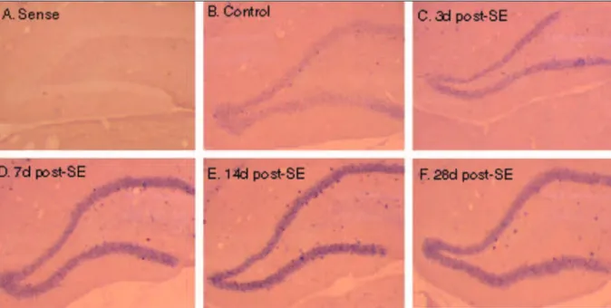
us-Fig 1. Expression MEF2C mRNA in dentate gyrus. (A) In situ hybridization of 14 days post-SE tissue with MEF2C sense riboprobe shows the representative lack of signal and minimal background staining. (B) In situ hybridization of control tissue with ME-F2C antisense riboprobe demonstrates the expression of MEME-F2C troughout the dentate granule cell layer. (C) At 3 days fol-lowing SE, the MEF2C expression appears to be slightly elevated as compared to the control group. The expression of MEF2C troughout the dentate granular cell layer increases acutely within 7 days (D) and 14 days (E) following SE, remaining elevat-ed until 28 days post – SE (F).
ing the Genius RNA labeling kit (Roche Molecular Biochemicals) and puriied with Chromaspin-100 columns packed in DEPC wa-ter (Clontech Laboratories, Palo Alto, CA, USA).
RESULTS
Pilocarpine treatment induced the following sequence of behavioral changes: akinesia, facial automatisms, and limbic seizures consisting of forelimb clonus with rearing, salivation, and masticatory jaw movements and falling. This type of behaviour built up progressively into motor limbic seizures that recurred repeatedly and rapidly and developed into status epilepticus. After SE, animals were unresponsive to their environment and akinetic; behavior returned toward normal over a 3 to 5-day period2.
In situ analysis of MEF2C mRNAs at various times fol-lowing SE course indicates that this molecule is expressed during epileptogenesis. In situ hybridization of control tis-sue with MEF2C antisense riboprobe demonstrates expres-sion of MEF2C throughout the dentate granule cell layer (Fig 1B). The speciicity of the probe was documented using an MEF2C sense probe, with which no signal and minimal background staining were observed (Fig 1A). In addition, at 3 days following SE the MEF2C expression in adult dentate gyrus appears to be slightly elevated as compared to the control group (Fig 1C). The expression of MEF2C mRNA throughout the dentate granule cell layer increased acute-ly within 7 days (Figure 1D) and 14 days (Fig 1E) following SE, and remained elevated until 28 days post-SE (Fig 1F).
In situ analysis of MEF2C during development shows that MEF2C is expressed in a speciic pattern in the den-tate gyrus (Fig 2A). In situ hybridization of P0 tissue with MEF2C antisense riboprobe demonstrates expression of MEF2C in immature granule cells populations (Fig 2B). At P3 and P7 (Figs 2C and 2D respectively), MEF2C is distribut-ed throughout the developing dentate granule cell layer. MEF2C is expressed at highest levels in the granule cell lay-er at P14 (Fig 2E) and P28 (Fig 2F), when the entire innlay-er half of the granule cell layer has only recently been generated.
DISCUSSION
It has been known that MEF2C is expressed in a dy-namic pattern during development and in the mature mouse CNS, suggesting that the molecule may play an important role at different stages of neuronal differen-tiation and/or maturation4,10. Here it was demonstrated that MEF2C is up-regulated during development and in the adult rat dentate gyrus following SE, suggesting that MEF2C may also be involved in seizure-induced plasticity. Interestingly, the results in this report are not in agree-ment with the recent paper of Yoon and colleagues11. In their study, the authors have shown that MEF2C phos-phorylation was reduced immediately after
electrocon-vulsive shock. The discrepancies in these MEF2C activa-tion patterns may relect different epilepsy models and time points analyzed.
During development, four MEF2 genes (A, B, C, D) are expressed in spatially and temporally speciic patterns during brain development, suggesting that, similar to its expression pattern in striated muscle, MEF2 gene expres-sion in the CNS occurs in neurons exiting the cell cycle and entering differentiation10. In addition, human studies using immunocytochemical localization of MEF2C in hip-pocampal surgical specimens revealed immunoreactivity in nuclei of dentate granule cells and the hilus of dentate gyrus, supporting the concept of a role for MEF2C in post-mitotic neuronal differentiation4. Given that, the results of this study also showed that MEF2C is expressed in den-tate granule cells during speciic developmental stages, it seems quite possible that MEF2C is involved is in regulat-ing dentate gyrus development.
colleagues17 recently described antiapoptotic functions for MEF2C. However, in mature neurons exposed to ex-citotoxic injury or others forms of stress, an apoptotic effect18 mediated by caspases were demonstrated19. With these results, it is possible to support the hypothesis that MEF2C is involved with the mechanism of apoptotic de-generation following pilocarpine induced-SE.
Finally, the present results support previous evidence that MEF2C is expressed in areas of ongoing neural plas-ticity and raise, for the irst time, the possibility of a po-tential role for MEF2C molecules during epileptogenesis. Nevertheless, there are some limitations to this study. Firstly, the present data is only from in situ hybridization. A description of quantiication of MEF2C mRNA expres-sion in the dentate gyrus should be carried out. Secondly, to conirm the roles of MEF2C in epileptogenesis, an imu-nohistochemistry study should be realized to investigate the possible changes of MEF2C protein expression in the dentate gyrus. These future studies, which are in evalu-ation in our laboratory, are needed to gain a better un-derstanding of these and other possible mechanisms of MEF2C transcription factor during epileptogenesis.
ACknOwledgeMents – The authors would like to thank Dr. Dan-iel H. Lowenstein and Dr. Robert C. Elliott for their suggestions and helpful review of this manuscript and to Mr. Brian Kruegel for his help with technical procedures.
REFERENCES
1. Sutula TP. Epilepsy: a reappraisal from the perspective of neural plas-ticity. Int Rev Neurobiol 2001;45:355-386.
2. Cavalheiro EA. The pilocarpine model of epilepsy. Ital J Neurol Sci 1995; 16:33-37.
3. Lowenstein DH. Structural reorganization of hippocampal networks caused by seizure activity. Int Rev Neurobiol 2001;45:209-236. 4. Leifer D, Li Y, Wehr K. MEF2C expression in fetal mouse brain
devel-opment. J Mol Neurosci 1997;8:1-13.
5. Molkentin J, Olson EN. Combinatorial control of muscle development by basic helix-loop-helix and MADS-box transcription factors. Proc Natl Acad Sci 1996;93:9366-9373.
6. Schwab MH, Bartholomae A, Heimrich B, et al. Neuronal basic he-lix-loop-helix proteins (NEX and BETA2/Neuro D) regulate terminal granule cell differentiation in the hippocampus. J Neurosci 2000;20: 3714-3724.
7. Pleasure SJ, Collins AE, Lowenstein DH. Unique expression patterns of cell fate molecules delineate sequential stages of dentate gyrus de-velopment. J Neurosci 2000;20:6095-6105.
8. Elliott RC, Khademi S, Pleasure SJ, Parent JM, Lowenstein DH. Differ-ential regulation of basic helix-loop-helix mRNSa in the dentate gyrus following status epilepticus. Neuroscience 2001;81:10679-10688. 9. Edmondson DG, Lyons GE, Martin J, Olson E. MEF2 gene expression
markers the cardiac and skeletal muscle lineages during mouse em-bryogenesis. Development 1994;20:1251-1263.
10. Leifer D, Kranic D, Yu YT. MADS/MEF2-family transcription factor expressed in a laminar distribution in cerebral cortex. PNAS 1993;90: 1546-1550.
11. Yoon SC, Ahn YM, Jun SJ, et al.. Region-speciic phosphorylation of ERK5-MEF2C in the rat frontal cortex and hippocampus after electro-convulsive shock. Prog Neuropsychopharmacol Biol Psychiatry 2005; 29:749-753.
12. Kuhn HG, Dickinson-Anson H, Gage FH. Neurogenesis in the dentate gyrus of the adult rat: age-related decrease of neuronal progenitor pro-liferation. J Neurosci 1996;16:2027-2033.
13. Black BL, Ligon KL, Zhang Y, Olson EN. Cooperative transcriptional activation by the neurogenic basic helix-loop-helix protein MASH1 and members of the myocyte enhancer factor-2 (MEF2) family. J Biol Chem 1996;271:26659-26663.
14. Sloviter RS, Dean E, Sollas AL, Goodman JH. Apoptosis and necrosis in-duced in different hippocampal neuron populations by repetitive per-forant path stimulation in the rat. J Comp Neurol 1996;36:6516-6533. 15. Simonian NA, Getz RL, Leveque JC, Konradi C, Coyle JT. Kainate
in-duces apoptosis in neurons. Neuroscience 1996;74:675-683.
16. Ekdahl CT, Mohapel P, Elmer E, Lindvall O. Caspase inhibitors increase short-term survival of progenitor-cell progeny in the adult rat dentate gyrus following status epilepticus. Eur J Neurosci 2001 4:937-945. 17. Okamoto S, Krainc D, Sherman K, Lipton SA. Antiapoptotic role of the
p38 mitogen-activated protein kinase-myocite enhancer factor 2 tran-scription factor pathway during neuronal differentiation. PNAS 2000; 99:7561-7566.
18. Kikuchi M, Tenneti L, Lipton SA. Role of p38 mitogen-activated pro-tein kinase in axotomy-induced apoptosis of rat retinal ganglion cells. J Neurosci 2000;20:5037-5044.