Scriptaid Treatment Decreases DNA
Methyltransferase 1 Expression by Induction
of MicroRNA-152 Expression in Porcine
Somatic Cell Nuclear Transfer Embryos
Shuang Liang1,2, Ming-Hui Zhao1,2, Jeong-woo Choi1,2, Nam-Hyung Kim1,2*, Xiang-Shun Cui1,2*
1Department of Animal Science, Chungbuk National University, Cheongju, Chungbuk, 361–763, Republic
of Korea,2Brain Korea 21 center for Bio-Resource Development, Cheongju, Chungbuk, 361–763, Republic
of Korea
*[email protected](NHK);[email protected](XSC)
Abstract
Abnormal epigenetic reprogramming of donor nuclei after somatic cell nuclear transfer (SCNT) is thought to be the main cause of low cloning efficiencies. A growing body of evi-dence has demonstrated a positive role of Scriptaid, a histone deacetylase inhibitor (HDACi) that belongs to an existing class of hydroxamic acid-containing HDACis, on the development competence of cloned embryos in many species. The present study investi-gated the effects of Scriptaid on the development of porcine SCNT embryosin vitroand its mechanism. Treatment with 300 or 500 nM Scriptaid for 20 h after activation significantly increased the percentage of SCNT embryos that developed to the blastocyst stage and the total number of cells per blastocyst and significantly decreased the percentage of apo-ptotic cells in blastocysts. Scriptaid treatment significantly increased the level of histone H3 acetylated at K9 and the conversion of 5-methylcytosine into 5-hydroxymethylcytosine and significantly decreased the level of histone H3 trimethylated at K9 at the pronuclear stage. As a potential mechanism for the DNA methylation changes, our results showed that the expression of DNA methyltransferase 1 was frequently down-regulated in Scriptaid-treated embryos in comparison with untreated embryos and was inversely correlated to endoge-nous microRNA-152 (miR-152). Taken together, these findings illustrated a crucial func-tional crosstalk between miR-152 and DNMT1. Meanwhile, mRNA and protein levels of POU5F1 and CDX2 were increased in Scriptaid-treated embryos. mRNA levels of Cas-pase3, andBaxwere significantly decreased and that ofBcl-xLwas significantly increased in Scriptaid-treated embryos. In conclusion, these observations would contribute to uncover the nuclear reprogramming mechanisms underlying the effects of Scriptaid on the improve-ment of porcine SCNT embryos.
OPEN ACCESS
Citation:Liang S, Zhao M-H, Choi J-w, Kim N-H, Cui X-S (2015) Scriptaid Treatment Decreases DNA Methyltransferase 1 Expression by Induction of MicroRNA-152 Expression in Porcine Somatic Cell Nuclear Transfer Embryos. PLoS ONE 10(8): e0134567. doi:10.1371/journal.pone.0134567
Editor:Qing-Yuan Sun, Institute of Zoology, Chinese Academy of Sciences, CHINA
Received:March 28, 2015
Accepted:July 12, 2015
Published:August 11, 2015
Copyright:© 2015 Liang et al. This is an open access article distributed under the terms of the Creative Commons Attribution License, which permits unrestricted use, distribution, and reproduction in any medium, provided the original author and source are credited.
Data Availability Statement:All relevant data are within the paper and its Supporting Information files.
Funding:This work was supported by Next-Generation BioGreen 21 Program (PJ011126 and PJ01104103), Rural Development Administration, Republic of Korea.
Introduction
Although several mammalian species have been successfully cloned using somatic cell nuclear transfer (SCNT) technology [1–3], its success rate remains extremely low, especially in pigs [4]. This low efficiency is mostly attributed to defective epigenetic reprogramming, including geno-mic methylation. Histone tail modifications may result in abnormal epigenetic modification and gene expression in SCNT embryos [5–8]. Hence, epigenetic modifications might be key to improving the success of cloning. Recent studies showed that specific histone deacetylase inhibitors (HDACis) enhance somatic cell genomic reprogramming and repair epigenetic abnormalities, such as acetylation of histone H3 at K9 (H3-acK9)[9].
HDACis induce specific changes in gene expression and influence a variety of processes, including growth arrest, differentiation, cytotoxicity, and induction of apoptosis [10]. HDACis may also damage embryos [11]. Thus, avoidance of the side-effects of HDACi treatment is probably important to improve the cloning efficiency and to modify aberrant genomic reprogramming.
Histone deacetylases (HDACs) are divided into five categories: class I (HDAC 1–3 and 8), class IIa (HDAC 4, 5, 7, and 9), class IIb (HDAC 6 and 10), class III (SIRT 1–7), and class IV (HDAC 11) [12]. Scriptaid can inhibit classes I and IIa/b HDACs [12,13], belongs to an exist-ing class of hydroxamic acid-containexist-ing HDACis, is less toxic than other HDACis, and causes increases in the global acetylation of histones, transcriptional activity, and protein expression [14].
HDACs and microRNAs (miRNAs) have a complex relationship that is not fully under-stood, but may be of critical importance. miRNAs can regulate HDACs and influence histone acetylation, and HDACs can regulate miRNA expression. Thus, a careful balance between HDACs and miRNAs is important to maintain their appropriate levels in the cell [15]. HDACis can alter the expression profiles of miRNAs in some cells [16,17]. In our previous study, aber-rant epigenetic reprogramming of imprinted mir-127 was observed in cloned mouse embryos [18]. Thus, understanding of how HDACs and miRNAs influence each other and affect biolog-ical pathways is of great interest.
Scriptaid treatment significantly improves the development of cloned pig embryosin vitro
andin vivoand restores the correct expression of specific aberrantly expressed genes [19]. Fur-thermore, Scriptaid treatment of porcine SCNT embryos improves the cloning efficiency and nuclear reprogramming of inbred miniature pigs [19–21]. Similar results were obtained with embryos of several mammalian species [22,23]. These data indicate that Scriptaid is a promis-ing candidate to improve thein vitrodevelopment of porcine embryos; however, the mecha-nism underlying its effects needs to be elucidated.
In general, embryos producedin vitrocontain fewer cells and have a higher apoptotic index than embryos producedin vivo[24]; this is particularly true of cloned embryos [25]. An increased apoptotic rate might contribute to embryo mortality or fetal anomalies that can trig-ger early abortions [26]. Thus, further studies are required to evaluate factors that affect the quality of SCNT embryos as well as apoptosis in these embryos.
This study investigated the mechanism by which treatment of Scriptaid on the reprogram-ming of somatic nuclei following SCNT and on the in vitro development of SCNT embryos.
Materials and Methods
this study were purchased from Sigma Chemical Company (Sigma, St. Louis, MO, USA), unless otherwise indicated. Scriptaid was dissolved in dimethyl sulfoxide, prepared as a 2000× stock solution of 500μM, aliquoted, and stored in the dark at−20°C. The stock solution was
added to porcine zygote medium (PZM)-5 culture media at various concentrations using serial dilution.
Collection of porcine oocytes and
in vitro
maturation
Ovaries from pre-pubertal gilts were collected from a local slaughterhouse (Farm story dodarm B&F, Umsung, Chungbuk, Korea) and transported to the laboratory at 37°C in saline supple-mented with 75 mg/ml penicillin G and 50 mg/ml streptomycin sulfate. Follicles that were 3–6 mm in diameter were aspirated. Cumulus-oocyte complexes (COCs) that were surrounded by a minimum of three cumulus cells were selected for culture [27]. In brief, the COCs were washed three times in TL-HEPES supplemented with 0.1% polyvinyl alcohol (PVA, w/v) and 0.05 g/L gentamycin. Then the COCs were washed three times in maturation medium (TCM-199 supplemented with 0.1 g/L sodium pyruvate, 0.6 mM L-cysteine, 10 ng/mL epidermal growth factor, 10% porcine follicular fluid, 10 IU/mL luteinizing hormone, and 10 IU/mL folli-cle-stimulating hormone) and were then transferred to maturation medium. Maturation was performed by culturing approximately 50 COCs in 500μL of maturation medium in 4-well
dishes. The medium was covered with mineral oil and the plates were incubated at 38.5°C in a humidified atmosphere of 5% CO2for 44 h. The pH and osmolality of the maturation medium were 7.35 and 290 mmol/kg, respectively.
SCNT procedure
After 44 h ofin vitromaturation, cumulus cells were removed from the oocyte by gentle pipett-ing in TL-HEPES supplemented with 1 mg/mL hyaluronidase and 0.1% PVA, collected in a 1.5-ml Eppendorf tube, washed by centrifugation, maintained at 4°C, and used as donor cells.
For enucleation, only oocytes with an excellent morphology and that had extruded the first polar body, were used for SCNT. Denuded oocytes were incubated for 5 min in manipulation medium (calcium-free TL-HEPES supplemented with 0.1% PVA) containing 5μg/mL Hoechst
33342, washed twice with fresh manipulation medium, and transferred to a drop of manipula-tion medium containing 5μg/mL cytochalasin B (CB). Oocytes were enucleated by aspirating
the polar body and MII chromosomes in a small amount (<15% of the oocyte volume) of
cyto-plasm using a 25-μm beveled glass pipette (Humagen, Charlottesville, VA, USA).
After enucleation using a fine injecting pipette, a single donor cell was inserted into the peri-vitelline space of the enucleated oocyte. Donor cell-oocyte complexes were equilibrated with 280 mM mannitol solution (pH 7.2) containing 0.15 mM MgSO4, 0.01% PVA (w/v), and 0.5 mM HEPES for 2–3 min and transferred to a fusion chamber containing two electrodes overlaid with 280 mM mannitol solution. Membrane fusion was induced by applying an alter-nating current field of 2 V cycling at 1 MHz for 2 sec, followed by a 20-μsec direct current
(DC) pulse at 2 kV/cm using a cell fusion generator (LF201; Nepa Gene, Chiba, Japan). Follow-ing fusion, the reconstructed embryos were placed in bicarbonate-buffered PZM-5 containFollow-ing 0.4 mg/mL bovine serum albumin (BSA) for 1 h prior to activation.
Activation and in vitro culture
Reconstructed embryos were activated by two DC pulses of 120 V for 60μsec in 297 mM
pseudo-second polar body. Following culture, the reconstructed embryos were thoroughly washed and cultured in bicarbonate-buffered PZM-5 supplemented with 0.4 mg/mL BSA in 4-well dishes for 7 days at 38.5°C in 5% CO2in air without changing the medium. The develop-ment of the reconstructed embryos into blastocysts was examined on day 7 after activation.
Real-time reverse transcription-polymerase chain reaction (RT-PCR)
with SYBR green for mRNA analysis
mRNAs from SCNT embryos were isolated using a Dynabeads mRNA Direct Kit (Dynal Asa, Oslo, Norway), according to the manufacturer’s instruction. First-strand cDNA was synthe-sized by RT of mRNA using the Oligo(dT)12-18primer and SuperScript TM III Reverse Tran-scriptase (Invitrogen Co., Grand Island, NY). time RT-PCR using the CFX96 Touch Real-time RT-PCR Detection System (Bio-Rad) was performed in a final reaction volume of 20μL
with SYBR Green, a fluorophore that binds all double-strand DNA. The PCR conditions were as follows: 5 min at 95°C followed by 45 cycles of 10 sec at 95°C, 10 sec at 60°C, and 15 sec at 72°C. Finally, gene expression was quantified using the 2-ddCt method, with normalization to the mRNA expression of porcine ribosomal protein L19 (Rpl19). The primers used to amplify each gene are listed inTable 1. Each experiment was repeated at least three times, with five embryos per repeat.
Real-time RT-PCR with TaqMan for microRNA analysis
All primers, including those used for specific miRNA and cDNA synthesis and PCR amplifica-tion, and the kit used for miRNA analyses were purchased from Applied Biosystems (Bedford, MA, USA). For relative quantification of the expression of mir-29b, mir-148a, mir-152, and U6 snoRNA, RT reactions were set up containing 5μL RNA, 3μL stem-loop RT primer, 1× RT
buffer, 0.25 mM of each dNTP, 3.33 U/mL MultiScribe reverse transcriptase, and 0.25 U/mL RNase inhibitor. The 15-μL reactions were incubated for 30 min at 16°C, followed by 30 min at
Table 1. Primers used for real-time RT-PCR.
Gene Primer sequences (5'-3') Annealing temperature (°C) Product size (bp)
Bcl-xL F: CTTACCTGAATGACCACCTAGAGC 60 182
R: CCGACTGAAGAGCGAACCC
Bax F: CGGGACACGGAGGAGGTTT 60 189
R: CGAGTCGTATCGTCGGTTG
Casp3 F: ACTGTGGGATTGAGACGG 55 110
R: GGAATAGTAACGAGGTGCTG
Dnmt1 F: GGCAGACCACCATCACATC 55 165
R: GGAGCAGTCCGGCAACT
Dnmt3a F: GGACAAGAATGCCACCAAATCA 60 196
R: CTTGCCGTCTCCGAACCA
Dnmt3b F: GGGTGGAAAGACACGGGAT 60 243
R: TAGGAGCGTAGAAGCAAGGAA
Pou5f1 F:GCTCACTTTGGGGGTTCTCT 60 228
R:TTGCCTCTACTCGGTTCTC
Cdx2 F: GCAAAGGAAAGGAAAATCAACAA 60 120
R: GGGCTCTGGGACGCTTCT
Rpl19 F: GCTTGCCTCCAGTGTCCTC 60 179
R: GGCGTTGGCGATTTCAT
42°C and 5 min at 85°C, and were then held at 4°C to convert miRNA into cDNA. Real-time RT-PCR was performed in 20-μL reactions that included 4μL RT product, 1× TaqMan
Universal PCR Master Mix, and 1μL of 20× real-time solution containing TaqMan probe and
primers. The amplification parameters used for real-time RT-PCR were as set out in the manu-facturer’s protocol. miRNA expression was quantified using the same method as that described for mRNA expression, with U6 snoRNA used as an internal control. Each experiment was repeated at least three times, with five embryos per repeat.
Immunostaining and quantification of fluorescence intensity
Scriptaid-treated and non-treated embryos were collected at the pronuclear stage. Embryos were fixed for 30 min in 3.7% (w/v) paraformaldehyde, permeabilized for 1 hour with 0.2% Triton X-100 prepared in PBS-PVA, and then blocked for 30 min at room temperature in 1% BSA prepared in PBS-PVA. Next, the embryos were incubated with a rabbit polyclonal anti-body against H3-acK9 (Abcam, ab10812, Cambridge, UK) diluted 1:100, a rabbit polyclonal antibody against Dnmt1 (Santa Cruz Biotechnology, sc-20701, CA, USA), a rabbit polyclonal antibody against H3-m3K9 (Abcam, ab8898, Cambridge, UK), a rabbit polyclonal antibody against 5mc and 5hmc (Abcam, ab124936 and ab106918, Cambridge, UK), a rabbit polyclonal antibody against POU5F1 (Santa Cruz Biotechnology, sc-9081, CA, USA), or a rabbit poly-clonal antibody against CDX2 (Santa Cruz Biotechnology, sc-134468, CA, USA), which were all diluted 1:200 overnight at 4°C. After washing extensively with 0.2% Tween-20 prepared in PBS, embryos were incubated with Alexa Fluor-594-labeled goat anti-rabbit IgG (Invitrogen, Carlsbad, CA, USA) diluted 1:200 for 1 h at 37°C. The embryos were mounted onto slides using mounting medium containing 10μg/mL Hoechst 33342 to stain DNA for 5 min. Images
were captured using a laser scanning confocal microscope (Zeiss LSM 510 and 710 META, Oberkochen, Germany) and the appropriate excitation wavelength and exposure time. Images of embryos were analyzed using Image Pro Plus 6.0 software (Media Cybernetics, USA). In brief, target signal was recognized as (region of interest (ROI) by the software and fluorescence intensity per pixel were measured by the software automatically. The mean fluorescence inten-sities of H3-acK9, H3-m3K9, 5mc, 5hmc and Dnmt1 labeling were calculated and compared among the groups. Each experiment was repeated at least three times, with 10–15 embryos per repeat.
Counting the number of nuclei per blastocyst
Blastocysts at day 7 were collected from each group and fixed in 3.7% paraformaldehyde pre-pared in PBS-PVA for 30 min at room temperature. The embryos were mounted onto slides using mounting medium containing 10μg/mL of Hoechst 33342. After rinsing with PBS-PVA,
stained blastocysts were mounted onto glass slides beneath a coverslip, and the number of cells per blastocyst was examined under an inverted epifluorescence microscope (Nikon Corp., Tokyo, Japan).
Terminal deoxynucleotidyl transferase-mediated dUTP nick-end labeling
(TUNEL) assay
Germany) in the dark for 1 hour at 37°C. After being incubated with 10μg/mL Hoechst
33342 and 50 mg/mL RNase A for 1 hour at 37°C to label all nuclei, embryos were washed in PBS-PVA, mounted with slight coverslip compression, and examined using laser scanning con-focal microscope (Zeiss LSM 510 and 710 META, Oberkochen, Germany).
Experimental design
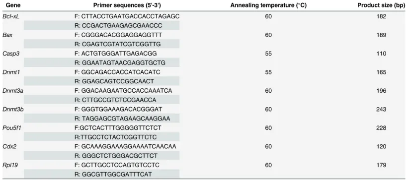
In Experiment 1. Reconstructed embryos treated with various concentrations of Scriptaid were examined to determine the optimal concentration. After activation, reconstructed embryos were cultured in PZM-5 medium supplemented with 0, 100, 300, or 500 nM Scriptaid for 20 h, and were then transferred to medium lacking Scriptaid. The percentage of embryos that developed to the blastocyst stage and the total number of cells per blastocyst were exam-ined in each group.
From this, 300 nM was selected as the optimal concentration of Scriptaid (Fig 1A and 1B). Reconstructed embryos were cultured with 300 nM Scriptaid for various amounts of time to determine the optimal duration of treatment. After activation, reconstructed embryos were cultured in medium supplemented with 300 nM Scriptaid for 0, 10, or 20 h, and were then transferred to medium lacking Scriptaid. The percentage of embryos that developed to the blas-tocyst stage and the total number of cells per blasblas-tocyst were examined in each group.
Experiment 2. Examined the effects of Scriptaid treatment on the number of apoptotic cells, the total number of cells, and the expression of apoptosis-related genes in blastocysts.In vitro-cultured embryos obtained as described in Experiment 1 were harvested at the blastocyst stage on day 7 and subjected to the TUNEL assay. The total number of nuclei per blastocyst was also counted. Blastocysts were washed in PBS and stored at−80°C until RT-PCR analysis.
In Experiment 3. To determine the levels of histone acetylation, histone methylation, and DNA methylation in SCNT embryos treated with Scriptaid, reconstructed embryos treated with or without 300 nM Scriptaid for 20 h were collected at the pronuclear stage (15 h). There-after, the fluorescence intensities of labeling for the epigenetic markers H3-acK9, H3-m3K9, 5hmc, 5mc andDnmt1were determined.
In Experiment 4. To investigate the effects of Scriptaid treatment on the relationship between DNA methylation and miRNA expression, the expression levels of DNA methyltrans-ferases (Dnmt1,Dnmt3a, andDnmt3b) and miRNAs (mir-29b, mir-148a, and mir-152) were determined in Scriptaid-treated and non-treated SCNT embryos.
In Experiment 5. To investigate the effects of Scriptaid treatment on mRNA and protein expression during early embryonic development, the expression levels of development-related genes and proteins (Pou5f1andCdx2) were determined in Scriptaid-treated and non-treated SCNT embryos.
Statistical analysis
Each experiment was repeated at least three times. All embryos were randomly allocated to a treatment group. Data were analyzed with a one-way analysis of variance and Tukey’s least sig-nificant test using GraphPad Prism 6 software. p<0.05 was considered significant.
Results
embryos that developed to the blastocyst stage was significantly higher among embryos treated with 300 or 500 nM Scriptaid than among those treated with 100 nM Scriptaid and non-treated embryos (300 nM, 23.50%; 500 nM, 25.78%; 100 nM, 16.48%; non-treated, 15.26%;Fig 1A). However, the percentage of embryos that developed to the blastocyst stage did not significantly differ between those treated with 300 nM Scriptaid and those treated with 500 nM Scriptaid. Similar results were observed for the total number of cells per blastocyst (Fig 1B), which was significantly higher among embryos treated with 300 nM or 500 nM Scriptaid than among those treated with 100 nM Scriptaid and non-treated embryos.
Based on these results, 300 nM was chosen as the optimal concentration of Scriptaid. Next, the effects of treatment with 300 nM Scriptaid for various amounts of time (0, 10, and 20 h) on thein vitrodevelopment of SCNT embryos were analyzed. The percentage of SCNT embryos that developed to the blastocyst stage was significantly higher among those treated with
Fig 1. Effect of treatment with Scriptaid at various concentrations (a and b) and for various amounts of time (c, d, and e) on the development of porcine somatic cell nuclear transfer embryos to the blastocyst stage.The numbers of embryos examined in each experimental group are shown in the bars. *indicates P<0.05 compared with the control group. Values are the mean (±standard deviation of the mean)
of four independent experiments. In E, embryos were not treated or were treated with 300 nM Scriptaid for 20 h. Ctrl: no treatment; SCR: Scriptaid treatment. Magnification, × 100.
Scriptaid for 20 h (24.90%) than among those treated with Scriptaid for 0 h (15.19%) or 10 h (17.02%) (Fig 1C and 1E). The total number of cells per blastocyst was significantly higher among Scriptaid-treated embryos (10 or 20 h) than among non-treated embryos (Fig 1D).
Effects of Scriptaid treatment on apoptosis-related gene expression in
the blastocysts
DNA fragments generated by apoptotic nicking of genomic DNA were measured in individual embryos using the TUNEL assay. The percentage of apoptotic cells in blastocysts was signifi-cantly lower in Scriptaid-treated embryos than in non-treated embryos (Fig 2A and 2B). The effects of Scriptaid treatment on expression of the apoptosis-related genesB-cell lymphoma-extra large (Bcl-Xl),Bcl-2-associated X protein (Bax), andCaspase3(Casp3) were determined in SCNT embryos at the blastocyst stage. In comparison with non-treated blastocysts, mRNA expression ofCas3andBaxwas significantly lower and mRNA expression ofBcl-xLwas signif-icantly higher in Scriptaid-treated blastocysts (Fig 2C).
Effects of Scriptaid treatment on histone acetylation, histone
methylation, and DNA modification
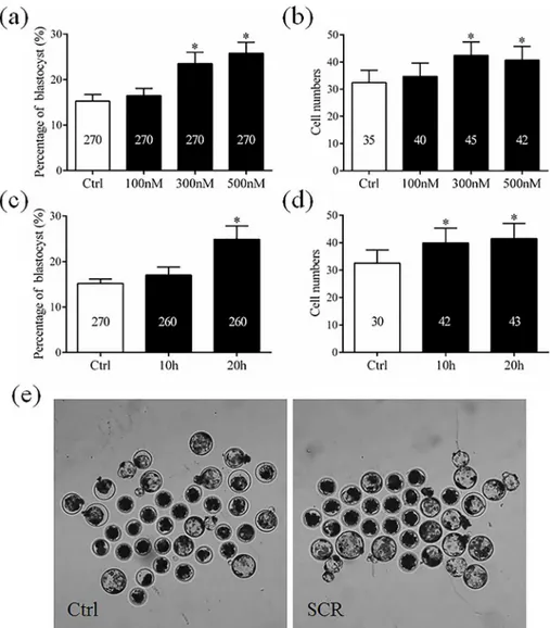
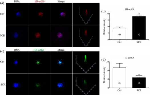
To analyze the mechanism underlying how Scriptaid treatment increases the developmental competence of SCNT embryos, levels of H3-acK9, H3-m3K9, 5mc, and 5hmc were examined in Scriptaid-treated SCNT embryos at the pronuclear stage. Scriptaid treatment significantly increased the level of H3-acK9 (Fig 3A and 3B) and significantly decreased the level of H3-m3K9 (Fig 3C and 3D) at the pronuclear stage. Meanwhile, conversion of 5mc into 5hmc was increased in Scriptaid-treated embryos (Fig 4A and 4B). Based on quantification of the fluorescence intensity ofDnmt1staining (Fig 5A and 5B), the level ofDnmt1at the pronuclear
Fig 2. Laser scanning confocal microscopy images of nuclei and fragmented DNA (×400, a and b) and relative mRNA expression levels ofBcl-xL,Bax, andCaspase3(Cas3) (c) in porcine blastocysts after 7 days ofin vitroculture, with or without Scriptaid treatment.The numbers of embryos examined in each experimental group are shown in the bars.*indicates P<0.05 compared with the control group. Values are
the mean (±standard deviation of the mean) of four independent experiments. Ctrl: no treatment; SCR:
Scriptaid treatment.
and blastocyst stage significantly decreased in Scriptaid-treated embryos. Furthermore, mRNA expression ofDnmt1was significantly lower in Scriptaid-treated embryos than in non-treated embryos (Fig 6A). To determine whether miRNAs are involved in downregulation of DNMT1, we compared the expression of miR-29b, miR-148a and miR-152 between Scriptaid-treated embryos and non-treated embryos. We found that the expression of 152, but not miR-29b and miR-148a, was significantly increased in Scriptaid-treated embryos, compared with non-treated embryos (Fig 6B).
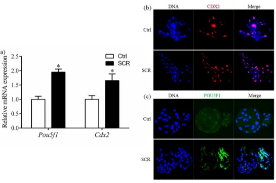
Effect of Scriptaid treatment on expression of POU5F1 and CDX2
mRNA and protein expression of two pluripotency-related genes, POU domain, class5, tran-scriptionfactor-1 (Pou5f1) and caudal type homeo box transcription factor 2 (Cdx2), was deter-mined in SCNT embryos at the blastocyst stage. mRNA expression ofPou5f1andCdx2was significantly higher in Scriptaid-treated embryos than in non-treated embryos (Fig 7A). Con-sistent with the mRNA results, protein expression of POU5F1 and CDX2 at the blastocyst stage was higher in Scriptaid-treated embryos than in non-treated embryos (Fig 7B).Discussion
Scriptaid is a novel HDACi that belongs to an existing class of hydroxamic acid-containing HDACis. In this study, we performed Scriptaid treatment after activation to improve epigenetic modifications in SCNT embryos and to increase their developmental competence.
Abnormal epigenetic reprogramming of donor nuclei after SCNT is thought to be the main cause of low cloning efficiencies. There are dynamic interactions between histone acetylation, histone methylation, and gene transcription [28–30]. Furthermore, previous studies have
Fig 3. Laser scanning confocal microscopy images and quantification of levels of histone H3 acetylated at K9 (H3-acK9, a and b) and histone H3 trimethylated at K9 (H3-m3K9, c and d) in embryos at the pronuclear stage, with or without Scriptaid treatment.Embryos were labeled for H3-acK9 (red), H3-m3K9 (green), and DNA (Hoechst 33342, blue). The numbers of embryos examined in each experimental group are shown in the bars.*indicates P<0.05 compared with the control group. Values are the mean (±standard deviation of the mean) of four independent experiments. Scale bar, 20μm. Ctrl: no treatment; SCR: Scriptaid
treatment. Magnification, × 400.
demonstrated that HDACi treatment could inhibit the histone deacetylase and results in hyperacetylation, which can expose DNA active binding sites and decrease DNA methylation that are important for gene activation [31]. K9 of histone H3 is an important site for both acet-ylation and methacet-ylation [32,33]. In the present study, treatment with 300 nM Scriptaid for 20 h enhanced the level of H3-acK9 and reduced the level of H3-m3K9 in SCNT embryos at the pronuclear stage. Hypoacetylation of K9 of histone H3 is one of the major causes of the low
Fig 4. Laser scanning confocal microscopy images (a) and quantification (b) of the relative levels of 5-methylcytosine (5mc) and 5-hydroxymethylcytosine (5hmc) in early embryos at the pronuclear stage, with or without Scriptaid treatment.5mc and 5hmc were detected using 5mc (red) and anti-5hmc (green) antibodies, respectively.*indicates P<0.05 compared with the control group. Values are the
mean (±standard deviation of the mean) of four independent experiments. Ctrl: no treatment; SCR: Scriptaid treatment. Magnification, × 400.
doi:10.1371/journal.pone.0134567.g004
Fig 5. Laser scanning confocal microscopy images (a and b) and quantification (c and d) of the relative levels ofDnmt1in early embryos at the pronuclear and blastocyst stage, with or without Scriptaid treatment.The numbers of embryos examined in each experimental group are shown in the bars. *indicates P<0.05 compared with the control group. Values are the mean (±standard deviation of the mean)
of four independent experiments. Ctrl: no treatment; SCR: Scriptaid treatment. Magnification, × 400.
cloning efficiencies of SCNT embryos in various species, including cattle [34], rabbits [35], and mice [23]. Our results suggest that Scriptaid treatment can significantly reduce the level of H3-m3K9 by increasing acetylation of K9 of histone H3. In addition, Scriptaid treatment increased conversion of 5mc into 5hmc.
In cloned embryos, the failure of DNA to be demethylated and remethylated causes incom-plete nuclear reprogramming [36]. DNA methyltransferaseDnmt1is important for the mainte-nance of DNA methylation in mammalian cells [37,38]. Accumulated evidence has been demonstrated thatDnmts mediated transcriptional silencing in mammalian cells [39].Dnmt1
is also required to maintain patterns of DNA methylation and histone acetylation in cloned
Fig 6. Relative mRNA levels of DNA methyltransferases (Dnmt1,Dnmt3a, andDnmt3b; a) and microRNAs (mir-29b,mir-148a, andmir-152; b) in porcine blastocysts after 7 days ofin vitroculture, with or without Scriptaid treatment.*indicates P<0.05 compared with the control group. Values are the
mean (±standard deviation of the mean) of four independent experiments. Ctrl: no treatment; SCR: Scriptaid treatment. Magnification, × 400.
doi:10.1371/journal.pone.0134567.g006
Fig 7. Relative mRNA expression levels (a) and laser scanning confocal microscopy images of immunostaining (b, c) for POU5F1 and CDX2 in porcine blastocysts after 7 days of in vitro culture, with or without Scriptaid treatment.*indicates P<0.05 compared with the control group. Values are the mean (±standard deviation of the mean) of four independent experiments. Ctrl: no treatment; SCR: Scriptaid
treatment. Magnification, ×400.
embryos; overexpression ofDnmt1causes abnormal embryonic development [40]. MicroRNAs (small non-coding RNAs, miRNA) play a critical role in maintenaning the pluripotent cell state and in the regulation of early mammalian development [39,41]. Recent studies have been indicating that miRNAs involved into the regulation of pluripotency, not only in the repro-gramming of embryos and somatic cells, but also participating the differentiation of stem cells [42,43]. In our previous study, we showed that the expression levels of mir-127 and mir-136 between normal fertilized mouse embryos and cloned embryos are different [18]. In addition, deletion and knockdown of Xist, a non-coding RNA that control X-chromosome inactivation in female mammals, can increase cloning efficiency greatly, and correct epigenetic reprogram-ming errors [44]. These findings suggested that miRNAs played an important role during reprogramming of somatic cell. A recent study reported that miRNAs are involved in DNA methylation via their regulation of Dnmts [45]. mir-29a [46,47], mir-148a, and mir-152 [48, 49] modulate Dnmt1 in cancer cells. In present study, we characterized the role of miR-152 in the regulation of DNA methylation in Scriptaid-treated porcine SCNT embryos for the first time. Our data showed that high level of miR-152 was associated with low Dnmt1 expression in Scriptaid-treated porcine SCNT embryos. Dnmt1 inhibition could be related with the over-expression of miR-152, meanwhile, resulting the conversion of 5mc into 5hmc to prevent hypermethylation. These findings indicated that miR-152 may have an indirect effect in DNA methylation. Therefore, we presume that Scriptaid treatment improves nuclear reprogramming in cloned embryos and its improvement of cloned embryo development might be owing to the correction of gene expression, including those encoding miRNAs.
Histone acetylases and deacetylases modulate the transcriptional activities of specific pro-moters by locally perturbing the chromatin structure, and increased histone acetylation can increase the transcriptional activities of genes [30]. In addition, methylation of H3K9 is caus-ally linked to the formation of heterochromatin and long-term transcriptional repression [29]. Therefore, decreased methylation of K9 of histone H3 and DNA might improve the transcrip-tional activities of genes. To determine whether Scriptaid activates the expression of pivotal genes through histone acetylation in SCNT embryos, we examined the mRNA and protein expression of two genes that play important roles during development, namely,Pou5f1and
Cdx2. Scriptaid treatment significantly increased the mRNA and protein levels of these two genes in porcine SCNT blastocysts, indicating that Scriptaid treatment can increase the tran-scriptional activities of genes in SCNT embryos. From our study, it could be implied that miR-152, which involved in the process of DNA demethylation of cloned embryos, will also initiate the expression ofPou5f1andCdx2. Further supports the idea that Scriptaid treatment improves the developmental capacity and nuclear reprogramming of SCNT embryos.
Apoptosis occurs frequently during early embryonic development and has a marked impact on embryo development [50]. Therefore, we investigated the expression of three apoptosis-related genes, namely,Bcl-xL,Bax, andCas3.Bcl-xl, which inhibits apoptosis, andCas3and
Bax, which promote apoptosis, belong to theBcl2family. At least 11Caspasegenes have been identified that mediate protein cleavage and induce apoptosis. Among these,Cas3executes apoptosis. In this study, expression ofCas3andBaxat the blastocyst stage was lower in Scrip-taid-treated embryos than in non-treated embryos. This indicates that Scriptaid treatment enhances the vitality of blastocysts. Moreover, expression of the apoptosis-inhibiting gene
Bcl-xLat the blastocyst stage was higher in Scriptaid-treated embryos than in non-treated embryos. TUNEL assay also showed that Scriptaid inhibited apoptosis in blastocyst. These result demonstrated that Scriptaid treatment improves the quality of the produced blastocysts.
embryos are suitable for use as control, as well as IVF embryos. However, there was extremely high possibility of polyspermic penetration during porcine IVF procedure [51,52]. It is unlikely to get monospermic embryos in blastocyst stage. Thus according to the methods from previous studies [19,21], non-treatment group was used as controls. We recommend treatment with 300 nM Scriptaid to improve the preimplantation development of porcine cloned embryos. Our findings demonstrate that this improvement is owing to enhanced epige-netic modification of somatic cells via Scriptaid-induced hyperacetylation and demethylation and upregulation of genes critical for SCNT embryonic development.
Acknowledgments
This work was supported by grants from the Next-Generation BioGreen 21 Program (PJ011126 and PJ01104103), Rural Development Administration, Republic of Korea.
Author Contributions
Conceived and designed the experiments: SL NHK XSC. Performed the experiments: SL JWC MHZ. Analyzed the data: SL XSC. Contributed reagents/materials/analysis tools: MHZ JWC. Wrote the paper: SL XSC. Discussed the results of the experiment: XSC NHK.
References
1. Wilmut I, Schnieke AE, McWhir J, Kind AJ, Campbell KH. Viable offspring derived from fetal and adult mammalian cells. Nature. 1997; 385(6619):810–3. doi:10.1038/385810a0PMID:9039911.
2. Wakayama T, Perry AC, Zuccotti M, Johnson KR, Yanagimachi R. Full-term development of mice from enucleated oocytes injected with cumulus cell nuclei. Nature. 1998; 394(6691):369–74. PMID:
9690471
3. Kato Y, Tani T, Sotomaru Y, Kurokawa K, Kato J-y, Doguchi H, et al. Eight calves cloned from somatic cells of a single adult. Science. 1998; 282(5396):2095–8. PMID:9851933
4. Onishi A, Iwamoto M, Akita T, Mikawa S, Takeda K, Awata T, et al. Pig cloning by microinjection of fetal fibroblast nuclei. Science. 2000; 289(5482):1188–90. PMID:10947985.
5. Ohgane J, Wakayama T, Senda S, Yamazaki Y, Inoue K, Ogura A, et al. The Sall3 locus is an epige-netic hotspot of aberrant DNA methylation associated with placentomegaly of cloned mice. Genes to Cells. 2004; 9(3):253–60. PMID:15005712
6. Kang YK, Koo DB, Park JS, Choi YH, Chung AS, Lee KK, et al. Aberrant methylation of donor genome in cloned bovine embryos. Nat Genet. 2001; 28(2):173–7. doi:10.1038/88903PMID:11381267.
7. Dean W, Santos F, Stojkovic M, Zakhartchenko V, Walter J, Wolf E, et al. Conservation of methylation reprogramming in mammalian development: aberrant reprogramming in cloned embryos. Proc Natl Acad Sci U S A. 2001; 98(24):13734–8. doi:10.1073/pnas.241522698PMID:11717434; PubMed
Cen-tral PMCID: PMC61110.
8. Santos F, Zakhartchenko V, Stojkovic M, Peters A, Jenuwein T, Wolf E, et al. Epigenetic marking corre-lates with developmental potential in cloned bovine preimplantation embryos. Current biology: CB. 2003; 13(13):1116–21. PMID:12842010.
9. Wang F, Kou Z, Zhang Y, Gao S. Dynamic reprogramming of histone acetylation and methylation in the first cell cycle of cloned mouse embryos. Biology of reproduction. 2007; 77(6):1007–16. doi:10.1095/
biolreprod.107.063149PMID:17823087.
10. De Ruijter A, Van Gennip A, Caron H, Kemp S, van Kuilenburg A. Histone deacetylases (HDACs): char-acterization of the classical HDAC family. Biochem J. 2003; 370:737–49. PMID:12429021
11. Tung EW, Winn LM. Valproic acid increases formation of reactive oxygen species and induces apopto-sis in postimplantation embryos: a role for oxidative stress in valproic acid-induced neural tube defects. Molecular pharmacology. 2011; 80(6):979–87. doi:10.1124/mol.111.072314PMID:21868484
12. Blackwell L, Norris J, Suto CM, Janzen WP. The use of diversity profiling to characterize chemical mod-ulators of the histone deacetylases. Life sciences. 2008; 82(21):1050–8.
13. Chuang D-M, Leng Y, Marinova Z, Kim H-J, Chiu C-T. Multiple roles of HDAC inhibition in neurodegen-erative conditions. Trends in neurosciences. 2009; 32(11):591–601. doi:10.1016/j.tins.2009.06.002
14. Glaser KB, Staver MJ, Waring JF, Stender J, Ulrich RG, Davidsen SK. Gene expression profiling of multiple histone deacetylase (HDAC) inhibitors: defining a common gene set produced by HDAC inhibi-tion in T24 and MDA carcinoma cell lines. Molecular Cancer Therapeutics. 2003; 2(2):151–63. PMID:
12589032
15. Bourassa MW, Ratan RR. The interplay between microRNAs and histone deacetylases in neurological diseases. Neurochemistry international, In Press. 2014.
16. Scott GK, Mattie MD, Berger CE, Benz SC, Benz CC. Rapid alteration of microRNA levels by histone deacetylase inhibition. Cancer research. 2006; 66(3):1277–81. PMID:16452179
17. Zhou R, Yuan P, Wang Y, Hunsberger JG, Elkahloun A, Wei Y, et al. Evidence for selective microRNAs and their effectors as common long-term targets for the actions of mood stabilizers. Neuropsychophar-macology. 2008; 34(6):1395–405. doi:10.1038/npp.2008.131PMID:18704095
18. Cui X-S, Zhang D-X, Ko Y-G, Kim N-H. Aberrant epigenetic reprogramming of imprinted microRNA-127 and Rtl1 in cloned mouse embryos. Biochemical and biophysical research communications. 2009; 379 (2):390–4. doi:10.1016/j.bbrc.2008.12.148PMID:19126398
19. Whitworth KM, Zhao J, Spate LD, Li R, Prather RS. Scriptaid corrects gene expression of a few aber-rantly reprogrammed transcripts in nuclear transfer pig blastocyst stage embryos. Cellular reprogram-ming. 2011; 13(3):191–204. doi:10.1089/cell.2010.0087PMID:21548827.
20. Zhou Y, Huang Y, Xie W, Song Q, Ji Y, Zhang Y, et al. Scriptaid affects histone acetylation and the expression of development-related genes at different stages of porcine somatic cell nuclear transfer embryo during early development. Chinese Science Bulletin. 2013; 58(17):2044–52. doi:10.1007/
s11434-013-5827-x
21. Zhao J, Ross JW, Hao Y, Spate LD, Walters EM, Samuel MS, et al. Significant improvement in cloning efficiency of an inbred miniature pig by histone deacetylase inhibitor treatment after somatic cell nuclear transfer. Biology of reproduction. 2009; 81(3):525–30. doi:10.1095/biolreprod.109.077016PMID:
19386991; PubMed Central PMCID: PMC2731980.
22. Wang L-J, Zhang H, Wang Y-S, Xu W-B, Xiong X-R, Li Y-Y, et al. Scriptaid improves in vitro develop-ment and nuclear reprogramming of somatic cell nuclear transfer bovine embryos. Cellular Reprogram-ming (Formerly" Cloning and Stem Cells"). 2011; 13(5):431–9.
23. Van Thuan N, Bui HT, Kim JH, Hikichi T, Wakayama S, Kishigami S, et al. The histone deacetylase inhibitor scriptaid enhances nascent mRNA production and rescues full-term development in cloned inbred mice. Reproduction. 2009; 138(2):309–17. doi:10.1530/REP-08-0299PMID:19433501.
24. Rizos D, Fair T, Papadopoulos S, Boland MP, Lonergan P. Developmental, qualitative, and ultrastruc-tural differences between ovine and bovine embryos produced in vivo or in vitro. Molecular reproduction and development. 2002; 62(3):320–7. PMID:12112595
25. Hao Y, Lai L, Mao J, Im G-S, Bonk A, Prather RS. Apoptosis and in vitro development of preimplanta-tion porcine embryos derived in vitro or by nuclear transfer. Biology of reproducpreimplanta-tion. 2003; 69(2):501–7.
PMID:12700186
26. Brill A, Torchinsky A, Carp H, Toder V. The role of apoptosis in normal and abnormal embryonic devel-opment. Journal of assisted reproduction and genetics. 1999; 16(10):512–9. PMID:10575578
27. Zhao MH, Kwon JW, Liang S, Kim SH, Li YH, Oh JS, et al. Zinc regulates meiotic resumption in porcine oocytes via a protein kinase C-related pathway. PloS one. 2014; 9(7):e102097. doi:10.1371/journal. pone.0102097PMID:25019390; PubMed Central PMCID: PMC4096513.
28. Geiman TM, Robertson KD. Chromatin remodeling, histone modifications, and DNA methylation—how
does it all fit together? Journal of cellular biochemistry. 2002; 87(2):117–25. PMID:12244565
29. Saccani S, Natoli G. Dynamic changes in histone H3 Lys 9 methylation occurring at tightly regulated inducible inflammatory genes. Genes & development. 2002; 16(17):2219–24.
30. Struhl K. Histone acetylation and transcriptional regulatory mechanisms. Genes & development. 1998; 12(5):599–606.
31. Bui HT, Seo HJ, Park MR, Park JY, Thuan NV, Wakayama T, et al. Histone deacetylase inhibition improves activation of ribosomal RNA genes and embryonic nucleolar reprogramming in cloned mouse embryos. Biology of reproduction. 2011; 85(5):1048–56. doi:10.1095/biolreprod.110.089474PMID:
21753193.
32. Xiong X, Lan D, Li J, Zhong J, Zi X, Ma L, et al. Zebularine and scriptaid significantly improve epigenetic reprogramming of yak fibroblasts and cloning efficiency. Cellular Reprogramming (Formerly" Cloning and Stem Cells"). 2013; 15(4):293–300.
33. Chung Y, Bishop CE, Treff NR, Walker SJ, Sandler VM, Becker S, et al. Reprogramming of human somatic cells using human and animal oocytes. Cloning and stem cells. 2009; 11(2):213–23. doi:10.
34. Cui X-S, Xu Y-N, Shen X-H, Zhang L-Q, Zhang J-B, Kim N-H. Trichostatin A modulates apoptotic-related gene expression and improves embryo viability in cloned bovine embryos. Cellular Reprogram-ming (Formerly" Cloning and Stem Cells"). 2011; 13(2):179–89.
35. Yang F, Hao R, Kessler B, Brem G, Wolf E, Zakhartchenko V. Rabbit somatic cell cloning: effects of donor cell type, histone acetylation status and chimeric embryo complementation. Reproduction. 2007; 133(1):219–30. PMID:17244748
36. Bourc'his D, Le Bourhis D, Patin D, Niveleau A, Comizzoli P, Renard J-P, et al. Delayed and incomplete reprogramming of chromosome methylation patterns in bovine cloned embryos. Current Biology. 2001; 11(19):1542–6. PMID:11591324
37. Yokomine T, Hata K, Tsudzuki M, Sasaki H. Evolution of the vertebrate DNMT3 gene family: a possible link between existence of DNMT3L and genomic imprinting. Cytogenetic and genome research. 2005; 113(1–4):75–80.
38. Bönsch D, Lenz B, Fiszer R, Frieling H, Kornhuber J, Bleich S. Lowered DNA methyltransferase (DNMT-3b) mRNA expression is associated with genomic DNA hypermethylation in patients with chronic alcoholism. Journal of neural transmission. 2006; 113(9):1299–304. PMID:16463117
39. Luczak MW, Jagodziński PP. The role of DNA methylation in cancer development. Folia histochemica et cytobiologica. 2006; 44(3):143–2. PMID:16977793
40. Kurihara Y, Kawamura Y, Uchijima Y, Amamo T, Kobayashi H, Asano T, et al. Maintenance of genomic methylation patterns during preimplantation development requires the somatic form of DNA methyl-transferase 1. Developmental biology. 2008; 313(1):335–46. PMID:18048024
41. Pauli A, Rinn JL, Schier AF. Non-coding RNAs as regulators of embryogenesis. Nature Reviews Genet-ics. 2011; 12(2):136–49. doi:10.1038/nrg2904PMID:21245830
42. Liao B, Bao X, Liu L, Feng S, Zovoilis A, Liu W, et al. MicroRNA cluster 302–367 enhances somatic cell
reprogramming by accelerating a mesenchymal-to-epithelial transition. Journal of Biological Chemistry. 2011; 286(19):17359–64. doi:10.1074/jbc.C111.235960PMID:21454525
43. Miyoshi N, Ishii H, Nagano H, Haraguchi N, Dewi DL, Kano Y, et al. Reprogramming of mouse and human cells to pluripotency using mature microRNAs. Cell stem cell. 2011; 8(6):633–8. doi:10.1016/j.
stem.2011.05.001PMID:21620789
44. Matoba S, Inoue K, Kohda T, Sugimoto M, Mizutani E, Ogonuki N, et al. RNAi-mediated knockdown of Xist can rescue the impaired postimplantation development of cloned mouse embryos. Proceedings of the National Academy of Sciences. 2011; 108(51):20621–6.
45. Sun X, He Y, Huang C, Ma T-T, Li J. The epigenetic feedback loop between DNA methylation and microRNAs in fibrotic disease with an emphasis on DNA methyltransferases. Cellular signalling. 2013; 25(9):1870–6. doi:10.1016/j.cellsig.2013.05.013PMID:23707521
46. Pandey M, Sultana S, Gupta KP. Involvement of epigenetics and microRNA-29b in the urethane induced inception and establishment of mouse lung tumors. Experimental and molecular pathology. 2014; 96(1):61–70. doi:10.1016/j.yexmp.2013.12.001PMID:24361357
47. Qin H, Zhu X, Liang J, Wu J, Yang Y, Wang S, et al. MicroRNA-29b contributes to DNA hypomethyla-tion of CD4+ T cells in systemic lupus erythematosus by indirectly targeting DNA methyltransferase 1. Journal of dermatological science. 2013; 69(1):61–7. doi:10.1016/j.jdermsci.2012.10.011PMID:
23142053
48. Xu Q, Jiang Y, Yin Y, Li Q, He J, Jing Y, et al. A regulatory circuit of miR-148a/152 and DNMT1 in modu-lating cell transformation and tumor angiogenesis through IGF-IR and IRS1. Journal of molecular cell biology. 2012;no(0):1–11.
49. Azizi M, Teimoori-Toolabi L, Arzanani MK, Azadmanesh K, Fard-Esfahani P, Zeinali S. MicroRNA-148b and microRNA-152 reactivate tumor suppressor genes through suppression of DNA methyltrans-ferase-1 gene in pancreatic cancer cell lines. Cancer biology & therapy. 2014; 15(4):419–27.
50. Haouzi D, Hamamah S. Pertinence of apoptosis markers for the improvement of in vitro fertilization (IVF). Current medicinal chemistry. 2009; 16(15):1905. PMID:19442154
51. Nagai T, Takahashi T, Masuda H, Shioya Y, Kuwayama M, Fukushima M, et al. In-vitro fertilization of pig oocytes by frozen boar spermatozoa. Journal of reproduction and fertility. 1988; 84(2):585–91.
PMID:3199378
52. Funahashi H. Polyspermic penetration in porcine IVM–IVF systems. Reproduction, Fertility and